eISSN: 2377-4304


Case Report Volume 13 Issue 5
1Department of Gynecology and Obstetrics, OSI Bilbao-Basurto, Basurto University Hospital, Bilbao, Spain
2Department of Anesthesiology and Resuscitation. Basurto University Hospital, Bilbao, Spain
Correspondence: Angela Palenzuela Blasco, MD, Department of Gynecology and Obstetrics, OSI Bilbao-Basurto, Basurto University Hospital (Iturrizar Pavilion), Montevideo Avenue No. 18, 48013-Bilbao, Spain
Received: September 07, 2022 | Published: September 19, 2022
Citation: DOI: 10.15406/ogij.2022.13.00663
Anti-NMDA receptor antibody encephalitis usually develops as a characteristic syndrome with a multiphasic course and a broad differential diagnosis. This type of encephalitis may be associated with a tumor, and therefore be considered a paraneoplastic syndrome, predominantly affecting women, with ovarian teratoma being the most frequently involved tumor.
Clinical case: A 35-year-old patient with a prominent neuropsychiatric condition with a final diagnosis of limbic encephalitis due to anti-NMDAr antibodies secondary to ovarian teratoma, with complete remission of the condition after multimodal treatment.
Discussion: This type of pathology represents a true diagnostic-therapeutic challenge. It is important to know the epidemiology of this disease in order to avoid delays in diagnosis and potential curative treatment.
Keywords: ovarian teratoma, autoimmune encephalitis, NMDA, paraneoplastic, differential diagnosis
In 2007, a type of limbic encephalitis related to antibodies against the NMDA receptor (NMDAR) was discovered.1 This is a cell membrane receptor with cryptic roles in synaptic transmission and neuronal plasticity.2
The immunological attack on this receptor produces a characteristic clinical picture with symptoms that affect several systems and develop in phases in a predictable manner. Following a prodromal illness that may include headache, fever, and respiratory or digestive tract symptoms, patients develop prominent psychiatric symptoms (agitation, mania, hallucinations, paranoia) that usually precede seizures and progress to a rapidly deteriorating level of consciousness. mutism, catatonia, abnormal facial, trunk, or limb movements, and autonomic disturbances.3–5
Diagnosis is made by detection of antibodies against subunit 1 (NR1) in blood or cerebrospinal fluid.6
The syndrome usually affects young patients. The association with tumors depends on age and sex. It is more frequent in women (80%) between 18 and 45years old, who in 56% of cases present ovarian teratoma.4 In men, when this occurs, it is usually related to a testicular seminoma. Other types of tumors that have been linked to anti-NMDAr encephalitis include lung cancer, thyroid cancer, breast cancer, colon cancer, and neuroblastoma.
Ovarian tumors are common findings in women. Germinal neoplasms represent approximately 20-25% and teratomas are the most common type. They are classified into four categories: mature, immature, malignant and monoderm. Almost all are cystic and 10-15% are bilateral. They may contain embryonic tissue from all 3 layers (ectoderm, mesoderm, and endoderm). The detection of ovarian teratomas in these patients is age-dependent: approximately 50% of women over 18 years of age present unilateral or bilateral teratomas secreting antibodies against the NMDA receptor, while less than 9% of those under 14 years of age present this. tumor type. It is more common in black patients.
First-line treatment includes tumor resection, immunotherapy (steroids, immunoglobulins, plasmapheresis), or a combination of both.3 Resistant cases can be treated with cyclophosphamide or rituximab. If the patient does not respond favorably to conventional medical treatment, after ruling out infectious encephalitides, paraneoplastic causes should be considered. Symptoms respond to both tumor treatment, if present, and immunotherapy.3,7,8
Despite the seriousness and significant neurological deterioration, the condition is potentially reversible, with improvement of symptoms in reverse chronology to the phases of presentation.
A 35-year-old white woman, with 2 pregnancies and 2 eutocic deliveries without incident, and with no medical history of interest.
He went to the emergency room due to a sudden onset of insomnia, restlessness, racing thoughts, anxiety and delusional ideation. This symptomatology progressed into a picture of hallucinations and disinhibition, for which she was admitted to the Psychiatry service. After several days in which the agitation, dysautonomia, catatoniform syndrome and decreased level of consciousness progressed, the patient was transferred to the resuscitation service.
After the detection of anti-NMDA antibodies in CSF, the patient was diagnosed with autoimmune encephalitis due to anti-NMDAr antibodies, which under sedation and connection to mechanical ventilation is expressed as refractory status epilepticus. Treatment with immunotherapy (corticosteroids, immunoglobulins and plasmapheresis) was started.
After diagnosis, in accordance with the epidemiology of this entity, screening begins in search of the primary tumor.
The following imaging tests were performed, with the results described:
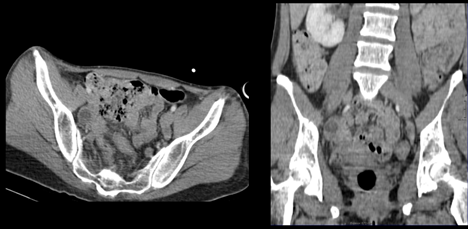
Figure 1 Abdominal-pelvic CT.
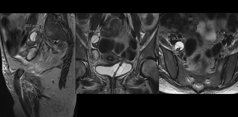
Figure 2 Pelvic MRI.
All this avascular compatible with a probable benign dermoid ovarian tumor. Given the findings and the progressive worsening of the patient despite first-line treatment, it was decided to perform an exploratory laparoscopy and act according to the findings. The surgical findings were normal, both the uterus and both adnexa as well as the rest of the abdomino-pelvic cavity. Based on the imaging tests and the high incidence of occult teratoma, it was decided to perform a right adnexectomy with intraoperative biopsy, which reported a mature cystic teratoma with a maximum dimension of 2.1cm.
Histopathological analysis of the teratoma showed a predominance of mature, dysplastic and dysmorphic neurons, most of them multinucleated and with a very high proliferation index (Figure 3).
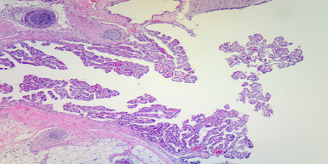
Figure 3 Predominance of neural tissue in the pathological study.
Days later, after surgical removal, the patient was discharged and transferred to a long-term care facility. She was discharged with no discernible neurological sequelae at 6 months.
Patients with suspected limbic encephalitis represent a true diagnostic challenge.5 On suspicion in young women.
The existence of an ovarian teratoma must be ruled out through imaging studies, always bearing in mind that gynecological ultrasound is the most accurate imaging test in adnexal lesions.
The role of the tumor in the production of anti-NMDAr encephalitis is not fully understood and is currently under investigation. What is known is that most tumors associated with anti-NMDAr encephalitis contain mostly neural tissue, and NMDA receptors, as our pathology analysis clearly showed.9 Antibodies that are initially formed against NMDA receptors are presumed to be found within tumors, and then attack similar-looking receptors in the brain, producing the symptoms and signs associated with anti-NMDAr encephalitis. It is important to know that the immune response to treatment does not depend on the size of the tumor but on the proportion of neural tissue in it.
The current recommendation, although not without controversy, is to perform bilateral laparoscopic adnexectomy if imaging tests are inconclusive in revealing the presence of a tumor in those patients who experience sustained deterioration.10 This is based on the relatively high frequency of ovarian teratomas in women with this type of encephalitis and the possibility of a microscopic tumor, such as the one we describe in this case. However, the treatment of this condition is specific to each case and clinical decisions must be made on an individual basis. Tumor removal within the first four months after the onset of symptoms is the main recovery factor.
The recurrence rate after conservative treatment is 15-25% and often occurs within the first year of primary treatment; therefore, postoperative follow-up should be close. Adequate gynecological follow-up is important for these patients that includes an annual transvaginal ultrasound for 5years in those patients who have undergone conservative surgery. Likewise, it is essential to maintain continuous monitoring in those patients who are undergoing immunosuppressive treatment (especially if they are receiving azathioprine or mycophenolate mofetil, both category D drugs in pregnancy). These patients must have a pregnancy test prior to treatment and hence consistent with the clinical correlation in the rest of the visits. For future pregnancies, it is recommended to wait at least six months after stopping treatment, especially due to the delayed effect of mycophenolate mofetil. LARC methods should be considered in these patients, especially those in whom cognitive disorders persist.
As a conclusion to this case, it is important to point out that the pathology that diagnosis in patients with suspected limbic encephalitis is difficult and that diagnosis is still usually delayed in most cases. The relative ignorance of the disease is only one of the causes. The clinical characteristics of its form of presentation, as well as the low specificity of the usual analytical and radiological tests, often lead to confusion. The delay in the diagnosis and treatment of our patient depended on the multidisciplinary approach of several specialists (psychiatry, anesthesia and resuscitation, radiology, pathological anatomy, rehabilitation, gynecology and obstetrics).
None.
The research work is financed by the Research Commission of the OSI BILBAO BASURTO.
Author declares there is no conflict of interest exists.

©2022 , et al. This is an open access article distributed under the terms of the, which permits unrestricted use, distribution, and build upon your work non-commercially.