eISSN: 2377-4304


This is a case report of a 31-year old woman who presented with an incidental finding of a large cervical fibroid in pregnancy. She was closely monitored antenatally. The cervical fibroid increased in size as the pregnancy progressed. Magnetic resonance imaging of the pelvis suggested low risk of malignancy despite the rapid growth of the cervical fibroid. She was managed conservatively throughout her pregnancy and subsequently underwent an elective caesarean section to deliver a baby girl. Postnatally, the cervical fibroid shrunk minimally and the patient underwent a laparotomy myomectomy 7 months later. The surgery was complicated by severe haemorrhage, which resulted in a total hysterectomy
Keywords: Cervical fibroid, Pregnancy, Myomectomy, Hysterectomy
MRI, Magnetic Resonance Imaging
Fibroids in pregnancy are increasingly becoming more common with a prevalence of approximately 10.7%.1 Cervical fibroids in pregnancy is rarer with a prevalence of <1%.2 Depending on the size of the fibroids, it may potentially cause complications in all three trimesters of pregnancy and in the antepartum, intrapartum and postpartum period. With this, there is a need for obstetricians to be vigilant throughout the entire pregnancy. Literature about cervical fibroids in pregnancy is limited to case reports and case series; as such there are no clear guidelines on how to manage these patients. We report a case of a woman who had an incidental finding of a cervical fibroid that grew rapidly in pregnancy and her outcomes postnatally.
A 31-year old primigravida booked early in her pregnancy at 13 weeks gestation. At her booking visit with her private obstetrician, the dating ultrasound showed that she had a large cervical fibroid 11.9 x7.8cm in size. Subsequently, the woman transferred her care and booked at our tertiary institution at 16 weeks gestation. At 18 weeks gestation, the cervical fibroid had increased to 15.0 x14.4 x8.9cm in size. Her fetal anomaly scan at 21 weeks gestation was otherwise normal, but the fibroid increased in size gradually during her antenatal visits. At 33 weeks gestation, Magnetic Resonance Imaging (MRI) pelvis was performed and ruled out sarcomatous changes in the fibroid; however the cervical fibroid was then 23.0 x14.2 x 23.2cm (Figure 1). Serial ultrasound scans indicated that the fetus’ growth was appropriate for gestation but the fetus remained in breech presentation most of her pregnancy. The woman remained asymptomatic except for one episode of non-specific abdominal pain in her second trimester, which was conservatively managed with oral analgesia.
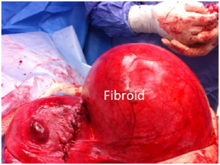
The woman was counseled regarding the risk of preterm labour and as such, an elective caesarean section was planned at 35 weeks and 5 days gestation. Her case was co-managed with the maternal fetal medicine specialists, neonatologists and anaesthetists and discussed at our multidisciplinary meeting. Intramuscular steroids were administered prior to surgery. Intra-operatively, a midline incision was made and a lower segment caesarean section was performed. A baby girl weighing 2460 grams with Apgar scores of 9 at 1 minute, 9 at 5 minutes was delivered via breech extraction. A large 23 cm cervical fibroid was seen occupying the pelvis and the uterus was pushed upwards towards the right side (Figure 2 & 3). The caesarean section was uneventful and she recovered well with no complications.

Figure 3 Image showing the uterus repaired in 2 layers with the large cervical fibroid pushing the uterus upwards and to the right side.
Post delivery, she had serial ultrasound surveillance of the cervical fibroid. At 2 months post-delivery, the fibroid size was 20.0 x18.2 x 13.5cm and 4 months later; there was no change in the size of the fibroid. MRI Pelvis was also repeated indicating that the fibroid was 19.4x10.5x21.4cm, which was slightly smaller than before (Figure 4). However, in view of the persistence in size of the cervical fibroid, the patient was counseled for open myomectomy 7 months post-delivery but was extensively counseled that a hysterectomy may occur due to the position of the cervical fibroid. Medical treatment was considered. However, the patient was keen for definitive management.
Intra-operatively, an open myomectomy was intended for a 20 x15cm cervical fibroid. However, there was profuse bleeding from the broad ligament and the left uterine artery, which was supplying the cervical fibroid(Figure 5). As such, the team proceeded with a total hysterectomy and bilateral salpingectomy to cease the haemorrhage. The estimated blood loss was 1500ml and she received 2 units of blood transfusion. She had an uncomplicated recovery otherwise. Histology revealed a degenerative benign leiomyoma weighing 2375g and measuring 21 x 19 x 13cm.
Cervical fibroids in pregnancy can be challenging for obstetricians to manage especially in our case where the fibroid had occupied almost the entire pelvis, which would not allow a vaginal delivery to be possible. Cervical fibroids may cause non-cephalic presentation of the fetus, obstructed labour, infection, pain, urinary or bowel symptoms and bleeding.3 It is important to anticipate the issues that could be faced in pregnancy and counsel patients adequately to seek for multidisciplinary help early. Most high risk pregnancies like this should be managed in a tertiary centre where blood products and interventional radiology services should be easily accessible.
Women who have fibroids should be treated conservatively antenatally. Close monitoring and assessment of the size of the fibroids will determine if a vaginal delivery will be suitable. It is also important to rule out malignancy if there is a solitary fibroid growing rapidly. MRI is a recommended tool to rule out any sarcomatous change and is also useful to delineate the location of the fibroid.1
Obstructed labour is one of the major concerns with fibroids in pregnancy. Depending on the size and the anatomy of the fibroid, a caesarean section should be advised accordingly. Advanced planning ahead of surgery will be ideal to prevent complications from arising. A hysterectomy may be considered if severe haemorrhage occurs, as the larger the fibroid, the higher the risk of bleeding may occur.4
Postnatally, the patient should be closely followed up to ensure that the fibroids have shrunk to its original size. However, as in our case, the fibroid did not shrink and surgery should be advised as cervical fibroid may decrease the chances of fertility due to the obstructed canal and if grows in size, can cause worsening pressure symptoms.
It is therefore important for obstetricians to be aware of the complications associated and the challenges faced with fibroids in pregnancy.
We would like to thank Dr Serene Thain for assisting in the photography of the figures for the article.
None.

© . This is an open access article distributed under the terms of the, which permits unrestricted use, distribution, and build upon your work non-commercially.