eISSN: 2377-4304


Background:Dermatofibrosarcoma protuberans (DFSP) is a low-to-intermediate grade sarcoma of dermal origin1-3 that rarely presents in the vulva.1,2 It typically presents on the trunk of young to middle-aged adults.4 Even though it has a low potential for distant metastases, it often recurs locally.5 Histopathologically, DFSP typically consists of spindle cells in a “storiform” pattern and stain positive for CD34, which can aid in establishing a diagnosis.1,6-8 The traditional treatment of choice has been wide surgical excision, but multiple resections are often required for clearance of the lesion.9 Mohs micrographic surgery (MMS) has recently been described as an alternative treatment option to help decrease the rate of recurrence.
Objective: To present a case of dermatofibrosarcoma protuberans of vulva in a young patient treated with excisional surgery with a systematic review of the condition.
Case:This patient is a 35-year-old female, G0P0, from The Republic of the Congo (Central Africa), who presented with an eight-year history of a “large left keloid of the mons pubis”. The patient stated the mass was non-tender and the size of a ping-pong ball that changed in size. The patient underwent surgical excision of the “large left keloid of mons pubis”. The specimen consisted of a 6 x 3 x 3 cm elevated portion of smooth tan skin with subcutaneous tissue. It was determined that the specimen was composed of extensive, ill-defined, firm, smooth, homogeneous tan tissue and was sent to pathology for further evaluation. The pathological specimen findings were “Dermatofibrosarcoma protuberans with positive margin”. A second surgery was performed to excise the positive margins of DFSP that remained. Review of an intraoperative frozen specimen found no tumor in the tissue margins. The patient had an uneventful postoperative course and was discharged two days after the surgery was performed.
Conclusion: Dermatofibrosarcoma protuberans (DFSP) infrequently involves the vulva and should be considered in the differential diagnosis of other spindle cell lesions presenting in this unusual site.10,11 The role of immunohistochemical staining with CD34 is imperative in establishing the diagnosis. The rate of local recurrence is high, but it rarely shows metastasis.12 Surgical excision is the treatment of choice and close follow-up to detect recurrence is imperative.1
Keywords: bednar tumor, CD34 positive, dermatofibrosarcoma, dermatofibrosarcoma protuberans (DFSP), factor XIIIa negative, vulvar carcinoma
Dermatofibrosarcoma protuberans (DFSP) is a low-to-intermediate grade sarcoma of dermal origin1-3 that rarely presents in the vulva.1,2 It is an uncommon soft tissue tumor that has a high local recurrence rate. The incidence of DFSP is 0.1% of all cancers and 1% of all soft tissue sarcomas.4 It most commonly appears on the trunk or proximal extremities of young to middle aged adults. Surgical excision is the treatment of choice and early recognition is extremely important because of the excellent prognosis following adequate excision.13 We report an unusual case of this tumor involving the vulva.1
This patient is a 35 years old female, G0P0, from The Republic of the Congo (Central Africa), who presented with an eight year history of a “large left keloid of mons pubis”. The patient stated the mass was non-tender and the size of a ping-pong ball with a changing size. The patient’s past surgical history was significant for two abdominal myomectomies in 2001 and in 2009 for symptomatic uterine leiomyomas. Her past medical history was significant for infertility, large uterine fibroids and oligomenorrhea after a myomectomy in 2009.
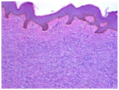
The patient underwent surgical excision of the “large left keloid of mons pubis”. The specimen consisted of a 6 x 3 x 3 cm elevated portion of smooth tan skin, with subcutaneous tissue. It was determined that the specimen was composed of extensive, ill-defined, firm, smooth, homogeneous tan tissue and was sent to pathology for further evaluation. The pathological specimen results came back as “Dermatofibrosarcoma protuberans with positive margin” (Figure 1). Subsequent excisional biopsy of the mass was performed.
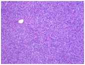
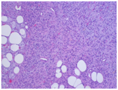
Histologic evaluation of the specimen showed a spindle cell lesion consisting of fibroblast-like cells arranged in a storiform pattern1,6-8 (Figure 2). On average, there were 2 to 3 mitotic figures per 10 high power field (HPF). The neoplastic cells showed extension into the surrounding fibro adipose tissue14,15 (Figure 3). Based on the morphologic staining pattern, a diagnosis of Dermatofibrosarcoma protuberans (DFSP) was rendered.1 The patient underwent a second resection of positive margin for Dermatofibrosarcoma protuberans of vulva (Figure 4). Wide local excision of tumor positive margin performed (Figure 5). An intraoperative frozen specimen was sent to pathology with negative margins (Figure 6). The patient had an uneventful postoperative course and was discharged on postoperative day two. Patient will be followed up in 6 months and then annually for revelation of possible recurrence.

Figure 1 Histopathologic Slide, epidermis, basal lay and Tumor cells, Hematoxylin and Eosin Stain (H&E stain). Low power view demonstrating the tumor, The epidermis appeared thinned.32

Figure 2 Histopathologic Slide, Tumor cells demonstrating a storiform pattern, Hematoxylin and Eosin Stain (H&E stain).

Figure 3 Histopathologic Slide, Tumor cells infiltrating into the subcutaneous fat, Hematoxylin and Eosin Stain (H&E stain).
There are a paucity of data describing voiding trial failure rates among patients undergoing minimally invasive sacrocolpopexy, and even fewer studies examining preoperative risk factors in this specific patient population. In our study of 56 patients, 8.9% of patients undergoing a minimally invasive sacrocolpopexy failed a backfill TOV on postoperative day 1. This is consistent with observed voiding trial failure rates following minimally invasive procedures in gynecologic literature (7.1% to 18.7%).11,12 To our knowledge, there exists only one other study specifically describing voiding trial failure rates after minimally invasive sacrocolpopexy.13 This retrospective cohort study of 290 patients at a single institution over a 3 year period reported a 27.2% failure rate, however, this cohort included patients who underwent concomitant mid-urethral slings, which are known risk factors for postoperative urinary retention (and were excluded from our study for this reason specifically). In terms of voiding trial failure rates after vaginal prolapse surgery, rates ranging from 29% to 32% have been observed; much higher than those seen in our study.4 This is potentially explained by increased edema and tissue destruction surrounding the urethra caused by aggressive dissection and manipulation required by extensive vaginal surgery. These studies also include patients with concomitant anti-incontinence procedures.
The main strength of this study was the focus on risk factors that could be identified at a preoperative visit, such that the patient provider could have the opportunity to teach self-catheterization techniques in order to mitigate the risks associated with an indwelling catheter. Risk factors for voiding trial failure, in general, are multi-factorial, including those occurring preoperatively, perioperatively, and postoperatively. Perioperative and postoperative risk factors, such as estimated blood loss, intravenous fluid administered, perioperative and postoperative analgesic and sedative medication administered, extensive surgical dissection resulting in disruption of nerve plexuses, and tissue edema causing obstruction of voiding, have all been described in the literature.2,5,14 As these factors are not readily available preoperatively, these were not examined in depth in this study. An attempt was made, however, to look at surrogate markers that may predict some of these risk factors. These include chronic pain syndromes (leading to need for higher doses of analgesic and sedative medication), a history of pelvic surgery and concomitant vaginal surgery, including anterior and posterior repair (contributing to tissue edema and nerve disruption from more extensive dissections or distorted anatomy secondary to scarring), planned concomitant hysterectomy (more surgical steps leading to higher blood loss and intraoperative complications), etc. Other preoperative risk factors examined included age, BMI, menopausal status, parity and prolapse stage, as these can reflect anticipated surgical difficulty.
Our study was unable to find any linkage between the patient variables analyzed and failure of a postoperative voiding trial. Concomitant vaginal surgery did closely approach significance, however, with a p-value of 0.058. Data are mixed regarding the effect of an anterior repair on voiding trial failure, with some studies noting an association3, while other have been unable to do so4. In the latter study, a higher prevalence of voiding dysfunction in patients with a high grade cystocele and the application of a posterior repair was also been observed.4 Geller et al.13 examined a wide variety of procedures including vaginal, abdominal, laparoscopic and robotic, while Hakvoort et al.4 examined exclusively vaginal procedures, thus, results may not be entirely generalizable to our patient population. In the aforementioned study by Turner et al.13 examining preoperative risk factors for voiding dysfunction in patients undergoing a minimally invasive sacrocolpopexy, none of the 290 patients who underwent a minimally invasive sacrocolpopexy had a concomitant vaginal surgery, thus the potential for this variable to cause postoperative voiding dysfunction was unable to be analyzed.
A significant proportion of our patients underwent preoperative urodynamic testing, and each was determined not to need a concomitant anti-incontinence procedure on this basis, as well as by history and physical exam. Unfortunately, this information was missing in enough patients to be deemed unsuitable to include in the analysis. While it is well documented that preoperative urodynamic abnormalities do not predict postoperative voiding dysfunction, preoperative voiding dysfunction is a known predictor, and by eliminating the subset of these patients with this a priori confounder, our results are strengthened.10,15,16
The major limitation of this study is small sample size. Any statistical significance is difficult to achieve with a small patient population, thus, repeating this pilot study with a larger cohort would be beneficial. In addition, as subjects undergoing concomitant anti-incontinence procedures were excluded, our results may not be generalizable to that specific patient population. Another inherent limitation of this study is the multi-factorial nature of postoperative voiding trial failure, as previously mentioned. Without the ability to accurately predict and quantify many perioperative and postoperative variables, it is very difficult to assess the impact of these variables on the results of our study.
Dermatofibrosarcoma protuberans (DFSP) infrequently involves the vulva and should be considered in the differential diagnosis of other spindle cell lesions presenting in this unusual site.37 The role of immunohistochemical staining with CD34 is imperative in establishing the diagnosis.14 The rate of local recurrence is high, but rarely are metastatic lesions present.38,39 The infrequent metastasis of fibrosarcoma associated with DFSP may be secondary to its superficial location, low cytologic grade, and small size.7 Surgical excision is the treatment of choice and close follow-up to detect recurrence is necessary.1

© . This is an open access article distributed under the terms of the, which permits unrestricted use, distribution, and build upon your work non-commercially.