Open Access Journal of
eISSN: 2575-9086


Research Article Volume 8 Issue 1
 Luciana Karen Calábria
Federal University of Uberlândia, Pontal Campus, Institute of Exact and Natural Sciences of Pontal, Ituiutaba, MG, Brazil
Correspondence: Luciana Karen Calábria, Federal University of Uberlândia, Pontal Campus, Institute of Exact and Natural Sciences of Pontal, Ituiutaba, MG, Rua 20, 1600, Tupã, CEP 38304-402, Ituiutaba, MG; Brazil
Received: November 25, 2025 | Published: December 9, 2025
Citation: Souza NCG, Rezende AAA, Calábria LK. Leptin and obesity: in silico genetic and biochemical characterization. Open Access J Sci. 2025;8(1):303-308. DOI: 10.15406/oajs.2025.08.00277
Obesity is a multifactorial chronic disease characterized by the expansion of adipose tissue, which performs an endocrine function by synthesizing and secreting leptin, a fundamental peptide hormone in the regulation of satiety through hypothalamic action. The present study aimed to describe the genetic and biochemical characteristics of leptin using bioinformatics databases and software, emphasizing its structure, function, signaling pathways, and its relationship with obesity. Leptin is encoded by the LEP gene (locus 7q32.1), translated into 167 amino acids, expressed in various organs and tissues, with a deposited three-dimensional structure and binding sites that determine its function. Its main signaling pathways include JAK-STAT and AMPK, which are involved in regulating energy homeostasis. The results corroborate leptin's central role in energy metabolism and highlight its potential as a therapeutic target for the treatment of obesity and its comorbidities.
Keywords: energy metabolism, hormonal signaling, bioinformatics
According to data from the World Health Organization (WHO), global obesity has been increasing considerably since 1975. In 2016, approximately 2 billion adult individuals worldwide were overweight, among them more than 650 million were already considered obese. Another alarming fact is that 39 million children, in the age group under 5 years old, were overweight or obese in 2020.1 According to Vigitel Brazil,2 more than 448 thousand Brazilians residing in capital cities, aged over 18, were overweight and more than 175 thousand were considered obese (Brasil, 2022).
Obesity is a disease that causes an accumulation of excess fat in adipose tissue, characterizing a dysfunction of energy metabolism.3 It goes far beyond aesthetic standards and numbers on the scale, as it is a chronic disease of a multifactorial nature, meaning it involves various individual or collective, political, socioeconomic, cultural, historical, psychosocial, and biological factors, affecting adolescents, adults, children, and the elderly.4
Recent advances in the areas of endocrinology and metabolism have shown that, contrary to what was thought a few years ago, adipocytes are not just energy-storing cells, but an endocrine tissue that synthesizes and releases various substances, leptin being one of them.5
Leptin is a protein that plays a regulatory role in various body systems, including the immune, respiratory, and reproductive systems, as well as energy balance via hypothalamic action.6 Since the discovery of leptin, studies have shown considerable progress in characterizing the mechanisms that control food intake by the hypothalamus, revealing complex systemic details and providing new perspectives on specific pharmacological treatments.
According to Sternson and Eiselt,7 understanding the neural pathways involved in appetite signaling is as important as the increase in obesity diagnoses. Furthermore, the authors highlight three interacting but completely distinct pathways: the Agouti-related protein (AGRP) expressed in neurons present in the hypothalamic arcuate nucleus (ARC) that are activated by hormones such as leptin and ghrelin; the feeding-related neurons of the lateral hypothalamus (LH); and the neurons of the parabrachial nucleus (PBN) involved with the calcitonin gene-related peptide (CGRP) which cause feeding suppression.
In a review, Chaput et al.8 highlight the impact of sleep and circadian rhythm dysregulation on the appetite hormones leptin, ghrelin, and peptide YY (PYY), related to obesity in humans, describing how the pathology of obesity and also overweight is multicausal and therefore complex to treat, emphasizing not only hypotheses related to leptin receptors but also to the integrity of the blood-brain barrier of obese individuals due to triacylglycerol levels.
The entry of leptin into the central nervous system (CNS) is conditioned by the presence of the blood-brain barrier (BBB), occurring through a mechanism of transcytosis mediated by leptin receptors (LEPR). In peripheral organs that are not protected by the BBB, such as circumventricular organs, the hormone accesses specific neurons directly. Leptin's intracellular signaling primarily depends on the JAK2/STAT3 pathway, in which the JAK2 kinase phosphorylates IRS proteins (insulin receptor substrate), activating several cascades, such as the PI3K/phosphatidylinositol 3-kinase related to insulin production. Besides the CNS, leptin receptors, including LEPR and its short and long isoforms, are expressed in peripheral tissues such as the liver, pancreas, heart, and gastrointestinal tract, indicating a systemic action of the hormone in metabolic regulation.9
Furthermore, obesity is classified as a risk factor for cardiovascular diseases, musculoskeletal disorders, diabetes, and some types of cancer, thus affecting not only the individual's physical health but also mental health, involving prejudice and discrimination that directly affect the emotional well-being of people who are overweight or obese.1
The diagnosis of overweight and obesity is still also made using the Body Mass Index (BMI), considering weight in kilograms and height in square meters (Kg/m²), with a BMI greater than or equal to 25 considered overweight for adults, and a BMI greater than or equal to 30 considered obesity.1
BMI has long been used as the main calculation basis for identifying overweight and obesity, but an ideal value is not always obtained, and this value is rarely necessary when aiming for an improvement in the individual's clinical condition. In this sense, specialists are proposing a change in the classification of obesity treatment, using the term “reduced or controlled obesity,” thus complementing the BMI calculation, focusing not only on an ideal BMI but considering the achievement of a healthy and controlled weight, thereby reducing patient frustrations.10
Experimental procedure
For the genetic and biochemical characterization of Homo sapiens leptin, as well as its physiological aspects, bioinformatics tools were used (Figure 1), including databases and open-access software. In the NCBI database (https://www.ncbi.nlm.nih.gov/gene/3952), it was possible to obtain information such as the protein's FASTA sequence, the location of the H. sapiens LEP gene on the chromosome, gene name, ID code, symbolism, RNASeq expression and number of exons.
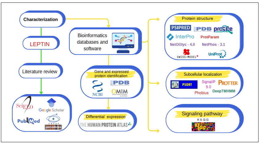
Figure 1 Methodological steps for in silico characterization of H. sapiens leptin
Source: Own authorship.
For mapping the expression of mRNA and leptin protein in all cells, tissues, and organs, THE HUMAN PROTEIN ATLAS database (https://www.proteinatlas.org/ENSG00000174697-LEP) was used, and phenotypes related to the LEP gene were retrieved from the OMIM database (https://www.omim.org/entry/164160). To visualize and obtain the image of the crystallized structure, possible variants, and entry code, the UNIPROTKB (https://www.uniprot.org/uniprotkb/P41159/entry), PDB (https://www.rcsb.org/structure/1AX8), SWISSMODEL (https://swissmodel.expasy.org/), and ALPHAFOLD (https://alphafold.ebi.ac.uk/entry/P41159) databases were used.
The physicochemical characteristics and quantification of the amino acids composing the protein were extracted from PROTPARAM (https://web.expasy.org/protparam/). The presence of the signal peptide was validated on the platforms
Phobius (https://phobius.sbc.su.se/) and SignalIP 5.0
(https://services.healthtech.dtu.dk/services/SignalP-5.0/). PSIPRED (http://bioinf.cs.ucl.ac.uk/psipred/) was accessed for visualization of secondary structure arrangements and amino acid composition.
Protein scanning to obtain sites was performed within SCANPROSITE (https://prosite.expasy.org/). Phosphorylation sites were validated using NetPhos (https://services.healthtech.dtu.dk/service.php?NetPhos-3.1), PhosphoSitePlus (https://www.phosphosite.org/homeAction) and O-glycosylation sites were identified using NetOGlyc (https://services.healthtech.dtu.dk/services/NetOGlyc-4.0/), both with a score greater than or equal to 0.600.
To obtain the family class to which the Homo sapiens leptin protein belongs, INTERPRO (https://www.ebi.ac.uk/interpro/) was used, while subcellular localization was obtained using PSORT (https://psort.hgc.jp/) and PROTTER (http://wlab.ethz.ch/protter/start/), indicating the presence of a signal peptide. DeepTMHMM (https://dtu.biolib.com/DeepTMHMM) was used for predicting the protein's transmembrane arrangements.
The leptin signaling pathways of Homo sapiens were visualized in KEGG (https://www.genome.jp/entry/K05424).
H. sapiens leptin is a protein containing 167 amino acids, playing an important role in energy homeostasis by reducing food intake and increasing energy expenditure. In H. sapiens, gene ID 3952 (LEP), which encodes the protein, is located on chromosome 7 (Figure 2), more specifically in the 7q32.1 region, featuring three exons and major mRNA expression in adipose tissue.11,12

Figure 2 Chromosomal location of the H. sapiens LEP gene
Source: PDB (2025).
The LEP gene is linked to obesity and associated factors, and leptin can be an agent contributing to the disease, either when there is a deficiency in leptin production or resistance to its action, leading to an imbalance between caloric intake and energy expenditure, and consequently to weight gain.13 The amino acid sequence of H. sapiens leptin in FASTA format (Table 1) has a methionine residue at the N-terminal end and a cysteine residue at the C-terminal end, with a prevalence of leucine (16.2%) and serine (10.2%).
|
Expressed protein |
NCBI access code |
Protein name |
Primary sequence in FASTA format |
|
Leptin precursor [Homo sapiens] |
NP_ 000221.1 |
Leptin |
MHWGTLCGFLWLWPYLFYVQAVPIQKVQDDTKTLIKTIVTRINDISHTQSVSSKQKVTGLDFIPGLHPIL TLSKMDQTLAVYQQILTSMPSRNVIQISNDLENLRDLLHVLAFSKSCHLPWASGLETLDSLGGVLEASGY STEVVALSRLQGSLQDMLWQLDLSPGC |
Table 1 Primary sequence of the H. sapiens leptin protein
Source: The author. Data extracted from NCBI (2025).
From the primary sequence, H. sapiens leptin was identified as belonging to the leptin family (Figure 3), information also revealed in its UniProtKB entry (P41159). Furthermore, via Interpro, the primary structure of the protein revealed six signature intervals that recognize the leptin sequence as an obesity factor.
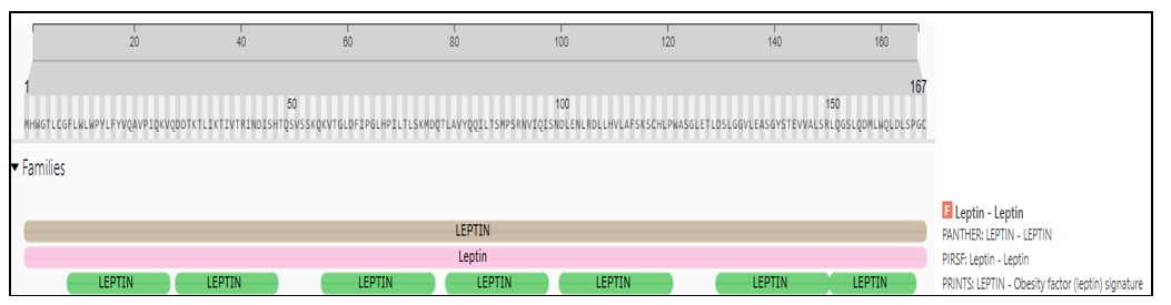
Figure 3 Identification of the H. sapiens leptin family from the primary sequence.
Source: Interpro (2025).
The crystallized structure of leptin verified by X-ray diffraction (Figure 4), extracted from the PDB (1AX8), contains two disulfide bonds at residues Cys96 and Cys146 and presents a shorter amino acid sequence, diverging from the structure deposited in AlphaFold (AF-P41159-F1-v4) and UniProtKB (P41159) where the deposits present the three-dimensional structure of leptin with disulfide bonds located at residues Cys117 and Cys167, besides being larger due to the presence of the signal peptide with 21 amino acid residues (range 1-21), being authenticated by Phobius and SignalIP 5.0 (score 0.9654).
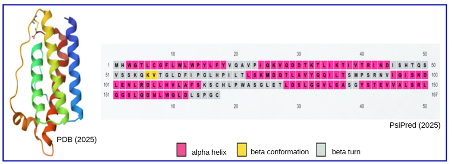
Figure 4 Secondary (right, arrangement prediction) and tertiary (left, obtained by X-ray diffraction) structures of the H. sapiens leptin protein.
Source: PDB (2025) and Psipred (2025).
The prediction of alpha-helix, beta-sheet, and beta-conformation arrangements is divergent when obtained by the Psipred software (six alpha-helices and one beta conformation), not coinciding with those present in the three-dimensional structure of the protein deposited in Alphafold (five alpha-helices and no beta conformation) and the PDB (five alpha-helices and no beta conformation).
Regarding the protein's post-translational modifications, myristoylation sites are restricted to the residue intervals 4-9 and 133-138, but they were not validated, while O-glycosylation sites, despite not being predicted by SCANPROSITE, were analyzed and indicated by NetOGlyc at Ser50, Ser52, and Ser58 residues.
Phosphorylation sites are described with their corresponding residues in Table 2, being revealed by SCANPROSITE and NetPhos, with a score greater than or equal to 0.600. The protein kinases involved in this modification are PKC and PKA, CK1 and CK2, and DNAPK. It is observed that only Ser52 and Ser153 residues were validated in both software programs. Furthermore, only Thr38 residue has been validated by PhosphoSitePlus. Phosphorylation on serine, threonine, and tyrosine residues is a fundamental post-translational mechanism for regulating protein function, due to the presence of a reactive hydroxyl group in the amino acid side chain. The enzymatic specificity of serine/threonine kinases ensures that phosphorylation occurs only at these targets.
|
Target protein kinase |
Predicted residues |
|
|
SCANPROSITE |
NetPhos |
|
|
Protein kinase C (PKC) |
Ser52 |
Thr33, Ser50, Ser52, Ser53, Ser71 e Thr78 |
|
Casein kinase II (CK2) |
Thr58, Ser73, Ser123 e Ser153 |
|
|
DNA-dependent protein kinase (DNAPK) |
|
Thr48 |
|
Casein kinase I (CK1) |
|
Ser130 |
|
Protein kinase A (PKA) |
|
Ser153 |
Table 2 Phosphorylation sites present in the primary sequence of H. sapiens leptin
Source: The author. Data extracted from ScanProsite (2025) and NetPhos (2025).
More than 300 different post-translational modifications have already been identified, including methylation, ubiquitination, myristoylation, acetylation, phosphorylation, and glycosylation. Among them, glycosylation is one of the most diverse, having the main types N-glycosylation linked to asparagine, O-glycosylation linked to serine or threonine, C-glycosylation linked to tryptophan, and glycosylphosphatidylinositol-anchored linkage.14 Of these, N-glycosylation and O-glycosylation are the most common types and contain most of the glycosylation machinery associated with the pathogenesis and progression of a disease.15
In leptin, glycans are linked via hydroxyl side groups on three serine residues of the protein through O-glycosylation. This type of glycosylation is mainly classified into two subtypes, O-GlcNAcylation and O-GalNAcylation, but based on the prediction performed in this study, it was not possible to characterize the linked monosaccharide. It is worth noting that there is a close connection between O-glycosylation and physiological processes, such as inflammatory response, immune evasion, viral infection, cell adhesion, metastasis, apoptosis, among others.15,16
Zhang et al.17 demonstrated that leptin regulation by O-glycosylation occurs at the transcription level, and many factors can be modified by O-GlcNAcylation.18 On the other hand, recognizing the regulation of leptin by O-GlcNAcylation, Gao et al.19 investigated the effects of iron on this post-translational modification and revealed that iron reduces leptin regulation by decreasing CREB glycosylation, resulting in increased phosphorylation of this transcription factor in the leptin signaling cascade.
It is noted that O-glycosylation and phosphorylation are often reciprocal, in some cases due to the sharing of modification sites. For example, in the case of leptin, protein kinases only target serine or threonine residues (Table 2). In the case of PKC, Dempsey et al.20 demonstrated that leptin has stimulating and inhibitory effects on this serine/threonine protein kinase implicated in various cellular events.
Besides its significant expression in adipose tissue, H. sapiens leptin is also present in other tissues and organs, such as the breast and female reproductive system, bone marrow and lymphoid tissues, skin, pancreas, gastrointestinal tract, as well as in trophoblastic cells. Leptin is secreted and is associated with pathologies, such as renal, rectal, ovarian, and mammary cancers, among others (Figure 5).
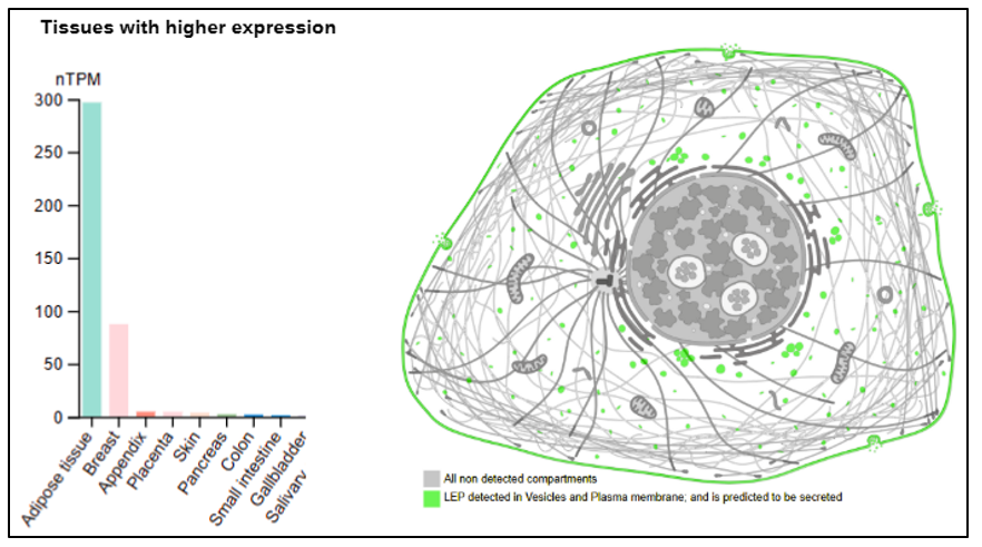
Figure 5 Expression of leptin from H. sapiens in different organs and tissues, and its subcellular localization (in green) in vesicles and plasma membrane.
Source: Adapted from The Human Protein Atlas (2025).
Through DeepTMHMM, it was possible to confirm that H. sapiens leptin has an extracellular location (Figure 6), validated by PSORT, and does not possess transmembrane domains, presenting a signal peptide corresponding to the amino acid residue interval 1 to 21, with variants located at residues 49, 94, 100, 105, and 110, validated in SwissModel.
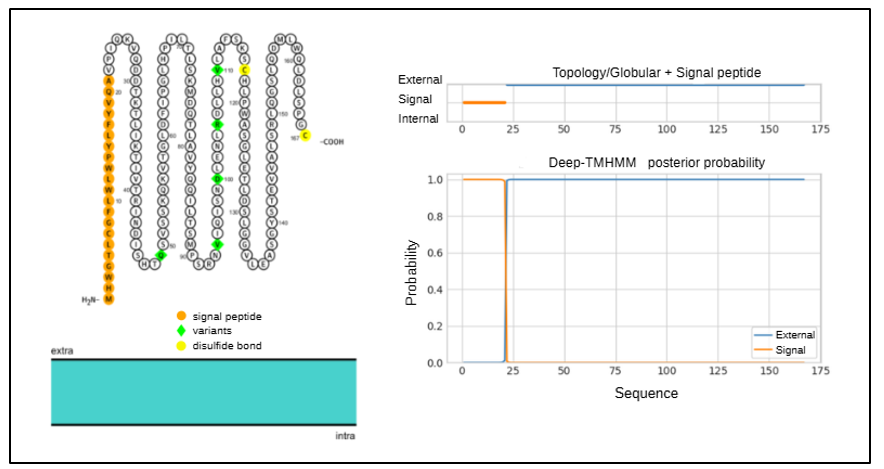
Figure 6 Location of the H. sapiens leptin protein in relation to the cell membrane, and the presence of a signal peptide, variants, and disulfide bonds.
Source: Adapted from PROTTER (2025) and DeepTMHMM (2025).
In UniProtKB,21 H. sapiens leptin presents 169 variants, two of which are reviewed and involved with obesity. The variant rs724159998 (VAR_075144) is classified as pathogenic, described as a leptin dysfunction where the protein cannot activate or bind to the LEPR receptor, while the variant rs104894023 (VAR_008094) is pathogenic, described as somatic with moderate impact, resulting in obesity due to congenital leptin deficiency.21
Leptin deficiency is related to its main signaling pathways, such as interaction with the cytokine receptor, interaction with the neuroactive receptor of AMPK signaling, JAK-STAT signaling (Figure 7), non-alcoholic fatty liver disease, and adipocytokine signaling (KEGG, 2023). Congenital leptin deficiency is a rare human syndrome, and the positive effects of its replacement in this condition have already been reported.22
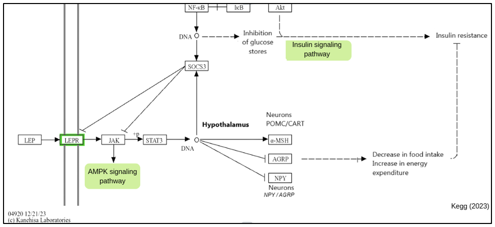
Figure 7 Map of the main leptin signaling pathway.
Source: Adapted from KEGG (2025).
In a study conducted with rat epithelial cells, Fazolini et al.23 reported that leptin was able to activate lipid droplet biogenesis, triggering inflammatory mediators, correlating leptin activity and oncogenic factors with breast, prostate, and colorectal cancers. This mechanism is due to the pro-inflammatory properties of leptin, which activate the maturation pathways of phagocytic cells, causing tissue infiltration and triggering processes that lead to injury and inflammation of tissues and organs.
The LEP gene, also known by the acronyms OB, OBS, or LEPD, is related to pathologies such as morbid obesity due to leptin deficiency.24 Typical obesity is more related to leptin resistance than to leptin deficiency. Saturated fatty acids, present in lipid-rich diets, activate Toll-like receptors (TLR), which in turn stimulate the transcription factor NF-kB to increase the production of pro-inflammatory cytokines. This process generates endoplasmic reticulum stress, which induces the overexpression of SOCS3 and PTP1-B proteins. Both acts as negative regulators of the tyrosine kinase JAK2, blocking leptin receptor signaling. Consequently, a compensatory increase in circulating leptin levels also occurs, characterizing hyperleptinemia.25
Leptin is known to cross the blood-brain barrier, where it acts primarily in the arcuate nucleus of the hypothalamus to regulate energy homeostasis, inhibiting neuropeptide Y (NPY) and agouti-related peptide (AgRP), which are orexigenic, increasing food intake, and stimulating pro-opiomelanocortin (POMC), which, in turn, activates anorexigenic factors, such as α-melanocyte-stimulating hormone (αMSH), which inhibits food intake. This action of leptin occurs via JAK kinase, STAT3 phosphorylation, and nuclear transcription effect. The binding of the leptin protein to its receptor LEPR activates the signaling pathway. Leptin receptor deficiency is a rare autosomal recessive condition that causes obesity and pituitary dysfunction.26–34
Obesity is a multifactorial chronic disease that can affect individuals across different life cycles. Its treatment is complex and involves changes in eating habits and physical activity, but it may also require drug therapy, which should only be prescribed by a healthcare professional. According to literature reviews, typical obesity is generally associated with leptin resistance, rather than its absence, being influenced by genetic variations in the LEP gene and consequently a change in its protein structure. Bioinformatics analysis of leptin revealed that its gene is located on chromosome 7 (region 7q32.1), and its protein expresses a high leucine content and features disulfide bonds that are crucial for its three-dimensional structure. Although primarily produced by adipose tissue, its expression also occurs in other organs, being related to various physiological functions and even cancer. Studies indicate that phosphorylation sites on the protein contribute to its regulation, while the lack of validation for myristoylation and glycosylation still requires clarification. The presence of a signal peptide and its extracellular location confirm its role as a secreted hormone. The in silico characterization of leptin, together with the scientific literature, reinforces its central function in metabolism and points to promising therapeutic possibilities for the treatment of obesity.
None.
The author declares there is no conflict of interest.

©2025 Souza, et al. This is an open access article distributed under the terms of the, which permits unrestricted use, distribution, and build upon your work non-commercially.