Open Access Journal of
eISSN: 2575-9086


Research Article Volume 8 Issue 1
 Maria Paula Carvalho Naves, Maria Eduarda Souza Alves, Mário Antônio Spanó, Alexandre Azenha Alves de Rezende
Institute of Exact and Natural Sciences of Pontal, Federal University of Uberlândia, Campus Pontal, Ituiutaba, Minas Gerais, Brazil
Correspondence: Alexandre Azenha Alves de Rezende, Institute of Exact and Natural Sciences of Pontal, Federal University of Uberlândia, Campus Pontal, 38304-402, Ituiutaba, Minas Gerais, Brazil
Received: May 20, 2025 | Published: June 11, 2025
Citation: Abu-Bakr AA, Naves MPC, Alves MES, et al. Citalopram and its enantiomers: analysis of the induction of mutation and recombination in wing somatic cells of Drosophila melanogaster. Open Access J Sci. 2025;8(1):91-99. DOI: 10.15406/oajs.2025.08.00249
The main goal of this research was to evaluate the mutagenic and recombinogenic effect of Citalopram and Escitalopram in somatic cells of Drosophila melanogaster and verify the influence of CYP450 complex enzymes in the induction of mutation and homologous recombination. Flies from the Standard (ST) and High Bio activation (HB) crosses were used for the Somatic Mutation and Recombination Test (SMART). The third instar larvae of D. melanogaster obtained from both crosses (ST and HB) were fed chronically with the antidepressant drugs Citalopram or Escitalopram (0.3125 to 10 mg/mL). Both drugs were toxic at higher concentrations. The results obtained from the crosses indicated that Citalopram was mutagenic and also recombinogenic only at the highest concentration tested in ST cross. However, it did not show any mutagenic effect when highly metabolized through CYP450 enzymes. Escitalopram was mutagenic and mainly recombinogenic in all ST treatments, but it was completely mutagenic in all HB treatments. More studies need to be done to better evaluate the DNA damage caused by these drugs.
Keywords: antidepressants, mutation, SMART, citalopram, escitalopram
SMART, somatic mutation and recombination test; ST, standard; S-DCT, S-desmethylcitalopram; S-DDCT, S-didesmethylcitalopram; SCE, sister chromatid exchange.
Depression is a mental disorder characterized by the gradual loss of interest in common activities, the feeling of sadness, and low self-esteem.1 Studies indicate the relationship between depression and heart disease, diabetes, and hypertension, in addition to being a common cause of suicides.68
According to the World Health Organization,68 more than 300 million people live with depression. Considering the increase in the number of cases and their social consequences, it constitutes a serious public health problem.
The discovery of antidepressant drugs in the late 1950s brought important advances in understanding the mechanisms involved in depressive disorders, as well as possibilities for pharmacological treatments.2,3 In general, these drugs produce an increase in the concentration of neurotransmitters in the synaptic cleft by inhibiting metabolism, blocking neuronal recapture, or by acting on presynaptic auto-receptors.2
Among the different classes of antidepressants, some belong to the group of selective serotonin/noradrenaline reuptake inhibitors (SSRI / SSIR), with representatives of Citalopram Hydrobromide (Figure 1A) and Escitalopram Oxalate (Figure 1B). These are preferred in the treatment of elderly patients who have two or more diseases simultaneously and in the treatment of patients with a comorbid anxiety disorder.2,4
Citalopram is a 1: 1 racemic mixture consisting of the R (-) and S (+) enantiomers, however, only the second one is therapeutically active, besides, it has a chemical structure different from other SSRIs and any other prescribed antidepressant drug.5 Its metabolism, in humans, occurs mainly in the liver via N-demethylation to its main metabolite, desmethylcitalopram through the enzymes CYP2C19 and CYP3A4.6 Other metabolites include didesmethylcitalopram via CYP2D6 and Citalopram N-oxide, via monoaminoxidase and aldehyde oxidase enzymes.6
Escitalopram is the S-Enantiomer of Citalopram. However, despite structural differences, their pharmacological activities are practically the same. Its metabolism also occurs in the liver by the same enzymes as the CYP450 complex, transforming it into S-desmethylcitalopram (S-DCT) and S-didesmethylcitalopram (S-DDCT).7
According to Battal et al.,8 drugs used in the prolonged treatment of depression can cause DNA damage. Most damage is usually repaired by enzymes in the repair system, however, this ability decreases over time.9 The accumulation of oxidative damage in DNA can lead to the development of degenerative diseases, such as cancer.10 Studies conducted in vitro with Escitalopram indicated that the drug causes sister chromatid exchange (SCE), however, no statistically significant increases in DNA damage and micronucleus formation (MN) were observed during treatment.11
Studies carried out in vivo by Hassanane and collaborators,12 with recommended concentrations of Amitriptyline used in the treatment of depression, indicated the increase in the appearance of numerical and structural chromosomal aberrations in somatic (bone marrow) and germ cells (spermatocytes), such effects also were seen in studies with Citalopram.13
There is a worldwide effort to reduce the use of mammals in toxicological and genetic research, especially concerning the development of alternative models.14 In this context, Drosophila melanogaster represents an insect widely used as an experimental model in scientific research, mainly in studies of genetics and developmental biology, having been appreciated for nearly a century.15 Besides, the fly stands out in the scientific community in studies that seek to track the toxicity and mutagenicity of different xenobiotics.16–18
Among the tests performed with D. melanogaster as a model organism, the Somatic Mutation and Recombination Test (SMART) stands out, considered one of the “gold standard” tests for the evaluation of mutagenicity / recombinogenicity.19 This test was developed by Graf and collaborators20 and improved by Graf and van Schaik.21 It is based on the detection of the loss of heterozygosis in marker genes, which alter the fly wing hair phenotype (Spanó & Graf, 1998).22
Considering the increase in the number of cases and their social consequences, depression is a serious public health problem, and, to date, there is little data regarding the induction of genetic damage through drugs used in treatment. Thus, it is necessary to assess the genotoxic, mutagenic, and recombinogenic potential of Escitalopram and Citalopram to collaborate with the scientific community on the possible adverse effects related to the use of these drugs.
Thus, the objective of the present work was to evaluate the mutagenic and recombinogenic potential of Citalopram and Escitalopram in somatic cells of D. melanogaster and also to verify the influence of enzymes of the CYP450 complex in the induction of mutant spots and homologous recombination.
Chemical compounds and culture medium
The Antidepressants, with pharmaceutical purity, used were: Citalopram Hydrobromhydrate (C20H21FN2O - CAS Number: 59729-33-8. Sandoz, Brazil) (Figure 1A) and Escitalopram Oxalate (C20H21FN2O - CAS Number: 128196- 01-0 Lexapro®, Lundbeck A / S, Denmark), (Figure 1B).
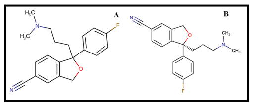
Figure 1 Citalopram Structural Formula (A) and Escitalopram Structural Formula (B).
We used Urethane (Ethyl Carbamate - CAS Number: 51-76-6, Buchs, Switzerland) as a positive control, and ultrapure water (18.2 MΩ), obtained by the MilliQ System (Millipore, Vimodrone, Milan, Italy), as the negative control. We used mashed potato as an alternative culture medium (Yoki Alimentos S.A. - São Bernardo do Campo, SP, Brazil).
Somatic mutation and recombination test (SMART) in drosophila melanogaster
Drosophila strains
Three different strains of mutant flies were used: (i) mwh males (mwh / mwh); (ii) females flr3 (flr3 / In (3LR) TM3, ri pp sep I (3) 89Aa bx34e and BdS); and (iii) ORR females; flr3 (ORR; flr3 / In (3LR) TM3, ri pp sep I (3) 89Aa bx34e and BdS). These mutant strains were provided by Prof. Dr. Mário Antônio Spanó, Institute of Biotechnology, Federal University of Uberlândia (Uberlândia, Brazil). Details of the strains used in the present work can be found in Graf et al.20 and Graf and Van Schaik.21 The mutant strains were kept in a B.O.D (Biologic Oxigen Demand) greenhouse (Tecnal - Equipamentos para Laboratórios Ltda., Piracicaba, SP), with a 12-hour light / dark photoperiod and a temperature of 25 + 1ºC.
Crossings and treatments
The crosses performed were: 1) Standard (Standard - ST) that uses male mwh and virgin female flr³ (Graf et al., 1989); and 2)23 Crossing of high metabolic bioactivation (High Bioactivation - HB), which uses mwh males and ORR virgin females; flr³,21 characterized by constitutive levels of gene expression of cytochrome P450 complex enzymes (CYP6A2). Both crosses (ST and HB) produce two types of offspring: marked trans-heterozygous flies (MH) and balanced heterozygous flies (BH), which were analyzed to assess the direct contribution of homologous recombination in the phenotypes observed after treatment.
Survival essay
To determine the toxicity of antidepressants in D. melanogaster, we transferred 40 third-stage larvae into vials containing alternative culture medium (1.5 g of mashed potatoes) hydrated with 5 mL24 of different concentrations of the antidepressants Citalopram and Escitalopram (0.3125; 0.625; 1.25; 2.5, 5 or 10 mg / mL). Consequently, we selected three non-lethal concentrations of the survival test.
After the hatching of the adult individuals, those were quantified, and the frequency of each treatment was compared to the frequency of the negative control, using the Chi-square test, for reasons of independent samples. As a negative control (CN), ultrapure water was used.25,16 Every treatment was performed in duplicate.
Slide preparation and analysis
After the treatment, emerging adults were collected and stored in bottles containing 70% ethanol. The wings were removed with the aid of entomological forceps and a stereomicroscope and aligned on a glass slide, with five pairs of female wings on the upper part and five pairs of male wings on the lower portion (totaling ten individuals per slide). We used Faure's solution (30 g of Arabic gum, 20 ml of glycerol, 50 g of chloral hydrate, and 50 ml of water) for the adhesion of the coverslip to the slide. The slides - after drying -, were subsequently analyzed using a light optical microscope with a final magnification of 400X.
Statistical analysis
To assess the genotoxic potential of the antidepressants Citalopram and Escitalopram, we compared the spots per fly frequency of each treated series with the negative control. For that, the conditional Binomial Test of Kastembaun and Bowman (1970) was used, following the procedure of multiple decisions according to Frei and Würgler (1988), which is applied on two hypotheses and allows to obtain four statistical diagnoses. In the null hypothesis (H0), is determined that there is no difference in the frequency of induced spots in the two groups, and in the alternative hypothesis (HA) a multiplier factor (m) is placed, in which it is stated that one of the groups has a frequency of spots m times larger or smaller than the other. As small single spots can have a high spontaneous frequency, the multiplier value m is fixed at 2. But, for large single and twin spots, which have a low spontaneous frequency, it is established in 5.26
Each hypothesis is tested to a significance level of 5%, with four possible decisions: 1) inconclusive (I): both hypotheses are accepted and as they cannot be simultaneously true, no conclusions can be made; 2) negative (-): accepts H0 and rejects HA; 3) positive (+): rejects H0 and accepts HA and 4) weak-positive (f +): both hypotheses are rejected. To confirm positive results and exclude outliers, the Wilcoxon, Mann, and Whitney U Test was performed.27 Based on the frequencies of clone induction by 105 cells, the recombinogenic activity can be estimated as mutation frequency (FM) = frequency of clones in BH flies / frequency of clones in MH flies; recombination frequency (FR) = 1 - FM. Total spot frequency (FT) = total spot observed in MH flies/number of flies.28 Statistical comparisons of survival rates between pupae and adults were performed using the X² test for proportions, for independent samples.
Citalopram is a racemic SSRI (1: 1) consisting of two enantiomers, an inactive R (-) and an active S (+), however, studies have shown that the R (-) enantiomer ends up suppressing the action of S (+) depending on the dose used, impairing its effectiveness in the treatment of depression.29,6 Therefore, to reduce side effects and enhance therapeutics, the active enantiomer was isolated and subsequently marketed as another drug called Escitalopram.30
We performed a survival assay to investigate the effects of drugs on the survival rates of the tested organisms. For this experiment, third-stage larvae from both the Standard (ST) and High Bioactivation (HB) crosses were exposed to varying concentrations of citalopram and escitalopram (0.3125, 0.625, 1.25, 2.5, 5, and 10 mg/mL). The findings demonstrated that a citalopram concentration of 10 mg/mL was toxic, leading to a significant decrease (p < 0.05) in the survival rates of individuals from both crosses (ST and HB) when compared to the negative control (Figure 2A and 2B, respectively). The Escitalopram concentration of 5 mg/mL demonstrated significant toxicity (p < 0.05) in individuals from the ST cross (Figure 3A). In contrast, no significant survival reductions were observed with other treatments, including all concentrations tested on the HB cross (Figure 3B). Therefore, the concentrations chosen for further analysis of the two antidepressants were 0.625, 1.25, and 2.5 mg/mL, as these levels did not significantly impact survival rates.
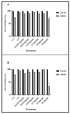
Figure 2 Survival Rate (%) of individuals from ST (A) and HB (B) crosses treated with different concentrations of Citalopram. * Concentrations with significant decrease in survival rate (p <0.05).
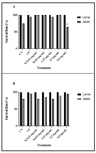
Figure 3 Survival Rate (%) of individuals from ST (A) and HB (B) crosses treated with different concentrations of Escitalopram. * Concentrations with significant decrease in survival rate (p <0.05).
Research has shown that Citalopram can lead to chromosomal abnormalities in the lung fibroblasts of Chinese rats and trigger reverse mutations in the TA98 and TA1537 strains of Salmonella typhimurium.31 These results imply that Citalopram possesses genotoxic and carcinogenic characteristics. Comparable effects have been noted with other SSRIs, including fluoxetine, sertraline, and clomipramine, which previous studies32 have also linked to DNA damage. On the other hand, findings from Wang et al.33 suggest that some antidepressants, notably SSRIs, may bolster cellular responses to DNA damage induced by neurotoxins, correct cell cycle disruptions, and act as neuroprotective agents.
From the information provided, there is no consensus on the potential impact of Selective Serotonin Reuptake Inhibitors (SSRIs) on DNA damage. Therefore, we employed the Somatic Mutation and Recombination Test (SMART) to assess the mutagenic and recombinogenic effects of Escitalopram and Citalopram in the wing cells of D. melanogaster, aiming to shed further light on this matter.
The base of the test is detecting the loss of heterozygosity in marker genes, which alter the fly wing hair phenotype and allows detecting point mutations, deletions, some chromosomal aberrations, and mitotic recombination (homologous recombination).20,34
The analysis of trans-heterozygous (MH) specimens from the ST cross treated with Citalopram showed that the drug can cause DNA damage, leading to a statistically significant increase (p < 0.05) in the overall frequency of mutant spots in flies exposed to a concentration of 2.5 mg/mL (Table 1). Conversely, the assessment of MH individuals from the HB cross indicated that none of the tested concentrations produced a significant rise in the total number of mutant spots (Table 2). Although mutant spot frequencies appear numerically similar across the two crosses, this finding is most likely linked to the inhibitory impact of Citalopram on the enzymatic activities of various components within the CYP450 complex.35,36,5 Studies on humans indicate that the metabolites of Citalopram are toxic to liver cells, causing DNA strand breaks and the formation of micronuclei.13,37
|
Spots per fly (number of spots); statistical diagnosesa |
|||||||||
|
Genotypes and Treatments (mg/mL) |
Nº of flies. (N) |
Small single spots (1-2 cells)b m = 2 |
Large single spots (>2 cells)b m = 5 |
Twin spots m = 5 |
Total spots m = 2 |
Spots with mwh clone (n) |
Frequency of clone formation/105 cells per divisiond |
Recombination (%) |
|
|
Observed |
Correct control |
||||||||
|
mwh/flr³ |
|||||||||
|
Negative Control |
40 |
0.45 (18) |
0.05 (02) |
0.00 (00) |
0.50 (20) |
18 |
0.92 |
||
|
Positive Control |
40 |
2.13 (85) + |
0.23 (09) + |
0.10 (04) i |
2.45 (98) + |
93 |
4.71 |
3.79 |
9.77 |
|
0.625 |
40 |
0.28 (11) - |
0.23 (09) + |
0.05 (02) i |
0.55 (22) - |
18 |
0.92 |
0 |
|
|
1.25 |
40 |
0.38 (15) + |
0.20 (08) i |
0.00 (00) i |
0.58 (23) - |
23 |
1.18 |
0.26 |
|
|
2.5 |
40 |
0.58 (23) i |
0.23 (09) + |
0.03 (01) i |
0.83 (33) + |
29 |
1.48 |
0.55 |
64 |
|
mwh/TM3, Bds |
|||||||||
|
Negative Control |
40 |
0.30 (12) |
0.05 (02) |
e |
0.35(14) |
14 |
0.72 |
||
|
Positive Control |
40 |
2.03 (81) + |
0.05 (02) i |
2.08 (83) + |
83 |
4.25 |
3.53 |
||
|
2.5 |
40 |
0.29 (09) - |
0.29 (09) - |
0.45 (18) i |
18 |
0.92 |
0.2 |
||
Table 1 Results obtained through the Somatic Mutation and Recombination Test (SMART) in Drosophila melanogaster wing cells, with trans-heterozygous (MH) and balanced heterozygous (BH) progeny from the standard cross (ST), treated with ultrapure water (negative control), urethane 10 mM (positive control) and different concentrations of Citalopram
Marker-trans-heterozygous flies (mwh/flr³) and balancer-heterozygous flies (mwh/flr³) were evaluated.
aStatistical diagnoses according to Frei e Würgler (1988; 1995): -, negative; i, inconclusive; +, positive. M: multiplication factor. Significance levels α = β = 0.05.
bIncluding rare flr3 single spots.
cConsidering mwh clones from mwh single and twin spots..
dFrequency of clone formation: clones/flies/48.800 cells (without size correction).
eOnly single mwh spots can be observed in heterozygous mwh / TM3 individuals since the TM3 balancing chromosome does not contain the gene flr3.
|
Spots per fly (number of spots); statistical diagnosesa |
||||||||
|
Genotypes and Treatments (mg/mL) |
Nº of flies. (N) |
Small single spots (1-2 cells)b m = 2 |
Large single spots (>2 cells)b m = 5 |
Twin spots m = 5 |
Total spots m = 2 |
Spots with mwh clone (n) |
Frequency of clone formation/105 cells per divisiond |
|
|
Observed |
Observed |
|||||||
|
mwh/flr³ |
40 |
0.58 (23) |
0.03 (01) |
0.03 (01) |
0.63 (25) |
24 |
1.23 |
|
|
Negative Control |
40 |
9.33 (373) + |
0.75 (30) + |
1.20 (48) i |
11.28 (451) + |
437 |
22.39 |
21.16 |
|
Positive Control |
40 |
0.28 (15) - |
0.18 (07) + |
0.05 (02) i |
0.60 (24) - |
22 |
1.13 |
-0.1 |
|
0.625 |
40 |
0.68 (23) - |
0.08 (03) i |
0.03 (03) i |
0.725 (29) - |
26 |
1.33 |
0.1 |
|
1.25 |
40 |
0.35 (14) - |
0.20 (08) + |
0.08 (03) i |
0.625 (28) - |
25 |
1.29 |
0.05 |
|
2.5 |
||||||||
Table 2 Results obtained through the Somatic Mutation and Recombination Test (SMART) in Drosophila melanogaster wing cells, with trans-heterozygous (MH) and balanced heterozygous (BH) progeny from the high bioactivation cross (HB), treated with ultrapure water (negative control), urethane 10 mM (positive control) and different concentrations of Citalopram
Marker-trans-heterozygous flies (mwh/flr³) and balancer-heterozygous flies (mwh/flr³) were evaluated.
aStatistical diagnoses according to Frei e Würgler (1988; 1995): -, negative; i, inconclusive; +, positive. M: multiplication factor. Significance levels α = β = 0.05.
bIncluding rare flr3 single spots.
cConsidering mwh clones from mwh single and twin spots..
dFrequency of clone formation: clones/flies/48.800 cells (without size correction).
eOnly single mwh spots can be observed in heterozygous mwh / TM3 individuals since the TM3 balancing chromosome does not contain the gene flr3.
In contrast to the results of the current study, Gürbüzel and colleagues38 presented findings on Citalopram that offer a differing perspective. The methodological differences between the two studies likely play a key role in explaining these variations. For instance, the earlier study used 1% Dimethylsulfoxide (DMSO) as the negative control, which produced only one mutant spot across the 20 individuals examined. Additionally, that study did not include a survival curve, which could have provided valuable insight into any potential toxicity related to DMSO use.
Nazir et al.39 highlighted the cytotoxic effects observed when DMSO concentrations exceed 0.5%. Likewise, Galvão et al.40 and Cvetković et al.41 recommended keeping DMSO concentrations below 1% when it is used as a solvent to dissolve drugs. They also suggested including a DMSO-free control group to assess the potential toxicity of the solvent (1% DMSO). Data obtained by our research group (Mendonça et al., 2019),16,25,42,43 along with findings from other studies using similar methodologies,44,45 show that the baseline frequency of mutant spots in D. melanogaster exposed only to water is not as low as previously reported in the study conducted by Gürbüzel et al.38
The assessment of MH progeny from the ST cross treated with Escitalopram demonstrated the drug's mutagenic properties, as it significantly increased (p < 0.05) the total number of mutant spots across all tested concentrations in comparison to the negative control (Table 3). Comparable results were obtained in the HB cross (Table 4). These findings imply that the elevated mutant spot count within the HB cross could be associated with an intensified metabolism of Escitalopram in organisms exhibiting higher expression levels of cytochrome P450 complex enzymes (CYP6A2).21
|
Spots per fly (number of spots); statistical diagnosesa |
|||||||||
|
Genotypes and Treatments (mg/mL) |
Nº of flies. (N) |
Small single spots (1-2 cells)b m = 2 |
Large single spots (>2 cells)b m = 5 |
Twin spots m = 5 |
Total spots m = 2 |
Spots with mwh clone (n) |
Frequency of clone formation/105 cells per divisiond |
Recombination (%) |
|
|
Observed |
Correct control |
||||||||
|
mwh/flr³ |
|||||||||
|
Negative Control |
40 |
0.43 (17) |
0.03 (01) |
0.00 (00) |
0.45 (18) |
17 |
0,87 |
||
|
Positive Control |
40 |
2.58 (103) + |
0.35 (14) + |
0.25 (10) + |
3.18 (127) + |
125 |
6,35 |
5.48 |
43.46 |
|
0.625 |
40 |
0.45 (18) - |
0.40 (16) + |
0.03 (01) i |
0.88 (35) + |
32 |
1,64 |
0.77 |
20 |
|
1.25 |
40 |
1.45 (58) + |
0.43 (17) + |
0.05 (02) i |
1.93 (77) + |
73 |
3,74 |
2.87 |
82 |
|
2.5 |
40 |
0.68 (27) i |
0.25 (10) + |
0.03 (01) i |
0.95 (38) + |
37 |
1.9 |
1.02 |
90 |
|
mwh/TM3, Bds |
|||||||||
|
Negative Control |
40 |
0.35(14) |
0.03 (01) |
e |
0.38 (15) |
15 |
0.77 |
||
|
Positive Control |
40 |
1.63 (65) + |
0.13 (05) + |
1.75 (70) + |
70 |
3.59 |
2.82 |
||
|
0.625 |
40 |
0.65 (26) + |
0.03 (01) i |
0.68 (27) + |
27 |
1.38 |
0.61 |
||
|
1.25 |
40 |
0.55 (22) i |
0.00 (00) i |
0.55 (22) i |
22 |
1.23 |
0.5 |
||
|
2.5 |
40 |
0.33 (13) i |
0.10 (04) + |
0.43 (17) + |
17 |
0.87 |
0.1 |
||
Table 3 Results obtained through the Somatic Mutation and Recombination Test (SMART) in Drosophila melanogaster wing cells, with trans-heterozygous (MH) and balanced heterozygous (BH) progeny from the standard cross (ST), treated with ultrapure water (negative control), urethane 10 mM (positive control) and different concentrations of Escitalopram
Marker-trans-heterozygous flies (mwh/flr³) and balancer-heterozygous flies (mwh/flr³) were evaluated.
aStatistical diagnoses according to Frei e Würgler (1988; 1995): -, negative; i, inconclusive; +, positive. M: multiplication factor. Significance levels α = β = 0.05.
bIncluding rare flr3 single spots.
cConsidering mwh clones from mwh single and twin spots..
dFrequency of clone formation: clones/flies/48.800 cells (without size correction).
eOnly single mwh spots can be observed in heterozygous mwh / TM3 individuals since the TM3 balancing chromosome does not contain the gene flr3.
|
Spots per fly (number of spots); statistical diagnosesa |
|||||||||
|
Genotypes and Treatments (mg/mL) |
Nº of flies. (N) |
Small single spots (1-2 cells)b m = 2 |
Large single spots (>2 cells)b m = 5 |
Twin spots m = 5 |
Total spots m = 2 |
Spots with mwh clone (n) |
Frequency of clone formation/105 cells per divisiond |
Recombination (%) |
|
|
Observed |
Correct control |
||||||||
|
mwh/flr³ |
|||||||||
|
Negative Control |
40 |
0.43 (17) |
0.05 (02) |
0.03 (01) |
0.50 (20) |
20 |
1,02 |
||
|
Positive Control |
40 |
13.93 (557) + |
1.98 (79) + |
1.08 (43) + |
16.98 (679) + |
656 |
34.8 |
33.78 |
35 |
|
0.625 |
40 |
0.75 (30) i |
0.40 (16) + |
0.13 (05) i |
1.28 (51) + |
51 |
2.61 |
1.59 |
44 |
|
1.25 |
40 |
0.88 (35) i |
0.33 (13) + |
0.03 (01) i |
1.23 (49) + |
46 |
2.36 |
1.33 |
26 |
|
2.5 |
40 |
1.05 (42) i |
0.05 (02) i |
0.03 (01) i |
1.13 (45) + |
45 |
2.3 |
1.28 |
11 |
|
mwh/TM3, Bds |
|||||||||
|
Negative Control |
40 |
0.40 (16) |
0.03 (01) |
e |
0.43 (17) |
17 |
0.87 |
||
|
Positive Control |
40 |
10.63 (425) + |
0.48 (19) + |
11.10 (444) + |
444 |
22.74 |
21.74 |
||
|
0.625 |
40 |
0.75 (30) + |
0.08 (03) i |
0.83 (33) + |
33 |
||||
|
1.25 |
40 |
0.85 (34) + |
0.03 (01) i |
0.88 (35) + |
35 |
||||
|
2.5 |
40 |
0.88 (35) + |
0.08 (03) i |
0.95 (38) + |
38 |
||||
Table 4 Results obtained through the Somatic Mutation and Recombination Test (SMART) in Drosophila melanogaster wing cells, with trans-heterozygous (MH) and balanced heterozygous (BH) progeny from the high bioactivation cross (HB), treated with ultrapure water (negative control), urethane 10 mM (positive control) and different concentrations of Escitalopram
Marker-trans-heterozygous flies (mwh/flr³) and balancer-heterozygous flies (mwh/flr³) were evaluated.
aStatistical diagnoses according to Frei e Würgler (1988; 1995): -, negative; i, inconclusive; +, positive. M: multiplication factor. Significance levels α = β = 0.05.
bIncluding rare flr3 single spots.
cConsidering mwh clones from mwh single and twin spots..
dFrequency of clone formation: clones/flies/48.800 cells (without size correction).
eOnly single mwh spots can be observed in heterozygous mwh / TM3 individuals since the TM3 balancing chromosome does not contain the gene flr3.
Based on experiments conducted with human cells by Sánchez and colleagues (2008), the liver metabolizes Escitalopram into S-desmethylcitalopram (S-DCT) and S-didesmethylcitalopram (S-DDCT), which exhibit a higher mutagenic potential than Escitalopram itself. Additionally, another study by Cobanoglu et al.11 demonstrated the cytotoxic effects and inhibition of mitosis caused by these metabolites.
The CYP450 enzyme complex plays a crucial role in detoxification within the body.46 Due to a variety of genetic polymorphisms, there are numerous isoforms of this complex.47 This implies that S-DCT and S-DDCT metabolites could be present in the studied organism, D. melanogaster. These metabolites, while not measured in this research, might potentially be associated with the mutation rates observed in the experiments (Table 3 and 4).
D. melanogaster is an excellent model organism for studying homologous recombination (HR) due to the pairing of homologous chromosomes in both somatic and pre-meiotic cells. As a result, double-strand break repair primarily relies on the homologous chromosome as the repair template.48 Additionally, in these Rad51-dependent HR systems, this pathway serves as the main mechanism for repairing double-strand breaks in mitotically dividing cells, such as the wing cells examined in the SMART assay.
Homologous recombination refers to a collection of interrelated processes that utilize a matching DNA sequence, such as the sister chromatid or a homologous chromosome, to mend serious lesions at the replication fork. This mechanism is specifically involved in repairing double-strand breaks and inter-chain cross-links.49–51 Nevertheless, HR repair can result in genetic alterations, including point mutations, deletions, translocations, chromosome loss, and non-disjunction.50
To assess the direct contribution of homologous recombination to the total number of spots (TM), an analysis of heterozygous-balanced descendants (BH) was performed, which due to multiple inversions of the TM3 chromosome, all recombination events are suppressed, leading the cell to apoptosis.20
The assessment of Citalopram-induced HR was performed exclusively at a concentration of 2.5 mg/mL in the ST cross, as this was the only dosage at which the drug demonstrated mutagenic activity. The results indicate that, at this concentration, the drug primarily exhibits a recombinogenic effect (Table 1).
Analysis of BH descendants treated with Escitalopram revealed distinct effects depending on the concentration and genetic cross. In the ST cross, the drug primarily exhibited recombinogenic activity at the two highest concentrations, while at the lowest concentration, its effects were predominantly mutagenic (Figure 4A). Conversely, in the HB cross, Escitalopram consistently showed a mutagenic profile across all tested concentrations (Figure 4B). Importantly, unlike the ST cross, the HB cross displayed a clear concentration-dependent increase in mutagenicity. These findings suggest that as Escitalopram metabolism intensifies through the CYP450 complex, the drug-induced pattern of DNA damage shifts, potentially reducing its capacity to induce double-strand breaks typically repaired through homologous recombination (HR). Additionally, the drug might disrupt or hinder the HR repair pathway, which could account for the observed increase in mutant patches following exposure.
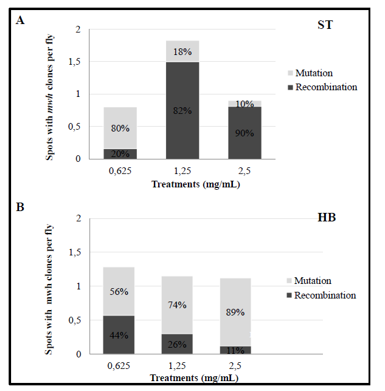
Figure 4 Contribution (%) of Recombination and Mutation by Total Frequency of Spots Observed in MH (ST) and HB (B) Crossover Individuals Treated with Different Escitalopram Concentrations.
Over the past few decades, extensive research has underscored the pivotal role of mutations and genomic instability in driving cancer development, originating from factors such as replication errors, environmental stresses, or defects in the repair of both endogenous and exogenous damage. Among these mechanisms, HR emerges as a significant contributor to genomic instability. For example, studies have shown that individuals whose cells display a heightened recombination frequency face a greater risk of developing cancer.53,54 On the other hand, mutations in critical recombination genes, such as BRCA1 and BRCA2, are tightly linked to an increased likelihood of developing certain cancers, particularly breast and ovarian tumors.49,55,56
HR events can cause loss of heterozygosis (LOH) through gene conversion, deletion, uneven crossing-over, and translocation. The loss of heterozygosis is the result of the loss of an allele in a cell, which becomes homozygous (if there is a gene conversion) or hemizygous (if an allele is simply lost) to the remaining allele.57
According to the results obtained in the present work, it was possible to observe that the drugs tested here were able to cause LOH in D. melanogaster wing cells, resulting in the appearance of cells with mutant phenotypes.
LOH events can lead to the development of cancers in humans, by allowing the expression of recessive alleles or by mutating tumor suppressors. Luo et al.58 demonstrated in vivo that the increase in the frequency of LOH from HR was the fundamental mechanism that causes tumor susceptibility in rats.
Previous work conducted by our group using D. melanogaster — although based on a different methodology — demonstrated that escitalopram at concentrations of 1.75 mg/mL and 3.5 mg/mL can promote tumor formation through LOH. Notably, these concentrations are comparable to those used in the present study.59
The findings of this study suggest that SSRIs have the potential to cause DNA damage. The results indicate that citalopram demonstrated mutagenic and recombinogenic effects only at the highest concentration in the ST cross, with no significant changes observed at lower concentrations, including in the HB cross. Conversely, the effects of escitalopram varied between the two crosses. In the ST cross, the damage noted was predominantly due to recombination, while in the HB cross, mutagenic processes were primarily responsible for the formation of mutant spots.60–71
It can be deduced that, despite being enantiomers with identical pharmacological effects, the drugs cause different types of DNA damage.7 Citalopram and its metabolic byproducts displayed a lower potential for mutagenicity (Table 1 and 2) in comparison to Escitalopram and its metabolites (Table 3 and 4). Moreover, the results suggest that the extensive metabolic conversion of Escitalopram might affect the type of damage sustained or potentially disrupt the HR repair system. Considering these effects contribute to tumor formation in various organisms, including humans, further investigation is essential to better comprehend the DNA damage linked to SSRIs.
None.
The authors declare that there are no conflicts of interest.

©2025 Abu-Bakr, et al. This is an open access article distributed under the terms of the, which permits unrestricted use, distribution, and build upon your work non-commercially.