eISSN: 2574-9927


Review Article Volume 2 Issue 6
1King Abdullah University of Science and Technology, Saudi Arabia
2Instituto Superior Técnico, Portugal
3Department of Physics, University of Strathclyde, United Kingdom
Correspondence: Roqan IS, King Abdullah University of Science and Technology, Physical Sciences and Engineering Division, Saudi Arabia
Received: July 31, 2018 | Published: November 21, 2018
Citation: Ajia IA, Miranda SMC, Franco N, et al. The optical and structure properties of high-quality InGaN/GaN epilayers grown on miscut sapphire substrate. Material Sci & Eng. 2018;2(6):193–197. DOI: 10.15406/mseij.2018.02.00056
InGaN/GaN, sapphire, optical, epilayer, temperature, X-ray, energy
We report a detailed optical properties and structural study of InGaN/GaN thin films, with a 0.46o misalignment between the surface and the (0001) plane, which were grown by metal-organic chemical vapor deposition (MOCVD) on 0.34o miscut sapphire substrates. Reciprocal space mapping was employed to determine the lattice parameters and strain state of the InGaN layers; Rutherford backscattering spectrometry with channeling measures their composition and crystalline quality with depth resolution. X-ray diffraction and x-ray reflectivity were used to ascertain the degree of miscut. No strain anisotropy is observed. Polarization-dependent photoluminescence spectroscopy was carried out to examine the effect of the miscut on the band edge emission of the epilayer. InGaN/GaN based light emitting diodes (LEDs) and laser diodes are pervasive due to their high luminescence efficiency. Understanding the effect of the induced strain of hetero structure based on InGaN materials on the optical properties still attracts the attention of researchers.1–5 Obtaining high quality surface morphology for a GaN or InGaN template grown on sapphire is significant to improving the performance of LEDs based III-nitrides quantum wells (QWs).6–8 Therefore, for growing III-nitrides on a foreign substrate, a miscut angle substrate has been introduced to enhance the surface morphology of the grown layers.9,10 As the c-parameter of the sapphire is different to that of GaN, the small difference in the length of the c-parameter of the substrate and that of the epilayer depends on the tilt angle of the c-plane of the epilayer, with respect to the substrate lattice plane.9 Therefore, the tilt angle of the miscut angle substrate should be optimal in order to obtain the desired surface. Kryśko et al.,8 found that the tilt of InGaN epilayer was introduced due to sapphire substrate miscutting.8 In addition, mis cutting also affected the In content.7 Shojiki et al.,7 reported that the In content increases as the miscut angle of the c-plane sapphire substrate around the a-axis increases.7 The effect of In contents on the quality of the luminescence properties of InGaN/GaN hetero structure and quantum wells grown on different angle of miscut substrate has been also investigated.11,12 There are still no reports showing the effect of the optimum substrate miscut on the strain isotropicity of the InGaN layers and their structural quality that can be used as a template for QW growth. In addition, there have been no studies reporting the effect of the tilt angle of InGaN/GaN layers on the energy anisotropicity of the InGaN band gap grown on miscut polar c-sapphire ([0001] Al2O3). The anisotropy of the band edge energy occurs for III-nitrides grown in semi-polar and non-polar directions by breaking the symmetry of the valence band (VB) wave function and induces the separation of the VB into a distinct maxima that causes a dependency of the near band edge (NBE) emission on the polarization angle of the excitation source.13,14 This energy anisotropy takes place when InGaN experiences an anisotropic compressive strain in growth directions that are non-parallel to the c axis. Such VB separation occurred at the Γ (k=0) point resembling the ([11-20]), ([1-100])and ([0001]) components, corresponding to the heavy hole (HH), light hole (LH), and crystal-field split-off hole (CH), respectively.15 A fully strained structure will experience a clear dissociation of the and type VB wave functions.16 In this work, we investigate the structural and optical properties of InGaN epilayers grown on a GaN buffer layer on a miscut polar sapphire substrate with the aim of obtaining a smooth surface morphology. We measured the slightly misaligned InGaN from the c direction and found that the miscut is initiated from the sapphire substrate and causes mis orientation of the c-planes of the GaN buffer layer with the surface. Thus, we can show the effect of the optimum miscut on the strain of the epilayers and their optical properties.
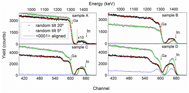
High quality thin InxGa1-xN/GaN bilayers (produced commercially by TopGaN)17 were grown by MOCVD on c-sapphire (0001) substrates. The targeted indium nitride fractions fall in the range 0.3% ≤ x ≤ 14% at growth temperatures varying from 920oC to 790oC as shown in Table 1. To determine the material quality, Rutherford backscattering spectrometry / channeling (RBS/C) measurements were performed using 2 MeV He+ ions and a Si surface barrier detector mounted in IBM geometry at a backscattering angle of 140o. ‘Random’ spectra were taken with tilt angles of 5 and 20o and fitted simultaneously using the NDF code.18 Furthermore, aligned spectra were acquired along the c-axis to assess the crystalline quality of the layers. To determine the lattice parameters, strain state, miscut of the sapphire substrates and the misalignement between the different layers of the samples, high resolution X-ray diffraction (XRD) and X-ray reflectivity (XRR) measurements were carried out using monochromated CuKα1 radiation on a Bruker-AXS D8 Discover system, employing a Göbel mirror and an asymmetric 2-bounce Ge(220) monochromator in the primary beam. XRD rocking cures (RC) were measured using the open scintillation detector. XRR as well as reciprocal space maps (RSM) around the 10-15 reciprocal lattice point were acquired using a 0.1mm slit in front of the detector in the secondary beam. Photoluminescence (PL) measurements were carried out using a monochromated 1000 W Xe arc lamp at 10K. The PL polarization and temperature-dependent PL measurements were carried out using a vertically polarized He-Cd laser operating at 325nm. The samples were mounted in a closed-cycle helium cryostat. The excitation light was incident at an angle of ~60o to the surface normal for the observation of polarization effects.19 For polarization angle-dependent measurements, the laser beam was first expanded and depolarized. The excitation light was subsequently repolarized with a Glan-Thomson prism mounted on a 360o adjustable stage with fine angular adjustment. The light was then focused on the samples in the cryostat using a plano-convex lens. The InN compositions and the layer thicknesses of 4 samples, labeled A-D, were measured using the random RBS spectra shown in Figure 1. The InN composition profile is shown to be uniform throughout the layer thickness for samples A, B, and C. However, Sample D (with high average InN content) shows enhanced InN incorporation near the surface, compared to that at the interface; such behavior is common for InN-rich layers.20 Table 1 summarizes the RBS results.
Sample |
Nominal Growth Temp |
χmin(%) |
InN content (%) (RBS) |
InN content (%) (XRD) |
Thickness (nm) (RBS) |
A |
920 |
2.0 |
0.4 |
n.a. |
40 |
B |
820 |
2.5 |
8.9 |
9.0 |
45 |
C |
780 |
7.0 |
15.8 |
15.9 |
50 |
D |
710 |
70 |
30 (Surface) |
23.9 |
29 (Surface) |
Table 1 The growth temperature, InN contents (measured by RBS and XRD) and InGaN layer thickness measured by RBS

Figure 1 Measured and simulated (solid lines) RBS/C spectra of InGaN layers grown at (a) 920oC (sample A) (b) 820oC (sample B) (c) 780oC (Sample C) and (d) 710oC (sample D). Random spectra were acquired with a tilt of 5ō and 20ō between the surface normal and the incoming beam. The χmin values have been calculated from the 5ō and the aligned spectra which have been acquired using the same integrated charge.
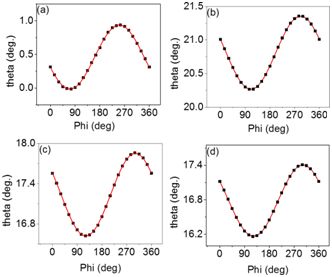
To evaluate the crystal quality of the samples, RBS/C was carried out. Crystal disorder and crystal quality can be quantified by the minimum yield χmin,21 the ratio of backscattering yields from the aligned spectrum to that from the random spectrum. RBS/C spectra in Figure 1 show that Sample A, B, and C have high crystal quality, similar to that of state-of-the-art GaN binary crystal22 with χmin= 2, 2.5 and 7%, respectively (Table 1). However, Sample D shows a significantly poorer χmin of 70%, indicating low crystal quality, as shown clearly in Figure 1. a and c lattice parameters of GaN buffer layers were determined using Bond’s methods yielding values typical of GaN grown on sapphire (a=3.184(1), c=5.189(1)).23 After the samples were accurately aligned using the GaN, diffraction peak the lattice parameters of the InxGa1-xN epilayers were extracted from the asymmetric (10-15) RSMs shown in Figure 2. To calculate the composition of the layers from these values (Table 1) biaxial strain was taken into account using Poisson’s equation. The strain-free a0 and c0 lattice parameters of GaN and InN are taken from Deguchi et al.,24 while their respective stiffness coefficient values C13 and C33 are adopted from extant work.25 Only the GaN diffraction signal was obtained in Sample A: the InGaN peak overlaps with the intense signal from the GaN buffer layer. (Note that the second peak above that corresponding to the GaN buffer layer is due to the Kα2 line, which is not completely suppressed by the monochromator.) The RSMs from Sample B and C show that the InxGa1-xN epilayers are pseudomorphically strained to the GaN buffer, as their Qx positions correspond with that of GaN. In addition, the high symmetry of the InxGa1-xN reflection signal from these samples, as shown in Figure 2B & Figure 2C, signifies high strain homogeneity across the films. The small broadening of the peak also suggests high crystallinity in agreement with the RBS/C results. As expected, the c parameter increases with increasing InN content (Table 2). In contrast, Sample D is found to be partially relaxed, which produces a very weak, and broad peak (figure not shown). Due to the poor crystal quality of sample D revealed by RBS/C and XRD, this sample was excluded from the optical polarization measurements to be described below. To determine the angles of miscut, X-ray reflectivity rocking curves (RC) were first measured as a function of the azimuthal angle Φ at fixed 2θ=0.5º. These measurements are sensitive to the sample surface position. Then, XRD RC of the 006 reflection of sapphire and the 002 reflection of GaN and InGaN (if distinguishable from the GaN peak) were acquired, equally as a function of Φ. The centers of the rocking curves (θmax) were then plotted as a function of Φ (Figure 3) and the experimental results fitted to the function:
(1)
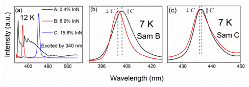
Here, B corresponds to the Bragg angle of the chosen reflection—or 0.25o in the case of x-ray reflectivity (XRR)—and the contribution of an inherent misalignment between the beam, the goniometer and the sample holder. A is the amplitude that is dependent on the angle between the surface in the case of XRR (diffraction planes in the case of XRD) and the sample holder plane on which the sample is mounted. The fitting parameters A and Y describe the vector corresponding to the normal to the measured plane from which the relative orientation of the different layers can be derived. Assuming that the sample surface is parallel to that of the sapphire substrates, for all samples, the sapphire shows a slight misalignment of ~0.34o, as shown in Table 3. In addition, the GaN 002 planes are tilted by ~0.13o from the 006 Al2O3 planes. The sum of these two values agrees well with the directly measured angle between the surface and (0001) planes of GaN. The GaN and InxGa1-xN layers are parallel to each other within experimental accuracy. The miscut analysis results are summarized in Table 3. However, no strain anisotropy is observed for these samples. Figure 4A shows PL spectra of the samples at 12K. Sample A, with 0.4% InN, exhibits only a spectral response typical of GaN, dominated by NBE emission at 357nm. Sample B, with 8.9% InN, has a prominent InGaN peak at 404nm, whereas sample C (15.8% InN) has a dominant peak at 442nm attributed to the InGaN NBE emission. In addition, spectra of both samples B and C include a small component of GaN NBE emission at 357nm.
Sample |
c [Å] aN |
a[Å] aN |
c [Å] InGaN |
a[Å] nGaN |
ε||[%] |
ε⊥ [%] |
A (only GaN detected) |
5.189(1) |
3.184(1) |
n.a. |
n.a. |
n.a. |
n.a. |
B |
5.190(1) |
3.185(1) |
5.262(1) |
3.186(1) |
-1.1 |
0.6 |
C |
5.189(1) |
3.184(1) |
5.324(1) |
3.185(1) |
-1.9 |
1.1 |
Table 2 GaN and InGaN lattice parameters and in-plane ε|| and out-of-plane ε⊥ biaxial strain

Figure 3 XRR and XRD rocking curve peak positions (symbols) and fits using equation1 (solid lines) of sample C, measured as a function of the azimuthal angle (Φ). (A) Sample surface (XRR), (B) sapphire (0001) plane (XRD), (C) GaN (0001) plane (XRD), and (D) InGaN (0001) plane (XRD). See text for details.

Figure 4 (A) Low temperature PL spectra for the samples taken at 12K, using 340nm monochromated Xe arc lamp. (B) and (C): Low temperature PL spectra showing energy blue-shift in the pseudomorphic InGaN films for samples B and C respectively.
Only samples B and C were used for the purpose of polarization PL measurements to investigate if there is an energy anisotropy, since fully compressive strain is a prerequisite for the occurrence of this anisotropy. As previously discussed, sample D is relaxed due to high InN content; hence, it is not reliable for the polarization study. It has to be considered that anisotropic polarization response can be induced for certain incident angles of the polarized excitation laser.19 Therefore, we carried such polarization measurements at low temperature as shown in Figure 4B & Figure 4C. We observed an energy shift of the NBE peak in the PL spectra of samples B and C for different polarized excitation light. In both samples, there is a small, but definite, blue-shift as the polarization angle of the excitation is switched from ∥c to ⊥c. On the other hand, we carried out the polarization measurement at the same incident angle on pure GaN grown on c-sapphire (not shown) and, there was no sign of energy shift of NBE peak with different angle of the polarized excitation laser. In Figure 5A, we show the angular response of sample B PL spectra taken at the low temperature of 7K. The degree of polarization, ρ, is determined using the equation;
(2)
Where I^ is the PL intensity of the sample excited with the laser polarized perpendicularly to the c-axis, and I∥ is the component of the PL intensity with polarization parallel to the c-axis. The polarization degree is highly energy dependent and peaked at 3.08eV, as shown in Figure 5A, corresponding to 31% polarization. The inset gives the angular dependence of the PL peak energy at 7K and 40K, indicating absence of temperature dependence. At these temperatures, as the polarization angle is changed from 180o (∥c) to 270o (⊥c), a gradual blue-shift in the band edge peak is observed (at 7K (40 K) the peak energy value rises from 3.06eV (3.05eV) to 3.07eV (3.06eV) with the increase in the polarization angle). Although polarization selection rules necessitate that the peak of the ^c component occupies the lowest energy,26 they were contradicted in the case of our InxGa1-xN thin epilayers. With polarization parallel to the basal axes (∥c) the samples exhibited higher peak energy than for ∥c. To understand this apparent incongruence, we used multiple Gaussian components peak fitting to resolve the PL spectra of the polarization vectors parallel (^c) and perpendicular (∥c) to the basal axes, as shown in Figure 5B. Both peaks are the cumulative superposition of transitions between two VB energy states, represented by P1 and P2, but with varying intensities. The average full width at half maximum (FWHM) values for P1 and P2 of the contributing peaks were 47.1 (± 2.8) meV and 97.7 (± 5.4) meV, respectively, which suggests that the respective Gauss components are from similar emission centers of heavy and light VBs with respect to both angles. The average shift between the peaks is ~14.24 meV. A similar observation, albeit with a higher (~40meV) shift between peaks, has been reported by Sun et al.,15 for InGaN grown on m-plane in agreement with the 31% polarization degree. However, this argument is not enough to prove the anisotropy as investigating the cause of such energy shift, measuring polarization dependence on InxGa1-xN layers with growth direction along c-axis is not an easy process. Hence, for further investigation, we carried out polarization measurements on the emitted light as the function of the angle of the incident laser with respect to the c-axis. No energy anisotropy was observed for the emitted light. Therefore, such energy shifts of the different polarized light cannot be due to strain anisotropy and valance band separation. Thus, the blue-shift of the peak wavelength due to the band-tail filling effect by carries,27 and observed on m-plane. Such band filling effects can be due to fluctuations of the band gap of inhomogeneous in distribution across the sample that creates localized states.28 In this case, when the laser intensity changes due to different polarization degrees, the filling of the low energy band-tail states occurs by the carries, causing such blue-shift in the NBE peak. In summary, we have carried out structural and optical analyses on slightly miscut InGaN thin film samples. Structural analyses showed that the miscut InGaN thin epilayer was pseudomorphically strained for low in content samples with a state-of-the-art GaN-like quality. We found that polarization dependent PL of excited light showed blue-shift energy with different excitations of light polarization, which may have been induced by the slight misalignment of the InGaN layers. The polarization measurements of the emitted light should not have an energy shift, indicating that energy anisotropy did not occur for the slight tilted InGaN from c-axis. Thus, we conclude that the blue-shift of the NBE emission with polarization is due to the band filling effect.
Sample |
Angles between planes (ō) |
|||
Surf*Al2O3 |
Surf*GaN |
Al2O3*GaN |
Al2O3*InGaN |
|
B |
0.31 |
0.43 |
0.13 |
n.a. |
C |
0.36 |
0.48 |
0.13 |
0.14 |
Table 3 Miscut angles estimated by XRD measurements.

Figure 5 (A) Solid lines are PL spectra of polarized intensities of sample B at angles parallel and perpendicular to the c-axis. The dotted line is the degree of polarization for the sample. (B) Parallel and perpendicular polarization intensities with their multiple Gaussian components at 3.051eV and 3.072eV, respectively.
The author thanks KAUST for the finance support
Authors declare that there is no conflict of interest.

©2018 Ajia, et al. This is an open access article distributed under the terms of the, which permits unrestricted use, distribution, and build upon your work non-commercially.