eISSN: 2574-9927


Research Article Volume 3 Issue 4
1Department of Technology and Metallurgy, University Ss Cyril and Methodius, Republic of Macedonia
2Department of Medicine, University Ss Cyril and Methodius, Republic of Macedonia
3Department of Optics and Spectroscopy, National Academy of Science of Ukraine, Ukraine
4University Ss Cyril and Methodius, Republic of Macedonia
5Institute of Public Health, Republic of Macedonia
Correspondence: Anita Grozdanov, Faculty of Technology and Metallurgy, University Ss Cyril and Methodius in Skopje, Ruger Bošković 16, 1000 Skopje, Republic of Macedonia
Received: July 14, 2019 | Published: August 27, 2019
Citation: Grozdanov A, Paunovi? P, Vasilevska-Nikodinovska V, et al. Structural analysis of x-ray irradiated carbon nanostructures. Material Sci & Eng. 2019;3(4):141-145. DOI: 10.15406/mseij.2019.03.00105
One of the new and promising applications of carbon nanostructures (CNs) are various dosimetry devices. Namely, these devices based on carbon nanostructures can provide the possibility for device miniaturization, lower costs and high scale manufacturing. So, very important factors for the device processing need a quantitative study of the effects of relatively lower doses of X-ray irradiation on the carbon nanostructures. In this study, we present the effects of relatively low doses of x-ray irradiation on the physical and chemical properties of three carbon based nanostructures (multiwall carbon nanotubes - MWCNTs, graphene - G, hybrid - G/MWCNTs). We have used a range of characterization techniques including scanning electron microscopy, Raman and FTIR spectroscopy, thermal and particle size analysis. Specifically, it was found that irradiation exposure results in a reduction in the sp2 nature of all three carbon based nanostructures.
Keywords: x-ray irradiation, carbon nanostructures, graphene, Raman, SEMEmerging nanotechnologies in which nuclear applications and radiations play key roles are: nano-electronics in environmental monitoring and remediation, electrode materials in hydrogen economy, polymer based nanocomposites in biotechnology, diagnostics and therapy. Radiation based technology using x-rays, e-beams and ion-beams is the key to avariety of different approaches. Due to the various ionizing irradiations, physical, chemical and biological properties of the materials can be significantly modified. Compared with conventional chemical reduction, the irradiation techniques are environmentally friendly, easily controlled, highly pure and less destructive. The most common defects induced by irradiation are vacancies and interstitials. Carbon based nanostructures with sp2-like hybridization, are exclusive due to the fact that its valence permitted researchers to engineer a large collection of molecular architectures. What makes all these structures truly phenomenal is that they are indeed built from the same component and they still can differ in shape and dimensionality. The most prolific irradiation-induced defects in graphenic carbon nanostructures are vacancies (single or multi - vacancies). These carbon sp2- nanostrucutres develop an extended reconstruction of the atomic network near the vacancy by saturating two dangling bonds and forming a pentagon. In graphene, single vacancies reconstruct, but in CNT the reconstruction is much stronger owing to the curvature and inherent nanoscale size of the system. It was found that for a CNTs to contract locally to "heal" the hole and thus saturate energetically unfavorable danging bonds. Thus, curved graphitic structures such as CNTs can be referred to as self-healing materials under irradiation. Some of the last experimental studies on the irradiation of MWCNTs reported a broad range of interesting phenomena such as surface reconstructions, modified mechanical properties, ion-irradiation induced changes in electrical coupling between nanotubes.1-3 Kis et al., have shown a strong stiffening of bundles of CNTs after electron irradiation.4 Last years, irradiation with γ-rays was studied as one of the clean and easy method for modification of carbon nanostructures. Namely, the effects of γ-irradiation strongly depend on the irradiation conditions, the materials type and the irradiation medium. Guo et al. observed a dramatic increase in the ID/IG of the Raman spectrum of γ-ray irradiated multi-walled CNTs (MWCNTs), which was attributed to the large presence of sp3-hybridized carbon atoms.5 This is opposite to the trend reported by Xu et al.,6 who noted an 8% decrease in ID/IG for MWCNTs irradiated to 20Mrad in air, signaling improved graphitic order.6 Also, it was found that γ-irradiation decreased the diameter of MWCNTs, increased their specific surface area and modified their oxygen functional groups.7 The graphitization of MWCNTs was improved with doses of 100kGy, while a higher dose of 150kGy induced structural damage.7 Regarding the graphene, γ-irradiation was used for the reduction of graphene oxide in different liquid media.8 Bardi et al.,9 studied x-ray irradiation induced structural changes on single wall carbon nanotubes.9 Based on the Raman and XPS measurements, they confirmed the modifications in the structure of the nanotube surfaces, and found that the degree of disorder in the CNTs structure correlates with the x-ray irradiation dose.9 Although a huge amount of theoretical works were done to understand the origin of various kinds of irradiated induced structural changes and defects in carbon nanostructures, very little is known experimentally. Thus, the present work is aimed to focus on the influence of X-ray irradiation on the structural identification of changes and defects formed in carbon based nanostructures (G, MWCNTs, hybrid G/MWCNTs).
Experimental irradiation treatment included exposure of carbon nanostructures to relatively low X-ray irradiation at 140keV of X-rays in air-atmosphere, for 30min. Three different carbon nanostructures: graphene - G, multi wall carbon nanotubes–MWCNTs and hybrid G/MWCNTs were used. All three carbon nanostructure’s were produced in the nano-lab of Faculty of Technology and Metallurgy, using the method of molten salt electrolysis. Pre- and post- irradiation characterization of all treated carbon nanostructures was performed using several techniques such as FTIR-ATR and Raman spectroscopy, Particle Size and Zeta potential measurement, SEM and TGA analysis. Thermal behavior was followed using TGA/DTA analysis PE-Dymond D7 in temperature range of 30-1000°C, in atmosphere of N2, at 20 K·min–1. The morphologies of pristine and irradiated CNs were observed by scanning electron microscopy (SEM) (JEOL model JT-FFX). Raman spectroscopic analysis of the examined CNs was performed using JobinYvon HR640 instrument, under ambient conditions. Zeta potential was measured using MALVERN Zetasizer NANO. 0,0025g of the functionalized CNs were mixed with 20ml of distilled water and sonicated for 1 h. Before the measurement, the samples were filtered through the 0,45mm membrane. All the measurements were performed at 25±0.10ºC. FTIR-ATR spectroscopy was performed using Thermo Nicolet iS50 FTIR-NIR spectrometer. FTIR spectra of all the studied samples were collected in ATR mode in the range from 400cm-1 to 4000cm-1.
In order to understand the influence and the effects of X-ray irradiation on the structure of MWCNTs, G and G/MWCNTs hybrid, applied at lower doses, several analytical techniques were used. Characteristic thermo grams obtained by TGA/DTA analysis are shown in Figure 1 & Figure 2. Due to the additional graphitization of carbon networks induced by X-Ray irradiation, the irradiated carbon nanostructures have demonstrated higher thermal stability and the main thermal peaks were shifted to higher values for all three tested carbon nanostructures (TdMWCNT=581,2°C; TdMWCNT-XRay=747,5°C). Raman spectroscopy has proven to be a powerful technique in the study of CNs and become a key tool to identify disorder in the sp2 network of different carbon nanostructures. This is a noncontact and nondestructive tool. Usually, in sp2-bonded carbon, the ratio of the intensity of the so-called “D-band” at around 135cm-1 to the intensity of the “G-band” at 1580cm-1 can be used as a parameter for estimating the amount of disorder. Characteristic Raman spectra of the studied MWCNTs, G and hybrid G/MWCNTs, before and after x-ray irradiation, are shown in Figure 3-5, while the calculated ID/IG ratio is given in Table 1. Evidently, due to the irradiation treatment structural changes were registered in the Raman spectra of the studied carbon nanostructures. For MWCNTs, it was found that G peak decreased and become wider. The same effect after irradiation was found and for the G/MWCNTs hybrid, G-band decrease while D band increase. For G/MWCNTs hybrid it was found also that 2D peak decrease. Smaller changes were registered for the irradiated G nanostructure (Figure 3-5) (Table 1). To evaluate the impact of the irradiation treatment, the ratio of the intensities of the D and G peaks was monitored before and after irradiation, as it is a useful indicator of long range order in carbon nanostructures and how great is the x-ray induced damage on the CNs.10 According the Raman theory, the ratio of intensities of D and G peaks (ID/IG) is the measure for orderness/disorderness of the carbon based nano-structure. The ratio of the D-peak (ID) height to the G peak height (IG) was also affected, but less markedly, changing from 0.21 to 0.18 for G (from 0.3 to 0.28 for MWCNTs and from 0.97 to 0.8 for the hybrid G/MWCNTs). These changes resulted in approximately 15% decrease of the ID/IG ratios which indicate the additional graphitization at these smaller doses. According the literature review, usually this ratio increased with total dose and decreased after annealing.11 Literature has shown that usually at lower doses of other type of irradiations (γ-irradiation or electron beam irradiation) this ratio increase which indicate structural disorders, but our results for x-ray irradiation effect is opposite. The FTIR spectra obtained in FTIR-ATR mode are shown in Figures 6-8. The FTIR spectra of x-ray irradiated carbon nanostructures at lower doses have shown that during the irradiation of carbon chains additional graphitization occurred.
Sample |
ID/IG |
G |
0,21 |
G/x-ray |
0,18 |
MWCNTs |
0,30 |
MWCNTs/x-ray |
0,28 |
G/MWCNT s |
0,97 |
G/MWCNTs/x-ray |
0,8 |
Table 1 ID/IG ratio of the raw and irradiated carbon nanostructures
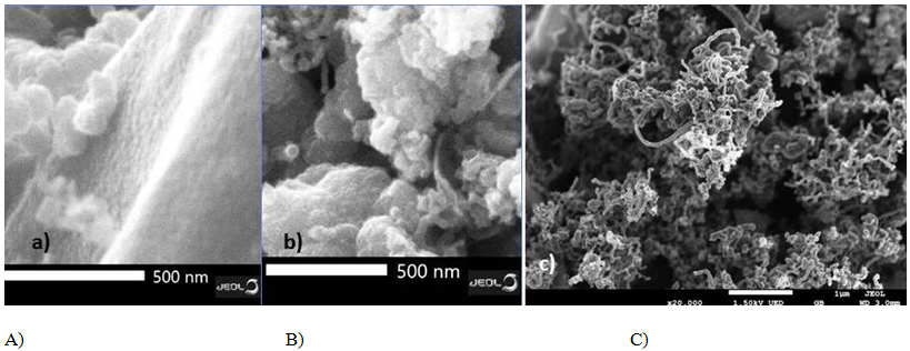
Namely, it was found that the position of all the prominent peaks remains unchanged after the irradiation except for the peak corresponding to C=C. It’s appeared at 1576cm–1 for an non-irradiated sample, and shifted to 1603 cm–1 upon irradiation. The shift in the peak may suggest a change in the carbon structure of G (Figure 7) and G/MWCNTs (Figure 8) nanostructures. The results of Zeta potential measurements are presented in Table 2. The obtained values for Zeta potential for all tested samples, pristine and x-ray irradiated ones are negative in pH ~7 (of distilled water) which means that the surface of the carbon nanostructures has negative charge. The measured values for the Zeta potential are changing from –16.8mV to –32.9mV in pH neutral media. The SEM images of the carbon nanostructures, before and after the x-ray irradiation, are presented in Figure 9 & Figure 10, to distinguish the varying morphologies. As it is shown in Figure 9A, graphene displays well-exfoliated nano-sheets, while after the irradiation; G (Figure 10A) shows crumpled layered structure which restrains the aggregation of G nano-sheet’s. For the hybrid G/MWCNTs (Figure 9B-before and Figure 10B-after the irradiation), where MWCNTs are incorporated into G layers, G/MWCNTs exhibited more crumpled, porous and loose architecture compared with G, indicating that MWCNTs alter the morphology distribution of G and impede the stacking of G to effectively enlarge accessible surface area. Pristine MWCNTs are shown in Figure 9C while the irradiated wrinkled MWCNTs are shown in Figure 10C.

Figure 10 SEM images of x-ray irradiated carbon nanostructures: A) graphene; B) G-MWCNTs hybrid; C) MWCNTs.
Sample |
Z[mV] |
St.Dev [mV] |
MWCNTs |
–32,4 |
6,27 |
MWCNTs/x-ray |
–20,4 |
8,11 |
G |
–32,5 |
6,61 |
G/x-ray |
–16,8 |
4,79 |
Hybrid G-MWCNTs |
–32,9 |
4,23 |
Hybrid G-MWCNTs/x-ray |
–26,0 |
4,64 |
Table 2 Zeta potential values of the raw and irradiated carbon nanostructures
Three carbon nanostructures (G, MWCNTs and hybrid G/MWCNTs) were treated by X-ray irradiation at lower doses. Morphological changes in all three carbon nanostructures were found by SEM. The TGA and Raman analysis have confirmed that additional graphitization occurred. TGA/DTA thermo-grams have demonstrated higher thermal stability and it was found that the main thermal peaks were shifted to higher values for all three tested carbon nanostructures, due to the additional graphitization of carbon networks induced by X-Ray irradiation. Raman analysis has shown that changes of the ID/IG ratios of irradiated carbon nano-structures have been resulted from the additional graphitization at smaller doses of irradiation. The results of Zeta potential measurements for all three types of carbon nanostructures, pristine and x-ray irradiated ones are negative in pH ~7 (of distilled water) which means that the surface of the carbon nanostructures has negative charge.
The author would like to thank you to the IAEA agency since this research work was supported by IAEA TC MAK1003 project.
Authors declare that there is no conflict of interest.

©2019 Grozdanov, et al. This is an open access article distributed under the terms of the, which permits unrestricted use, distribution, and build upon your work non-commercially.