MOJ
eISSN: 2374-6939


Case Report Volume 10 Issue 3
1Professor of Orthopedics, Department of Orthopedics, Jawaharlal Nehru Medical College, Datta Meghe Institute of Medical Sciences, India
2Resident Orthopedics, Department of Orthopedics, Jawaharlal Nehru Medical College, Datta Meghe Institute of Medical Sciences, India
Correspondence: Nareshkumar S. Dhaniwala, M2/2, Meghdoot Apartments, Paloti Road, Sawangi Meghe, Wardha, India
Received: January 29, 2018 | Published: June 11, 2018
Citation: Dhaniwala NS, Dhaniwala MN. Conservative management of acetabular fractures-case reports. MOJ Orthop Rheumatol. 2018;10(3):329-331. DOI: 10.15406/mojor.2018.10.00422
Acetabular fractures are common and complex injuries occurring due to high velocity trauma. There are proponents of both non-operative and operative management. Operative management requires proper training and planning. Long term follow up of conservative management have shown good to excellent functional and radiological outcome. The article describes two cases of acetabular fractures treated conservatively and showing good clinical outcome.
Keywords: Acetabulum fracture, conservative managementFracture acetabulum is a severe skeletal injury. It may be a part of pelvic injury or may present as a component of central fracture dislocation of hip. It occurs due to high velocity trauma such as road traffic accidents, vehicular collision and fall from height. High speed motor vehicle trauma has increased markedly the incidence of serious pelvic and acetabular fractures; however, the more widespread use of surgical stabilization has helped reduce the mortality and morbidity in patients with these injuries.
The treatment of acetabular fractures is a complex area of orthopedics that is being continually refined. It involves a definite learning curve, best documented by Matta & Merritt.1 The quality of acetabular fracture reduction is the single most important factor in the long term outcome of these patients, and such surgery should be undertaken only by surgeons with sufficient experience. Acetabular fractures usually are having associated injuries to other bones or the other system, such as abdomen, urethra, chest etc. Treatment of the patient should follow accepted Advanced Trauma Life Support protocol. In general, operative treatment of an acetabular fracture should not be performed as an emergency, except when it is part of open fracture management or is performed for a fracture associated with a dislocation of hip.
The term central fracture-dislocation of the hip is used to describe any acetabular fracture with medial dislocation of the femoral head. In a true central fracture dislocation, the femur head gets dislocated medially into the pelvis and may get locked between the fracture fragments. Closed reduction under general anaesthesia and fluoroscopic assistance may succeed in relocation of the head, but there is possibility of redisplacement into the pelvis if skeletal traction is not maintained.
The case series reports two cases of comminuted acetabular fractures which were treated successfully using skeletal traction along with mobilization.
A 26 years old male villager presented in the emergency service of tertiary care rural hospital with complaints of swelling over the right side of face, pain in right hip and inability to bear weight on the right lower limb for one day following road traffic accident the previous night. The patient was hit and thrown away by a motor vehicle. He had history of transient loss of consciousness. On examination, the GCS score was 15/15; there was diffuse swelling on the right side of face with tenderness over the zygomatic arch. The right hip was found to be in 10 degrees flexion attitude and there were abrasions on the right knee. There was tenderness in the right groin directly and on thumping at right greater trochanter. Besides, right clavicle was tender and irregular at the junction of medial two thirds and lateral one third. Right hip and right shoulder movements were grossly painful and restricted. There was no neuro-vascular deficit and the limb lengths were equal.
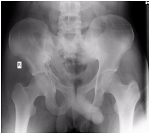
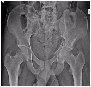
Radiograph of pelvis and right shoulder showed fracture right clavicle and comminuted fracture of right acetabulum with slight migration of the femoral head medially. The acetabulum floor was grossly comminuted and the fracture acetabulum was extending into the Ilium both anteriorly and posteriorly (Figure 1). It was extending to involve superior pubic ramus also. CT brain revealed extradural hemorrhage in the right temporal region with multiple facial bone fractures on the right side. The patient was applied right upper tibial pin for skeletal traction with 7kg weight. Foot end of the bed was elevated for counter traction. As the patient and relations refused any surgical management, traction was continued. Bed side X-ray pelvis with traction on was taken after 24 hours to check for femoral head position. The patient was explained quadriceps and knee exercises and was encouraged in rounds to do it properly. The pin tract was dressed daily with betadine. After 2 weeks reduction was confirmed again by x-ray and traction continued. Mobilization of the hip was encouraged gradually with traction on to help in the joint reformation and gain of movement. The skeletal traction was continued for 5 weeks, after which it was changed to below knee skin traction with 2kg weight. The patient had become painless by 4 weeks and regained 75% of hip movements by 6 weeks. His x-ray pelvis at 7 weeks showed uniting fracture floor of acetabulum with reduced femoral head and no evidence of true shortening (Figure 2). The patient was advised non-weight bearing ambulation with walker for three months. At three months follow up he was satisfied with function, had no shortening and was able to do his routine work. He continues to be in follow up.

Figure 1 X ray pelvis anteroposterior view on the day of trauma showing comminuted fracture floor of acetabulum extending into ilium.

Figure 2 X ray pelvis anteroposterior at 7 weeks showing uniting fracture acetabulum and ilium with femoral head in position.
A 45 years male presented with complaints of inability to bear weight on left lower limb and walk for two days following a fall of heavy object on his back. Examination revealed limb in neutral rotation, tenderness at left groin directly and on thumping at the greater trochanter. The left greater trochanter was less prominent than right side and all the hip movements were painfully restricted. X-ray pelvis showed comminuted fracture floor of acetabulum, with medial migration of the femur head. The patient was applied heavy (10kg) upper tibial pin traction with the thigh supported on a pillow. Lateral traction was applied on the proximal part of thigh with a thick cotton sheet sling having arrangement for traction at the end. Check x-ray after two days showed that the femoral head had pulled back to normal position in acetabulum and the displaced pieces of the floor of acetabulum came in acceptable position. Traction was continued after reducing the weight to 8kgs, for four weeks. Hip and knee mobilization was encouraged with traction on after three weeks. The patient regained fair range of movements by four weeks. Traction was changed to skin traction and maintained with less weight for further two weeks. He was kept non- weight bearing for three months, after which he resumed his activities gradually. At 12 months follow up he was painless, walking unsupported and carrying out his routine work satisfactorily.
Letournel & Judet’s2 classification of acetabular fractures is used widely. This divides acetabular fractures into two basic groups: simple fracture type and the more complex associated fracture type. Simple fracture types are isolated fractures of one wall or column along with transverse fractures. Associated fracture types have more complex geometries and include T-type fractures, combined fractures of one column and wall and both column fractures.
Longer follow up of operatively treated acetabular fractures has shown arthritis in cases of small residual incongruencies. On the basis of long term results indications for non- operative and operative treatment have been defined better. Indications for non- operative treatment are: Nondisplaced and minimally displaced fractures, fractures with significant displacement but in which the region of the joint involved is judged to be unimportant prognostically, secondary congruence in displaced fractures, medical contraindications to surgery, local soft tissue problem, elderly patients with osteoporotic bones, and refusal to accept surgical treatment.3 In a retrospective study, of 71 fractures in 69 patients, with mean age 38.6 years, Magu NK et al.,4 noted good to excellent function in all patients with posterior wall fracture, including four cases having more than 50% of broken wall. Good to excellent function was seen in 88.8% of both column fractures with secondary incongruence despite medial subluxation. Gansslen A et al.,5 also noted good hip function on long term follow up of five years. Secondary congruence of joint during conservative management and follow up is supposed to be associated with good function and minimal complications. Amarawati RS et al.,6 also found comparable congruent reduction in non- operatively treated acetabular fractures in their study.
The two cases reported herein are from the young age group from rural background. Both of them refused surgery and opted for conservative treatment by traction followed by mobilization and gradual weight bearing. Both were satisfied with the gain of hip function and the absence of pain in the short term follow up. In acetabular fracture without medial migration of hip longitudinal skeletal traction alone is sufficient for treatment. In case of medial migration of femoral head into pelvic cavity lateral traction is important for pulling back the femoral head in position. A larger and longer follow up study is needed to determine the long term results.
Conservative treatment in form of traction followed up with mobilization of hip can be tried with reasonable functional outcome in undisplaced/minimally displaced fracture acetabulum, moderately displaced fractures without involving weight bearing area of acetabulum, patients unfit for surgery and patients not consenting for surgery.
None
Author declares there are no conflicts in publishing the article.

©2018 Dhaniwala, et al. This is an open access article distributed under the terms of the, which permits unrestricted use, distribution, and build upon your work non-commercially.