MOJ
eISSN: 2574-8130


Case Series Volume 3 Issue 6
1University Pirogov Russian National Research Medical University (RNRMU), Russia
2N.N.Blokhin National Medical Research Center, Russia
Correspondence: M Gromova, Designation Assistant of the department, University Pirogov Russian National Research Medical University (RNRMU), Address 117997, Moscow, Ostrovityanov St, Russia, Tel 89056491356
Received: August 24, 2018 | Published: December 3, 2018
Citation: Gromova M, Tsurko V, Kashkadayeva A, et al. Early diagnostics of nefrosklerosis and urinary infection in patient with chronic tophaceous gout. MOJ Gerontol Ger. 2018;3(6):453-455. DOI: 10.15406/mojgg.2018.03.00165
Introduction: We report a case of renal failures diagnostics and urinary infection in the patient with chronic tophaceous gout and a premorbidal background.
Case Presentation: Man with obesity and arterial hypertension has gout. It is taped: hyperuricemia, azotemia, proteinuria; low clearance of a creatinine, stone of left kidney; hypoplasia of right kidney. Systemic examination of nephrourological status based on complex renal scintigraphy (SENS-CRS) has established symptoms of nephrosclerosis and urostasis in urinary tract against the background of an exacerbation of urinary tract infection.
Disucssion: The SENS-CRS technology allows to prognosticate the risk of a renal failure, to warn or slow down development of chronic kidney disease.
Conclusion: The SENS-CRS technology filled a disadvantage of control devices behind kidneys.
Keywords: diagnostics, gout, nephrosclerosis, infection of urinary tract
One of the frequent manifestations of gout is the gout nephropathy.1 For assessment of the urinary system functional reserves and the risk of renal failure routine analyses of urine in combination with a sonography are often not enough.2,3 Modern technology of the systemic examination of nephrourological status based on complex renal scintigraphy (SENS-CRS) was developed in the laboratory of radioisotope diagnosis in «N.N.Blokhin National Medical Research Center» and implementedas an automated workplace. SENS-CRS technology is designed for assessment of the functional reserves of the urinary system and the risk of renal failure at all macrostructural levels and allows lowest radiation doses (0.6 mSv for one patient).4,5 We report a case of renal failures diagnosticsand urinary infection in the patient with chronic tophaceous gout and a premorbidal background.
A 70-years old Caucasian male, weight of 95 kg, height of 170 cm. He complaints on frequent urination, pain in the lumbar region, pains, restriction of movements and edema of the right I metatarsophalangeal joint. From an anamnesis: the diagnosis of gout was established in December 2002, since January 2003, he has received allopurinol 100 mg per day. Rheumatologist has not observed the patient. The last gouty attack was about 2 weeks ago when the right knee joint has ached and has swelled up a little. He took the nonsteroid anti-inflammatory drugs (nimesulide 200 mg per day) not having long effect. The postponed diseases: arterial hypertension with arterial blood pressure up to 160/90 mm Hg. He receives anti-hypertensive drugs every day. When carrying out laboratory and tool inspections in blood tests uric acid rose up to 488 µmol/l, creatinine up to 163µmol/l, urea up to 12,9 mmol/l, cholesterin up to 6,7 µmol/l. In the general analysis of urine specific gravity was 1012, glucose isn't taped, protein -0,108g/l, leucocytes 4-5 under review, erythrocytes 3-4 under review. A 24-hours creatinine clearance test–79 ml/min, it corresponds to insignificant extent of reduction in the rate of glomerular filtration; canalicular reabsorption –99% (norm). At ultrasound examination of kidneys: left kidney - a parenchyma of 15 mm, sine condensed, stone of 12 mm; right kidney - parenchyma of 11 mm, sine dense; ultrasonic signs of diffuse changes of a right kidney, right kidney hypoplasia; fine cysts of both kidneys; good-quality hyperplasia of a prostate.
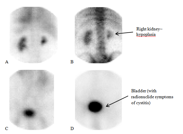
Data of SENS-CRS: The relative renal blood stream (QL:QR) is asymmetric: 30% in the right kidney, 70% in the left kidney (Table 1). 𝐴 [s]-an arterial index of renal parenchyma and 𝑉%] - a venous index of renal parenchyma, it is slightly broken, in right–the expressed hemostasis (a parenchyma edema). In the left– depression of concentration function (Gren) from insignificant to moderate degree, in the right–moderated with a trend to appreciable degree (Table 1). Radionuclide symptoms of irreversible "nephrosclerosis" are taped; removal from a parenchyma of kidneys on transition at the “cortex-medulla” level (an indicator of the D %) is steadily reduced, especially on the right (Figure 1). On an exacerbation of cystitis specifies the high level of a residual urine in a bladder (Figure 1).; the permeability in edematous ostiums of ureters is reduced. High relativeurostasis (KPL, KPR), steady a reflux arrhythmia of outflow of urine from pelvis and on ureters are characteristic of the infection of urinary tract (Figure 2). Outflow disturbance (more inthe right), but removal reserve: urostasis in the pelvis decreased after reception of 350 ml of water at examination. Conclusion of SENS-CRS: cooperative function of kidneys is moderately reduced with a trend to appreciable depression, the transitional and unstable level of compensation. The expressed asymmetry in renal blood stream with depression of function of a right kidney. Radionuclide signs of an exacerbation of infection of urinary tract. II stage of the chronic kidneys disease.
Kidney |
Relative renal blood stream (QL:QR) |
Haemodynamics of kidneys |
Measured level of 99mTc-technephore in the renal parenchyma (Gren) |
Total prognostic index for the urinary system |
Left (L) |
70% |
Accelerated (by meds) |
16,0 |
Transitional and unstable compensation with signs of an exacerbation of an infection of urinary tract is moderately reduced with a trend to appreciable degree (FSS=2b). The poliurine state against the background of hypotensive therapy. |
Right (R) |
*30% |
the expressed hemostasis |
*11,2 |
Table 1 Data SENS-CRS with99mTctechnephore
Note* The size of parameter went beyond norm

Figure 1 Scintigrams at the SENS-CRS basic test with99mTctechnephore (A–back, B-a forward projection) and the delayed examination (C–back, D-a forward projection). Symptoms of nephrosclerosis of kidneys are visualized: the picture of "a light rim" around the localized accumulation in pelvis is, presumably, caused by a sclerosis of interlobar arteries.
The probability of a nephrosclerosis and exacerbation of infection of urinary tract at gout is high. Unaccounted in time or undertreated disturbances of outflow of urine considerably complicate work of kidneys. The SENS-CRS technology allows to prognosticate risk of a renal failure, to warn or slow down development of chronic kidney disease. The detailed differential analysis of urodynamics according to the data of SENS-CRS made it possible to reveal a pronounced picture of exacerbation of latent infection of urinary tract in patient, as well as a lack of assistance in the treatment of prostatic disease.
The SENS-CRS technology provides the quantitative assessment of kidney blood cleansing from 99mTctechnephore and concentrational function of parenchyma as well as unique quantitative indicators of urodynamic delays in all parts of urinary tract. This kind of functional diagnostics allows to monitor parenchima and urinary tract condition promptly and with lowest radiation doses, apply therapeutic measures to prevent more severe kidney dysfunction and refer the patient who was regularly receiving treatment concerning gout, objective indications to treatment and dynamic observation not only to rheumatologists, but also to nephrologists, urologists.
None.
The author declares no conflicts of interest.

©2018 Gromova, et al. This is an open access article distributed under the terms of the, which permits unrestricted use, distribution, and build upon your work non-commercially.