Journal of
eISSN: 2373-6410


Case Report Volume 5 Issue 2
Royal medical services, Neurosurgery department, Jordan
Correspondence: Alqroom Rami, Neurosurgery department, Royal medical services, Jordan
Received: August 11, 2016 | Published: October 24, 2016
Citation: Alqroom R, Abu Nowar H, Firas S, Amer AS, Wesam K (2016) Thecoperitoneal Shunt ‘Cast Away’. J Neurol Stroke 5(2): 00172. DOI: DOI: 10.15406/jnsk.2016.05.00172
Mechanical shunt complication accounts for more than half of all shunt failures. Ever since its introduction, the use of cerebrospinal shunting systems has led to complications. Nevertheless, complication, though fewer nowadays, persist. Such complications: mechanical failure, infections and migration. We are reporting a very rare case of thecoperitoneal shunt migration not only towards peritoneal cavity also towards third ventricle.
Keywords: Shunt migration, Thecoperitoneal shunt complication, Cerebrospinal fluid
CSF fistula/CSF leak is a pathological disorder that spans all ages. the primary clinical characteristic of this disorder is abnormal leakage of cerebrospinal fluid (CSF), from CSF containing compartments of the central nervous system (CNS) to the around space due to abnormal defect of the Dura matter and the skull,1 which typically appears as a clear watery leakage from nose (rhinorrhea) or ears (otorrhea). CSF which is the fluid produced and contained within the ventricular system and subarachnoid space, has many putative roles including mechanical protection, homeostasis of neural tissue.2
CSF Fistula as a result of laceration of Dura and arachnoid associated with skull defect, pathophysiologicaly classified: spontaneous 4%, from surgical procedure 16%, and the vast majority due to nonsurgical trauma 80%1-3
Presentation varied in time and clinical manifestation terms and could be subtle. as in cases of spontaneous fistula , diligence and high index of suspicion required to diagnose a positional clear watery drainage from nose on intermittent fashion, headache, generalized malaise or the salty taste in mouth….! In cases of traumatic fistula both the time and the history mostly leading to diagnosis.1,2
Diagnosis and localization of CSF fistula may be based on expectation as in post surgical procedure,3 also different methods introduced such as: fluid analysis, Beta-2 transferrin, high-resolution CT scan, or Cisternoghraphy.2
Untreated CSF leak can represent a potentially life threatening situation leading to brain infection, meningitis, tension Pneumocephalus, stroke, or death.
Management of this condition needs treatment strategy which devised taking into account the cause and the location of CSF fistula. Conservative management is a valid option consisting of strict bed rest and elevation of the head at 30˚C, patient advised to refrain from straining or Valsalva maneuvers.3 Also some advocated carbonic anhydrase inhibitor (Diamox) as we do in our center. The overall rate of cessation with conservative management 39.5%, 23 if conservative management extended to 7 days, resolution rates improve to 85% especially in posttraumatic cases.2 Surgical treatment includes: transcranial approach3,4 intracranial approach5 Trans nasal approach,6 and endoscopic transnasal7,8 or thecoperitoneal shunt.3,9,10
Thecoperitoneal shunt is a technique of CSF diversion from the lumber peritoneal sac to the peritoneal cavity, although generally a safe procedure, potential complications might happen such: bleeding, nerve irritation, paralysis, infection, post spinal headache, pneumocephalus, acquired Chiari malformation, shunt migration.11-16
Shunt migration is a less frequent complication of shunt surgery. It is known to migrate into all the possible sites in the body. Cranial migration is rare. Very few reports about the LP shunt exist in the literature.10 Thecoperitoneal shunt migration into third ventricle has not been reported. Such a condition is reported in our case.
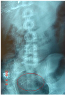
A 24 year old male patient today, developed traumatic CSF fistula (rhinorrhea) in 2001 after direct blow to head. Thin slices-CT scan showed cribri form plate defect. Surgical transcranial approach was opted, using fascia lata. The post-operative period was uneventful (this management was in other hospital, data according to his file. patient age was 11 years at that time). In 2006 patient presented to our center with intermittent watery clear discharge from the nose, especially when lying, family having in mind the previous history and the high suspicion was worrying from recurrence. Patient couldn’t recall any history of recent trauma. CSF analysis and beta-2 transfer in confirmed the diagnosis. No further evaluation required as family decision was "No transcranial approach!" Decision was taken to proceed with thecoperitoneal shunting as a valid alternative option. A thecoperitoneal shunt tube was inserted. Operation was incident free and post operative period was uneventful. Patient was discharged and for the following 3 visits he had no complaints at all; there were no symptoms or signs of raised intracranial pressure. 2 years later the patient presented to our out-patient clinic complaining of headache, he reported no associated symptoms also no history of trauma. As a routine evaluation in our center, after the clinical examination and fundoscopy which was normal, we ordered hardware X-ray Figure 1 to evaluate position, continuity or dislodgment of the CSF shunting system. Also a skull x-ray Figure 2 was ordered to evaluate possible pneumocephalus or other pathologies before proceeding to further images. It was very surprising, therefore, to see thecoperitoneal shunt tube in the intracranial cavity, although shunt hardware migration had been reported but not intracranially in such type of shunts. Immediately brain CT-scan performed Figure 3a & 3b which confirmed the diagnosis.

Figure 1 The distal end of the thecoperitoneal shunt dislodged and collected in lower peritoneal cavity.
Due to the fact that shunt tube is in eloquent area and relatively asymptomatic, the parents did not agree for any surgical intervention.
CSF fistula/ CSF leak is a pathological disorder that spans all ages. The primary clinical characteristic of this disorder is abnormal leakage of cerebrospinal fluid (CSF). Due to abnormal defect of the Dura matter and the skull.1 Which typically appears as a clear watery leakage from nose (rhinorrhea) or ears (otorrhea). CSF which is the fluid produced and contained within the ventricular system and subarachnoid space, has many putative roles including mechanical protection, and homeostasis of neural tissue.2 Untreated CSF leak can represent a potentially life threatening situation leading to bran infection, meningitis, tension pneumocephalus, stroke, or death. Management of this condition needs treatment strategy which devised taking into account the cause and the location of CSF fistula. Conservative management is a valid option and surgical treatment includes: transcranial approach3,4 intracranial approach,5 Trans nasal approach,6 and endoscopic transnasal7,8 or thecoperitoneal shunt.9,11,18
Lumboperitoneal shunt has an advantage of being completely extra cranial procedure. It is better in growing children as fewer revisions are required, it can be used in many conditions such as CSF fistula.
Ever since its introduction, the usage of cerebrospinal fluid shunt hardware has led to complications, so much so that in the early period it was abandond12-17,19 with the development of more user- friendly hardware system. Nowadays shunt systems regained their place in favor, nevertheless, complications, though fewer, persist. Shunt migration towards peritoneal cavity has been reported and can be due to the intestinal peristaltic movement, which create a pulling effect on shunt tube. But, what about the case of intracranial migration??!! Although the mechanism is not will understood several mechanisms have been reported to contribute in this complication: negative sucking intra-ventricular pressure, positive pushing intra-abdominal pressure.11,18 The majority of patients, like the present one, had been infants and children. Shunt migration is more frequent with hard and spring loaded shunt tubes. This complication is infrequent with flexible shunt systems, but they are more prone to obstruction due to bending and torsion. Migration may be in either direction. Distal migration of the shunt has often been reported.11
A rare complication of cranial migration of the catheter in the posterior fossa has been reported.17,11 Shunt migration into the 3rd ventricle has not been reported.20 The mechanism of shunt migration involves adhesion, necrosis, penetration, perforation; migration and extrusion, Pressure gradient between two cavities decide the direction of migration.21 It can be early or late and is assisted by tortuous subcutaneous tract and neck movements. Shunt chamber prevents shunt migration in cases of ventriculoperitoneal shunts.22 The shunt tube used in this child did not have a chamber. Migration of this type of shunts is prevented by locks and slip clips. In such cases, additional sutures could be placed around the tube to secure the shunt in place.23
Shunt migration usually presents as raised intracranial pressure. Absence of raised intracranial pressure signs in this case suggests equilibration of CSF pressure gradient. Diagnosis in such cases is incidental and the patient can be followed up expectantly. However presence of raised intracranial pressure would require removal of migrated shunt and implantation of a new shunt, preferably with a reservoir.
All authors certify that they have no affiliations with or involvement in any organization or entity with any financial interest (such as honoraria; educational grants; participation in speakers’ bureaus; membership, employment, consultancies, stock ownership, or other equity interest; and expert testimony or patent-licensing arrangements), or non-financial interest (such as personal or professional relationships, affiliations, knowledge or beliefs) in the subject matter or materials discussed in this manuscript.
Informed consent was obtained from participants included in the study. The patient has consented to submission of this case report to the journal.
None.
None.

©2016 Alqroom, et al. This is an open access article distributed under the terms of the, which permits unrestricted use, distribution, and build upon your work non-commercially.