Journal of
eISSN: 2373-6410


Review Article Volume 16 Issue 1
 Dhrubajyoti Chattopadhyay PhD1,2
1H. P. Ghosh Research Center, New Town, India
2Sister Nivedita University, India
Correspondence: Dr. Swarup K. Chakrabarti, PhD, H. P. Ghosh Research Center, HIDCO (II), EK Tower, New Town, Kolkata, West Bengal 700161, India
Received: November 25, 2025 | Published: January 13, 2026
Citation: Chakrabarti SK, Chattopadhyay D. Microplastics as drivers of neuroinflammation: a review. J Neurol Stroke. 2026;16(1):24-30. DOI: 10.15406/jnsk.2026.16.00647
Microplastics (MPs) and nanoplastics (MNPs) are emerging environmental pollutants that raise concerns about their effects on brain health. This review examines their neurotoxic potential, particularly their ability to cross biological barriers, cause oxidative stress, and lead to neuroinflammation—processes associated with neurodegenerative diseases. Research indicates that MPs/MNPs accumulate in the brain, disrupt cell function, and may contribute to diseases like Alzheimer’s and Parkinson’s and potentially affect addiction and behavioral health. However, there are still gaps in research concerning their exact neurotoxic effects and long-term impact. It is crucial to tackle challenges related to detection methods, exposure evaluation, and prevention strategies. With rising plastic pollution, urgent research and policy action are needed to mitigate the neurobiological risks of MPs and MNPs.
Keywords: microplastics, nanoplastics, neurotoxicity, oxidative stress, neuroinflammation, neurodegeneration, environmental pollutants
Microplastics (MPs) are one of the environmental factors that are widely present everywhere and exist in different ecosystems, like seas and oceans, freshwater bodies, air, soil, and also in human bodies.1–3 It is estimated that the release of MPs into the environment ranges from 10 to 40 million tonnes annually, and if current trends continue, this figure could potentially double by 2040.4. The breakdown of larger plastic fragments or the direct manufacture of small plastic particles leads to the emergence of MPs.1–3 Therefore, MPs are increasingly becoming more accessible to humans through the food chain, respiration, eating, and skin.5,6 Although the influence of MPs on the environment has been well detailed, their potential impact on human health, especially on neurohealth, attracted the researchers' attention very recently.7,8
The escalating global concentrations of MPs and nanoplastics (MNPs), which are now the leading cause of a public health emergency, have raised some important questions regarding their potential risk of damage to human health. MPs typically range in size from 0.1 to 5,000 μm, while MNPs are even smaller, ranging from 0.001 to 0.1 μm. These incredibly tiny particles could easily penetrate the body, causing more concern about their toxic impact on the body, especially on organs such as the brain that are particularly sensitive to the risk.9,10 The new findings show that MNPs reside in human tissues, kidneys, livers, and brains.11,12 Polyethylene, the most widespread polymer, has been recognized as the main ingredient of such particles in human bodies.13 Interestingly, polyethylene is found more in brain tissues than in liver or kidney tissues, and in the brain, MNPs are mostly seen as small sparkling dots at the nanoscale.14–16 This suggests that the brain may be particularly susceptible to MNP accumulation, which may lead to localized neurotoxicity. In addition, longitudinal studies show that in liver and brain tissues, there has been a marked increase in MNP concentrations from 2016 to 2024, meaning that exposure levels have risen over time.16 Alarmingly, the decedents with neurodegenerative diseases (NDs), predominantly dementia, had shown accumulation of MNPs in cerebrovascular structures and immune cells compared to others.16 This suggests a potential link between MNP exposure and neuroinflammation, necessitating more intense research regarding their link to neuroinflammatory processes and their contribution to the development of NDs.
Neuroinflammation is a key trait, or the hallmark, of many NDs, such as Alzheimer's, Parkinson's, and multiple sclerosis, with the activation of glial cells, specifically microglia.17–19 The main cause of inflammation is often environmental factors, such as toxins, infectious agents, or physical injury, which trigger the body’s immune response.20 The emerging research seems to suggest that MNPs cross the blood-brain barrier (BBB), potentially inducing neuroinflammation to augment neurological risk.21,22 Neuroinflammation, while an important defense mechanism in protecting brain health, when prolonged or dysregulated, leads to pathological consequences. In essence, neuroinflammation acts as a double-edged sword, providing initial protective responses to the assault but ending up damaging and dysfunctional neurons as it becomes dysregulated, thereby predisposing them to worsening progression of the long-term outcome of neuropathological conditions.17,23 Interestingly, in addition to entry through the BBB, MPs could enter the central nervous system (CNS) indirectly via the olfactory bulb or vagus nerve.24,25 Aside from neuroinflammation, in microplastics, the toxic components can also inhibit reward pathways and possibly make them addictive.26,27 The human data is scarce, but it can be seen from animal studies that microplastics are becoming a new threat to mental health and substance abuse.
Against this backdrop, this article intends to explore primarily the potential underlying mechanisms through which MNPs penetrate the brain and mediate neuroinflammation and their implications on brain health and NDs and finally address the challenges in the detection of MNPs in brain tissues, which might provide more impetus on critical research focused on the likely damages these environmental contaminants could cause. Importantly, the global significance of this issue has not been overlooked. International health agencies and environmental policy frameworks, including recent assessments by the World Health Organization and the United Nations, have emphasized the urgent need to evaluate the health impacts of plastic pollution and to establish coordinated strategies to reduce human exposure. Strengthening the scientific evidence base in this domain is therefore critical to guide regulatory measures and advance public health protection.
Humans encounter MPs and MNPs through several pathways, including ingestion, inhalation, and dermal contact.28,29 For example, various kinds of meat, like seafood and dairy, and, inherently, drinking water, are typically tainted with MPs. Moreover, salt, sugar, beer, and bottled water are additional food items that contain MPs. Additionally, MPs can be found in household items such as toothpaste as well as through inhalation of air filled with synthetic fibers, and the wear of tires contributes to the generation of synthetic particles, facilitating the entry and persistence of these plastic substances in the body.30,31 Once inside, MPs and MNPs can breach barriers that living organisms possess to access essential organs. The brain is among the key organs that MPs and MNPs are capable of infiltrating. This is why research has been conducted on the impacts of MPs and MNPs on the brain, as they are believed to be detrimental.16,32,33 The ability of MPs and MNPs to accumulate in brain tissues is largely due to their minute size, surface charge, and capability to interact with biological membranes.34
Several essential mechanisms aid their movement across biological barriers. The BBB, made up of endothelial cells linked by tight junctions, astrocytes, and pericytes, serves as a highly selective barrier that prevents harmful substances from entering the brain.35,36 Nevertheless, MPs and MNPs can penetrate this protective barrier through various routes. They can engage with the endothelial cells of the BBB, resulting in uptake via clathrin-mediated or caveolae-mediated endocytosis.37,38 Research involving Daphnia magna indicates that clathrin-dependent endocytosis and macropinocytosis may play a role in the translocation of MNPs within the intestine, and a similar uptake might occur in humans, especially when consuming lipids, which could promote the selective transmission of MNPs into the brain.39 Once internalized, these particles experience transcytosis and are moved across the endothelial layer, permitting them to enter the brain parenchyma. Furthermore, MNPs, due to their nanoscale size, can compromise tight junction integrity by inducing oxidative stress and inflammatory responses, thereby increasing BBB permeability and allowing MNPs to move through intercellular spaces.40,41 Additionally, MPs and MNPs can associate with lipoproteins, albumin, and other plasma proteins, forming a "protein corona" that aids their uptake by endothelial cells and facilitates transport across the BBB.42,43
Another direct path for MPs to access the brain is through the olfactory nerve, which runs from the nasal cavity to the olfactory bulb.24 When breathed in, airborne MPs can settle on the olfactory mucosa, where they may be absorbed by olfactory receptor neurons through endocytosis and moved via axonal transport methods to the olfactory bulb.24,44 Alternatively, they might navigate perineural spaces and reach deeper areas of the brain, completely avoiding the BBB. The vagus nerve, linking the gut to the brainstem, offers an extra indirect route for MPs.45–47 MPs ingested via food and water can interact with intestinal epithelial cells, where they may be absorbed through Peyer’s patches, unique immune structures found in the gut.48 These particles can then migrate to the enteric nervous system (ENS) and travel along vagal afferents, ultimately reaching the brainstem and spreading further into higher brain regions.49,50
Gaining insight into the intricate processes of MP movement into the brain is essential for evaluating their long-term effects on neurological well-being. Future studies should focus on defining the interactions between MPs and neural tissues, identifying biomarkers of MP-induced effects, and developing approaches to minimize their presence. Figure 1 illustrates the potential routes of entry and neurotoxic mechanisms of micro- and nanoplastics, highlighting how their systemic absorption and translocation across biological barriers can culminate in neuroinflammation and cognitive dysfunction.
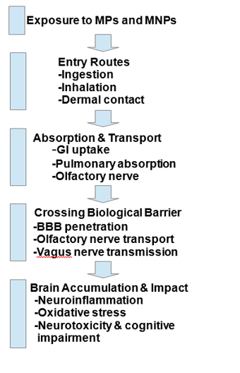
Figure 1 Pathways and Neurotoxic Effects of Micro- and Nanoplastics (MPs and MNPs) Exposure.
Diagrammatic illustration of the potential pathways and mechanisms by which MPs and nanoplastics MNPs access the human body and affect the brain. MPs and MNPs can enter the body through ingestion, inhalation, or dermal contact. Once inside, they may be absorbed and transported via gastrointestinal or pulmonary routes, or through the olfactory nerve. After internalization, MPs and MNPs can cross biological barriers such as the blood–brain barrier (BBB) by direct penetration, olfactory transport, or via the vagus nerve. Their accumulation in brain tissue can induce neuroinflammation, oxidative stress, neurotoxicity, and cognitive impairment.
There are several ways by which MPs can induce neuroinflammation. First, MPs enter the brain via direct cellular interaction. They cluster within neural cells to cause mechanical damage to cell membranes and structures, which then results in the release of damage-associated molecular patterns (DAMPs). DAMPs subsequently activate brain immune cells (microglia), which release pro-inflammatory cytokines and other immune signals, further propagating the inflammatory cascade in the CNS.51–53 Based on the accumulated findings, MPs, upon entering the brain, result in the generation of reactive oxidative species (ROS) that can directly affect cellular components such as membranes, proteins, and DNA.54–56 This leads to oxidative stress, which puts neurons in jeopardy and activates both microglia and astrocytes, critical cell types for sustaining overall brain health. However, that activation of glial processes can worsen inflammatory responses and amplify neuroinflammation, thereby creating a vicious cellular damage cycle.57,58 A major in vitro study was conducted to examine the effects of MNPs on key CNS cell types—astrocytes, oligodendrocytes, neurons, and neural stem cells (NSCs).59 Using polystyrene (PS) latex beads (50 nm and 500 nm, 0–1000 µg/mL), cell viability, uptake, and transcriptomic changes were assessed. Astrocytes exhibited the highest sensitivity to PS-MNP toxicity, with prolonged (7-day) exposure inducing greater cytotoxicity than 24-hour exposure. Both astrocytes and NSCs readily internalized PS beads, and transcriptomic analysis revealed inflammatory gene activation in astrocytes, leading to astrogliosis. These findings suggest that sustained MNP exposure may drive neuroinflammation, potentially disrupting CNS homeostasis. Another study looks into MPs in the brain in vivo using advanced imaging techniques.33 The study focuses on blood-borne MPs absorbed by brain cells and then blocking capillaries in the cortical area. These obstructions resemble blood clots that develop, restricting blood flow and ultimately causing the mice's brains to be damaged. This disruption in blood flow can potentially induce neuroinflammation, which worsens the brain's health.
Second, MPs induce BBB malfunctioning. MPs catastrophically affect this barrier via direct mechanisms or via indirect mechanisms, mostly involving inflammation and oxidative stress. By compromising the barrier, toxic substances, immune cells, and inflammatory mediators are allowed to enter the brain and induce neuroinflammation and subsequent neuronal damage.24,33,60,61 Third, MPs can induce specific molecular signaling pathways that are responsible for neuroinflammation. One of these pathways entails that of NF-kB (nuclear factor-kappa B), the activation of which by MPs in neural cells causes pro-inflammatory cytokines like TNF (tumor necrosis factor)-α and IL (interleukin)-6 to be secreted, which maintain the inflammatory state.62,63 Furthermore, they can also form inflammasomes, which are protein complexes that stimulate IL-1β and IL-18 release together with propagating inflammation and neuronal damage.64,65
Fourth, the endocrine system is another target through which MPs affect neural function.66 Many MPs bear additives toxic in nature, for example, bisphenol A (BPA) and phthalates, that are endocrine-disrupting chemicals (EDCs) themselves.67 Such chemicals can interfere with hormonal signaling in the brain and thus trigger neuroinflammation.67,68 When the functions of hormonal signaling are disrupted, it can interfere with the functions of neurotransmitters and exacerbate inflammation; thus, the brain can become susceptible to injury in the long run.69,70 Lastly, the neurotoxic problems posed by plastic additives are another primary ingredient in the equation. Substances such as flame retardants, stabilizers, and plasticizers, which are commonly found in MPs, are neurotoxic.26,71,72 These substances leach from MPs into the brain, where they can directly injure neuronal tissue and stimulate inflammatory responses.73,74 This consequently leads to an already aggravated inflammatory terrain, which further enhances neuroinflammation.21,22
The cumulative effect of these mechanisms ends up establishing a state of chronic neuroinflammation. MPs might result in a continually sustained inflammatory condition occurring in the brain over time, which in turn could later lead to NDs, including Alzheimer's and Parkinson's.17–20 In this respect, Figure 2 depicts the mechanistic cascade of microplastic-induced neurodegeneration, illustrating how neural injury, glial activation, and BBB disruption collectively drive chronic neuroinflammation and neuronal damage. Combating neurotoxins, MPs can negatively affect brain health through accompanying acids, heavy metals, and even persistent organic pollutants (POPs).26 Therefore, one of the necessary steps to mitigate the long-term impacts of microplastics on neurological health is to reduce exposure to them and their harmful additives.
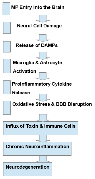
Figure 2 Mechanistic Cascade of Microplastic-Induced Neurodegeneration.
A diagram illustrating the sequence of events following microplastic (MP) exposure in the brain. MPs cause neural cell injury, triggering the release of damage-associated molecular patterns (DAMPs). These signals activate microglia and astrocytes, leading to the release of proinflammatory cytokines. The resulting inflammation induces oxidative stress and disrupts the blood–brain barrier (BBB). Increased BBB permeability allows toxins and immune cells to enter the brain, causing chronic neuroinflammation and neurodegeneration
MNPs are increasingly acknowledged as a growing risk to brain health, with numerous studies now associating their widespread presence in the human body with neurological impairments and NDs.17–20 The identification of MNPs in human soft tissues - and in the brain- demonstrates their ability to infiltrate the CNS and raises significant concerns regarding their long-term health impacts. MNPs have also been found in the placenta and fetal tissues, suggesting potential neurodevelopmental issues, while research in rodents indicates that MNPs enter brain tissue within hours of being ingested or inhaled.75–77 This alarming information necessitates urgent consideration of the implications of MNP exposure on cognitive abilities and the progression of NDs.
Cognitive impairment is a significant concern regarding MNP exposure.33,78,79 Research conducted on animals in vivo or in human cell culture in vitro has shown that the buildup of MNPs in the brain can lead to memory deficits, challenges in learning, and behavioral changes.10 As a result, these cognitive issues closely resemble symptoms commonly associated with NDs such as Alzheimer’s and Parkinson’s, implying that prolonged exposure to MNPs may exacerbate or even accelerate the symptoms and progression of these conditions.17–20,80,81 The exacerbation of NDs' symptoms owing to environmental exposure to MNPs poses an urgent need to evaluate their long-term influence on aging populations. Additionally, experiments analyzing animal behavior under MNP conditions indicated heightened anxiety-like responses, reduced cognitive flexibility, and impaired motor coordination, tending to indicate that the effects of MNP exposure are widespread neurologically.82,83 Importantly, MNPs influence pathways in the brain, including the gut-brain axis (GBA), through which MNPs can induce the development of NDs.51,84 Disruption of normal gut microbiome composition, or dysbiosis; heightened intestinal permeability; mood alterations; cognitive impairment; and neuroinflammation have been shown to be connected to MNP exposure.85–87 Considering the strong interconnections between the gut and the brain, it is reasonable to conclude that alterations in the gut microbiome resulting from MNP exposure can significantly impact mental health and increase the risk of NDs.
While these results are quite concerning, human epidemiological research on neurological disorders related to MNPs has largely been lacking thus far. Nevertheless, long-term exposure to low levels of MPs and MNPs seems likely due to their widespread occurrence in food, water, and air. With growing evidence indicating that MNPs are building up in brain tissue and resulting in cognitive and neurological difficulties, it is essential that more research be performed to accurately measure the effects. This environmental health risk calls for immediate attention to develop regulations that reduce exposure and lessen long-term impacts on brain health
At present, significant challenges about the true impact of MNPs on human health remain with regard to the concern of the health risks of MNPs. One major challenge simply relates to uncertainty regarding MNP distribution or accumulation within the body. Unlike "classic" toxicants, MNPs exhibit a variety of physicochemical properties, which vary with size, shape, composition, or surface charge—all of which influence biological interaction and biodistribution. As a true and proven principle of toxicology, framed by Paracelsus's thought, the poisonousness or toxicity of any agent is determined by how much one is forced to take into the body in the form of a substance.88 This principle indicates that injury by a given substance will depend on the amount or dose subjected to injury. While it sounds self-evident that with increasing doses of MNP would come greater damage, the actual internal dose left to reach the target organ, in the case of the brain, remains as poorly defined. A major hurdle is understanding how these MNPs move through different bodily compartments after ingestion, inhalation, or dermal exposure.
While there are some studies indicating that the particles can translocate across the gut and lung barriers and reach circulation, we do not have precise information on their long-term accumulation within human tissues, especially sensitive organs like the brain. Thus, the lack of clarity on this issue obfuscates the interpretation of controlled exposure studies, which tend to introduce known amounts of MNPs into experimental models and track their fates in the body to a lesser extent. Moreover, another complication arises when translating results from animal experiments into those in humans. In contrast to rodents, humans have blood volumes that are much larger, leading to the greater dilution of MNPs circulating in the blood and, in turn, potentially lower tissue concentrations than those recorded in animal studies.89 Such differences add to questions regarding the relevance of preclinical studies with respect to human risk assessment. There is also a complexity in the MNP interaction with biological systems. Aside from causing direct toxicity, MNPs can take up environmental pollutants, heavy metals, and endocrine disruptors, making the effects really hazardous. Its interference with crucial cellular processes, such as oxidative stress, mitochondrial dysfunction, and immune dysregulation, indicates a possibility for developing chronic diseases, mostly neurodegenerative and neurodevelopmentally impaired disorders. However, the exact mechanisms by which MNPs react with neural cells, cross the BBB, and contribute to neuroinflammation are still largely unexplained.
Recent studies suggest a link between MPs and neuroinflammation as well as dysfunction in the CNS due to oxidative damage, harm to mitochondria, and disruptions in immune function. Nevertheless, significant knowledge gaps still exist regarding the effects of prolonged exposure and their lasting impact: whether MPs accumulate in brain tissues, how they might affect neurodevelopment, and whether they could worsen existing NDs like Alzheimer’s and Parkinson’s disease (AD and PD). Long-term research is essential to observe how sustained exposure may bring cognitive impairment, degeneration of neural tissues, and risks across generations, especially and more importantly in vulnerable groups like children, the elderly, and people with occupational exposure. It should focus on how MPs induce activation of microglia, activate astrocytes, and disrupt synaptic plasticity and the balance of neurotransmitters and neuronal electrical activity.
Importantly, grasping the GBA in the context of MP-induced neurotoxicity is vital, as MPs may change gut microbiota, increase gut permeability, and cause systemic inflammation that can initiate neuroinflammatory responses. Large-scale epidemiological studies are crucial to establish a connection between the levels of MPs in biological samples and the occurrence of NDs, ultimately helping to identify potential risk factors and mechanisms behind these conditions. Although this holds true, it is essential to also establish strategies to reduce the impact, such as using alternative biodegradable microplastics, introducing stricter regulations on plastic manufacturing and waste management, implementing measures to decrease consumption, and improving mechanisms for detoxification to protect the brain.90,91 A collaborative approach involving toxicology, neuroscience, epidemiology, and environmental science is vital for grasping the full scope of the neurotoxic repercussions of MPs and for informing public health policies and treatments that can alleviate their influence on neurological well-being.
Lastly, accurate detection and quantification of MNPs in neural tissues remain highly demanding analytical challenges. Neural matrices are inherently complex and lipid-rich, making complete isolation and nanoscale differentiation of particles without contamination or loss extremely difficult.92 Traditional techniques such as Fourier-transform infrared (FTIR) and Raman spectroscopy are valuable but lack the spatial resolution required to identify particles in the low nanometer range.93 More sensitive modalities, including scanning electron microscopy (SEM), transmission electron microscopy (TEM), and mass spectrometry (MS)–based methods, offer improved detection capabilities but typically require destructive tissue preparation that compromises sample integrity.94,95 Furthermore, distinguishing synthetic polymers from endogenous biomolecules remains a significant obstacle, underscoring the need for standardized, contamination-free protocols and the development of appropriate reference materials.96,97 Overcoming these technical barriers is essential to validate MNP detection in neural tissues and to establish reliable dose–response relationships critical for assessing human neurotoxicity.
MPs and MNPs are increasingly threatening brain health. They can penetrate the brain, causing oxidative stress and neuroinflammation - factors linked to NDs. Although there are concerns about their connection to Alzheimer's and Parkinson’s, and addiction and behavioral health, more research is needed to establish a direct cause. It's essential to improve detection methods, understand the mechanisms involved, and develop strategies to mitigate their effects. With plastic pollution rising, immediate action is necessary to evaluate and reduce its impact on brain health through better regulations and innovative solutions. A coordinated multidisciplinary collaboration involving toxicology, neuroscience, environmental science, and public health is needed to understand and mitigate the impacts on brain health using stringent regulations and innovative interventions.
This research is supported by Bandhan, India.
The authors do have anything to declare.
Conceptualization: S. K. C.; Formal analysis: S. K. C.; Original draft preparation: S. K. C.; Writing—review and editing: S. K. C. and D. C.; Supervision: S. K. C.; Project administration: S. K. C.; Funding acquisition: S. K. C.

©2026 Chakrabarti, et al. This is an open access article distributed under the terms of the, which permits unrestricted use, distribution, and build upon your work non-commercially.