Journal of
eISSN: 2373-6410


Clinical Images Volume 5 Issue 2
Servidor Público Estadual de São Paulo Hospital, Neurology Department, Brazil
Correspondence: Bruno Gonzales Miniello, Hospital do Servidor Público Estadual de São Paulo, Neurology Department, Rua Pedro de Toledo, 1800 - Moema, São Paulo - SP, 04029-000, Brazil, Tel +55 (11) 98777-2942
Received: October 13, 2016 | Published: October 21, 2016
Citation: Miniello BG, Cunha JLM, Sekeff-Sallem FA, Azevedo DSMS, Melo ACP (2016) Meningeal, Parenchymal and Pituitary Involvement due to Lung Cancer in a Young Patient. J Neurol Stroke 5(2): 00170. DOI: 10.15406/jnsk.2016.05.00170
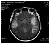
A 42-year-old female was admitted to the emergency department with pressure-type headache, weight loss, fever and night sweats. She had a history of smoking, around 27 years/pack. On Neurologic Examination, there were bilateral horizontal rotatory nystagmus, upbeat nystagmus, ataxic gait and dysarthria. Radiography and computed tomography (CT) of the chest revealed a large, superior right lobus lung mass. The cerebral spine fluid (CSF) presented pleocytosis, high protein and malignant cells. The brain MRI revealed a hyperintense suprasellar nodular lesion with intense contrast enhancement, highly suggestive of hypophyseal metastasis, along with diffuse leptomeningeal thickening over both brain and cerebellum. There was vasogenic edema on both temporal lobes with intense diffusion restriction, but without parenchymal metastasis. The patient died after 10 days. Meningeal carcinomatosis can occur in 9 to 25% of lung cancer patients,1,2 and parenchymal involvement may occur. Furthermore hypophysis metastasis are more frequently found in lung and breast cancers and in older patients with diffuse malignancy (Figure 1-4).

Figure 1 RM Axial Flair - There is vasogenico edema on both temporal lobes with intense diffusion restriction, but without parenchymal metastasis.
None.
None.

©2016 Miniello, et al. This is an open access article distributed under the terms of the, which permits unrestricted use, distribution, and build upon your work non-commercially.