Journal of
eISSN: 2373-6410


Case Report Volume 8 Issue 3
Departamento de Doenças Tropicais e Diagnóstico por Imagem, Universidade Estadual de São Paulo(UNESP), Brazil
Correspondence: Lee Van Diniz, Departamento de Doenças Tropicais e Diagnóstico por Imagem, Faculdade de Medicina de Botucatu- Universidade Estadual de São Paulo, Av. Professor Mário Rubens Guimarães Montenegro s/n, Botucatu, 18618687, São Paulo, Brazil, Tel +55(14)3880-1291
Received: January 18, 2018 | Published: June 19, 2018
Citation: Diniz LV, Junior LAJ, Rodrigues LP, et al. Intraventricular craniopharyngioma: a case report. J Neurol Stroke. 2018;8(3):185-188. DOI: 10.15406/jnsk.2018.08.00306
Craniopharyngiomas are tumors that emerge from remnants of Rathke’s pouch. Intraventricular craniopharyngiomas (IVCP) are uncommon and usually diagnosed in older patients. In this case report, we present a 36years old woman that started with holocranial headache, vomiting and bilateral papilledema. A cerebral angiotomography showed a mass inside the third ventricle. On Magnetic Resonance Imaging (MRI), a heterogeneous expansive lesion inside the third ventricle was found, in addition to hydrocephalus. The tumor was surgically removed and sent to pathology analysis that revealed an IVCP grade I.
Keywords: craniopharyngioma, intraventricular craniopharyngioma, adult craniopharyngioma, rathke cleft tumor, rathke's cleft, rathke's pouch tumor, papillary craniopharyngioma
CSF, cerebrospinal fluid; IVCP, intraventricular craniopharyngioma; CT, computerized tomography; MRI, magnetic resonance imaging; EVD, external ventricular derivation; WHO, world health organization; MIP, maximum intensity projection
Craniopharygiomas are rare and usually benign tumors that generally involve the anterior part of the third ventricle. Intraventricular Craniopharyngiomas (IVCP) are even more infrequent, accounting for 0.71 to 11%2 of all craniopharyngiomas and tend to be diagnosed in adults. In this report, we present a case of this brain neoplasm on a 36year-old woman with severe headache, vomiting and bilateral papilledema.
A 36years old woman arrived at the Emergency Room with incapacitating holocranial headache with photophobia, phonophobia and vomiting not preceded by nausea, without signs of infection. A quick eye fundoscopy identified bilateral papilledema.
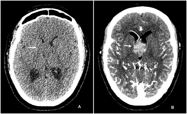
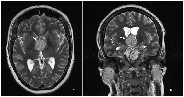
A cerebral angiotomography showed a round not calcified enhancing mass inside the third ventricle (Figure 1A) (Figure 1B). The Magnetic Resonance Imaging (MRI) revealed mild hydrocephalus and a heterogeneous, lobulated, expansive enhancing lesion inside the third ventricle, with intermediate T2 signal intensity, small cystic areas and microbleeding (Figure 2‒4). The patient underwent External Ventricular Derivation (EVD) with improvement of the initial symptoms.
Three days after admission, a surgery was performed to remove the tumor. Pathology analysis showed a well-defined, non-keratinized, squamous epithelium, with nodular formation and fibrovascular stroma in a papillary architecture (Figure 5), compatible with craniopharyngioma World Health Organization (WHO) grade I.

Figure 1 Axial CT scan without intravenous contrast (A) and Maximum Intensity Projection (MIP) reconstruction with intravenous contrast (B) showing a enhancing round mass inside the third ventricle lumen (arrow).

Figure 2 Axial (A) and coronal (B) T2W images show a round, well defined, heterogeneous lesion with intermediate signal intensity (arrows).
Cerebral ventricles are communicating cavities that contain cerebrospinal fluid. The brain has four ventricles: two lateral ventricles (right and left), the third ventricle and the fourth ventricle.3
The third ventricle may be affected by numerous lesions, mainly extrinsic ones. These can be classified according to the location they arise. There are five groups lesions: anterior, posterior and inferior masses, the ones located at foramen of Monro and intraventricular masses.4
Craniopharyngiomas are relatively benign tumors that account for 2 to 4% of primary brain neoplasms. Most of them are part of the anterior group, and only 0.71 to 11%2 of all craniopharyngiomas arise inside the third ventricle. Craniopharyngiomas emerge from remnants of Rathke's pouch and its location is determined by the path traced as the Rathke’s pouch migrates through the sphenoid bone, reaching the sellar and suprasellar regions during embryogenesis. The pial membrane will serve as a barrier preventing Rathke's pouch cells from coming into direct contact with the brain vesicle (the precursor of the infundibulum and the third ventricular floor). Without the protection of a mature pial membrane, Rathke's pouch cells may end up implanting on the neuroectoderm of the developing cerebral vesicle. If these cells develop into a tumor, it will be a purely intraventricular craniopharyngioma.5
Intraventricular craniopharyngiomas (IVCP) are rare, both in infants and adults,1 but they are usually diagnosed in older patients. Two reasons explain this: low rate of tumor replication and location in the third ventricle. These characteristics might delay the invasion of adjacent structures and, therefore, defer the onset of symptoms for a long period. When the tumor becomes larger, it blocks the passage of CSF and lead to symptoms.6
On case report of six patients with pure IVCP by Behari et al.,6 high intracranial pressure and papilledema were present in all of them. Preoperative diagnosis of pure IVCP was made by MRI. After surgery, all signs of increased intracranial pressure, including papilledema and visual disturbance were solved. An 8 to 36month follow-up was performed, resulting in no evidence of tumor recurrence or regrowth.6 It is common of suprasellar craniopharyngioma to present with endocrinological and visual disturbance, but these findings are rare in IVCP.7
Migliori & colleagues8 defined the radiologic criteria for differentiating IVCP from the suprasellar tumor that invaginates the floor of the third ventricle. Image findings in IVCP are: patent suprasellar cistern, no sellar or pituitary gland abnormalities, and intact/ballooned third ventricular floor. Suprasellar craniopharyngiomas usually have internal calcifications in 50 to 80% of patients, but these are rare in IVCP. IVCP may appear as solid masses or predominantly cystic lesions, but generally presents as a heterogeneous mass of varying intensity, often hyperintense in T1 and hypointense to mildly hyperintense compared to gray matter on T2. The location of intraventricular craniopharyngioma is more accurately determined by MRI.. It is important to determine the exact location of the tumor by MRI because of surgical approach.9 A wide variety of other pathologies can affect the third ventricle. Among the differential diagnoses it must considered: colloid cysts, ependymoma, choroid plexus papilloma, astrocytoma and meningioma.10
Intraventricular third ventricle masses are most often lesions of the choroid plexus, vascular malformations, metastasis or infectious lesions. Cases of IVCP are rarely reported in medical literature. CT and mainly MRI characteristics of the tumor are a useful and reliable to suggest IVCP diagnosis and ascertain its topography, which is extremely important to choose the surgical approach.
None
The author declares that there is no conflict of interest.

©2018 Diniz, et al. This is an open access article distributed under the terms of the, which permits unrestricted use, distribution, and build upon your work non-commercially.