Journal of
eISSN: 2373-6410


Short Communication Volume 8 Issue 2
1Institute of Higher Nervous Activity and Neurophysiology of RAS, Russia
2NN Burdenko National Medical Research Center of Neurosurgery of the Ministry of Health of the Russion Federation, Russia
3National Research Nuclear University MEPhI (Moscow Engineering Physics Institute), Russia
Correspondence: EV Sharova, Institute of Higher Nervous Activity and Neurophysiology of RAS, Russia
Received: February 13, 2018 | Published: March 16, 2018
Citation: Sharova EV, Mukhina TS, Boldyreva GN,et al. FMRI analysis of the motor network functional connections at rest and with motor load in healthy people and patients with STBI. J Neurol Stroke. 2018;8(2):91–92. DOI: 10.15406/jnsk.2018.08.00287
Severe traumatic brain injury (STBI) is accompanied by motor disorders in 2/3 of the victims - often in combination with cognitive impairment and depression of consciousness.1 An objective diagnosis of the motor sphere in patients with SBI at different severity of motor disorders is among the actual ones.
To investigate the possibility of the motor corticospinal tract network’s functional activity in normal and STBI estimating based on a comparison of fMRI connectivity in the motor act and at rest.
Observation groups: 18 patients with SТBI (main) and 15 healthy volunteers (control). Movement defect in the form of hemiparesis were evaluated on a scale of muscle strength.2 3T fMRI recorded at rest and passive right hand finger clenching (by experimenter). The validity of this motor test using is shown in our previous publications.3,4 The CONN program carried out a group analysis of the cortico-spinal tract’s motor network brain connectivity (ВС) - between the regions of interest (ROI) in the motor cortex of the left (1) and right (2) hemispheres, supplementary motor cortex (3), and cerebellum right hemisphere ( 4).
Three of these brain regions are involved in the fMRI response, to both the active and the passive movement of the arm (Figure 1).3,4 The intensity of ВС between the selected points was assessed, as well as the level of their significance.
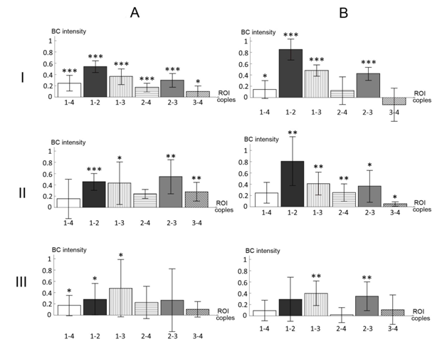
In Figure 2 shows histograms of the ВС intensity distribution in predetermined motor fMRI network of healthy people and patients with various degrees of right-sided hemiparesis after STBI in passive motor test (Figure 2A) and at rest (Figure 2B). Both the features of similarity and the differences in their spatial organization are noted. In both tests close order of the BC intensity values in the majority of ROI pairs of the same name was observed, as well as the tendency to weaken them from normal to rough hemiparesis.
At rest these changes are practically linear in most pairs analyzed (4 of 6). Less evident this orientation for the connections of the left and right motor cortex with the right hemisphere of the cerebellum. In the passive motor test for only one connection from 6 (between left and right motor cortex, 1-2) values decrease linearly with increasing hemiparesis. BC of the supplementary motor cortex with right motor, as well as with cerebellum, are the highest in easy hemiparesis in comparison with other samples of observations, and the connections of the right and left motor cortex with the cerebellum, on the contrary, are weakened. The nonlinearity of these changes correlates with the previously identified topographic features of motor fMRI responses at different degrees of posttraumatic hemiparesis,5 reflecting, in our opinion, the variational mechanisms of motor defect compensation.

Figure 2 Histograms of the fMRI motor network’s BC intensity in healthy people and patients with post-traumatic hemiparesis.
A -a passive motor test, B –resting state, I – norm (N=15), II - easy hemiparesis (N=8), III - rough hemiparesis (N=10).
Horizontal axis - the ROI couples. Vertical axis - BC intensity.
BC significance level by one-sample t-test: * 0,05<p£0,15; ** 0,01<p£0,05; *** p<0,01.
Both in rest and in a passive motor sample, changes of the BC between the symmetrical motor cortical zones are associated with the severity of posttraumatic hemiparesis most clearly. Evaluation of the motor network fMRI paretic arm at rest has a greater diagnostic potential. In the passive movement of the hand intergroup changes of the motor network BC spatial organization as hemiparesis increases can more reflect the compensatory possibilities of the studied system.
Supported by RFFI № 18-013-00355
The authors declare there is no conflict of interest.
None.

©2018 Sharova, et al. This is an open access article distributed under the terms of the, which permits unrestricted use, distribution, and build upon your work non-commercially.