Journal of
eISSN: 2373-6410


Clinical Report Volume 11 Issue 2
1FSBI Federal Scientific Clinical Center for Medical Radiology and Oncology, Dimitrovgrad, Russia
2Republican Scientific and Practical Center for Mental Health, Russia
3NN Alexandrov National Cancer Center of Belarus, Russia
4FSBI Russian Scientific Center of Roentgenoradiology, Russia
Correspondence: Elena Slobinam, FSBI Federal Scientific Clinical Center for Medical Radiology and Oncology, Dimitrovgrad, Russia
Received: March 05, 2021 | Published: April 24, 2021
Citation: Slobinam E, Dokukina T, Khlebokazov F, et al. Evaluation of the effectiveness of the combined treatment of symptomatic pharmacoresistant epilepsy by the 18-FDG PET/CT. J Neurol Stroke. 2021;11(2):53-55. DOI: 10.15406/jnsk.2021.11.00456
The problem of epilepsy is one of the most relevant in modern neurology and psychiatry. Although the number of antiepileptic drugs available in the last 20 years increased significantly, about a third of patients remain resistant to treatment.1 New diagnostic methods such as 18-FDG PET/CT and the possibility of neuroimaging against the background of normal brain MRI data are gaining great importance in the visualization of functional states of the brain, such as epilepsy.2–4
To present the possibilities of clinical application of modern neuroimaging such as 18-FDG PET/CT at the example of a clinical case of treatment of symptomatic pharmacoresistant epilepsy and to evaluate the feasibility, tolerance of this type of imaging.
In March 2017 patient, 24 years old, about generalized seizures (since 2014 after fright from explosion of firecrackers) continued to be held anticonvulsant therapy, which led to a decrease in seizure frequency from 8 to 13 in month. Taking into account the progressive course of the disease, when any a combination of 2-3 essential anticonvulsants, including the latest, did not have a noticeable effect on the frequency and severity of attacks, the patient is treated by combination therapy, including antiepileptic drugs and autologous bone marrow stem cells (the AMSC CM transplantation).
Electroencephalogram (EEG) showed epileptiform activity. An irregular alpha rhythm with a peak frequency of 10.5 Hz is recorded on the patient's EEG. The frequency-spatial structure of the alpha rhythm is pathologically distorted. The structure of the maximum values of average coherence is represented as a triangle in fronto-central departments, which is characteristic of an active epileptic process. A moderate increase in low diffuse beta and theta activity is noted. Against this background, in the left frontotemporal region, epileptiform activity in the form of spikes is recorded in large numbers, the frequency of which reaches 30 per minute. Rare discharges of high, sharp, bilaterally synchronous alpha waves with a frequency of 1-2 per min (Figure 1 & 2).
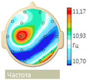
Figure 1 Pathological inversion of the frequency-spatial structure of the alpha rhythm (the minimum frequency values are located not in the frontal, but in the parietal regions).
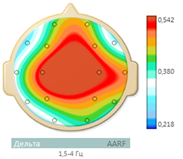
Figure 2 The maximum values of the mean coherence are presented in the form of a triangle in the frontal-central regions.
MRI of the brain showed diffuse cerebral subatrophy. The lateral ventricles are slightly enlarged, asymmetric S>D. Hippocampi are asymmetric (D> S), the volume on the left is reduced. Deformation of the soft tissue of the skull on the left (Figure 3).
PET/CT examination of the brain with F18 - fluorodeoxyglucose (18-FDG) after the first course of the introduction of AMSC CM revealed a pattern of increased metabolic active-STI of the left hemisphere of the brain. Significant a new increase in fixation of the radiopharmaceutical in the left frontal, parietal, temporal and occipital lobes (Figure 4).
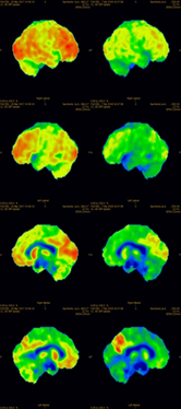
Figure 4 Results of brain examination of patient, 24 years old, by PET/CT method with F18 fluorodeoxyglucose radiopharmaceutical after the first course of administration of autologous mesenchymal bone marrow stem cells.
On an EEG, after the first course of stem cell administration, significantly decreased the amount of epileptic formal activity (Figure 5). The frequency of spikes in the left temporal region STI decreased from 30 to 2 per min, the frequency of bilateral synchronization discharges of acute alpha waves remained at the level of 1-2 in minutes. At a frequency of alpha rhythm of 10.5 Hz, normalization occurred of its frequency-spatial structure, but the structure maximum mean coherence values are still was presented in the form of a triangle, which indicated the presence of an active epileptic process.
On the EEG, after the second course of stem cell administration, paroxysmal activity was not registered (Figure 6).
On the EEG, a disorganized alpha rhythm was maintained with a peak frequency of 12.5 Hz with a diffuse increase in beta activity. Its frequency-spatial structure is largely normalized. A reduction in the pathological structure of the maximum values of mean coherence in the form of a triangle was also noted (Figure 7 & 8).
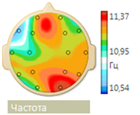
Figure 7 Frequency-spatial structure of the alpha rhythm with a tendency to normalization (the alpha rhythm of the maximum frequency is located mainly in the occipital region, and the minimum frequency - in the frontal region).
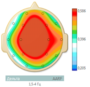
Figure 8 Reduction of the pathological structure of coherence in the form of an "epileptic triangle" (the maximum values of the average coherence are presented in the form of a diamond).
In order to assess the functional state of the brain according to metabolic activity carried out repeated PET/CT with FDG after 5 months after the second course of the introduction of AMSK KM (Figure 9). The obtained data on the metabolism of the radiopharmaceutical in dynamics and when compared with relative reference regions testify in favor of stabilization (striving for reference values) of metabolic activity in the interested areas: in the left parietal, temporal and occipital lobes. Control PET/CT - study was performed under identical conditions, which is a prerequisite for assessing dynamics (the study was conducted on the same scanner, with the same entered activity, subject to variables of the research protocol).
Combining modern methods of NI including 18-FDG PET/CT are very useful tools in the evaluation of effectiveness of complex treatment for symptomatic pharmacoresistant epilepsy.5–8 The encouraging results of the evaluation of effectiveness of the proposed method of treatment of pharmacoresistant epilepsy on the clinical case of one patient have been obtained, which requires further study of the clinical application of the 18-FDG PET/CT on a more significant number of clinical cases in larger clinical trials.
None.
The authors declare no conflicts of interest.

©2021 Slobinam, et al. This is an open access article distributed under the terms of the, which permits unrestricted use, distribution, and build upon your work non-commercially.