Journal of
eISSN: 2373-6410


Case Report Volume 12 Issue 6
Centro Universitário UNIFACIMED Cacoal-RO, Brazil
Correspondence: Diego Bezerra Soares, Centro Universitário UNIFACIMED Cacoal-RO, Brazil
Received: October 04, 2022 | Published: November 21, 2022
Citation: Soares DB, Goebel GC, Manzoli IR, et al. Case report: left frontal extra axial tumor caused by epithelial neoplasm of thyroid follicular origin. J Neurol Stroke. 2022;12(6):191-194. DOI: 10.15406/jnsk.2022.12.00526
From antiquity to the present day, tumors have always aroused curiosity in the field of medicine and instigated science in an obsession to understand its mechanism of action and seek its cure. Neoplasms were initially described around 2600 years before Christ in an Egyptian papyrus containing reports of 48 cases of the disease carried out through the studies of the Egyptian physician Imhotep. It was only centuries later, with the Renaissance movement and the beginning of the modern age in the 18th century, that the first anatomical and histopathological descriptions were made thanks to the development of microscopy and the work of the German physician Virchow and the Belgian anatomist Andreas Versalius. In addition, with the advance of the scientific revolution in the 19th century, factors related to the heredity of tumors as well as their compression of genetic aspects were elucidated through analytical observations and categorical writings developed by the Austrian monk Gregor Mendel and later applied to the study of the genetic basis. Modern. Since then, with the advent of technology, hundreds of scientists have embarked on a mad dash to improve new techniques and innovative treatments that allow us to understand and treat the mechanisms of the disease. In this context, epithelial neoplasms are known to be tumors that can be classified as benign or malignant, originating from a disordered proliferation of follicular cells in tissues influenced by thyroid hormones. Benign follicular nodules are the most common types presented, which contain in their cytological analysis variable proportions of colloid and benign-looking follicular cells arranged as macrofollicles or macrofollicle fragments in some tissues of the body. Therefore, in the case report in question, a 50-year-old female patient was identified with the presence of a left frontal extraaxial tumor caused by an epithelial neoplasm of thyroid follicular origin.
Keywords: epithelial neoplasm, follicular origin, frontal tumor
From antiquity to the present day, tumors have always aroused curiosity in the field of medicine and instigated science in an obsession to understand its mechanism of action and seek its cure. Neoplasms were initially described around 2600 years before Christ in an Egyptian papyrus containing reports of 48 cases of the disease carried out through the studies of the Egyptian physician Imhotep who, driven by the development of medicine, described the clinical manifestations of patients with aggressive tumors. It was only centuries later, with the Renaissance movement and the beginning of the modern age in the 18th century, that the first anatomical and histopathological descriptions were made thanks to the development of microscopy and the work of the German physician Virchow and the Belgian anatomist Andreas Versalius. Furthermore, with the advance of the scientific revolution in the 19th century, the factors related to the heredity of tumors as well as their understanding of genetic aspects were elucidated through analytical observations and categorical writings developed by the Austrian monk Gregor Mendel, which allowed the discovery of the influence of the genetic trait and the transport of these genes to the chromosomes, being later applied to the study of the modern genetic basis. Since then, with the advent of technology, hundreds of scientists have embarked on a maddening race to improve new techniques and innovative treatments that make it possible to understand and treat the mechanisms of the disease. In this context, epithelial neoplasms are known to be tumors that can be classified as benign or malignant, originating from a disordered proliferation of follicular cells in tissues, influenced by thyroid hormones.1 Benign follicular nodules are the most common types found in cases of neoplasms. epithelial cells presenting in their cytological analysis variable proportions of colloid and benign-looking follicular cells arranged as macrofollicles or macrofollicle fragments in some tissues of the body.2,3 Therefore, in the case report in question it was identified in a female patient with 50 years old, there is the presence of a left frontal extraaxial tumor visible in the appearance of an expansive mass caused by epithelial cells of thyroid follicular origin.
Patient arrived at the service of the Cacoal Regional Emergency and Emergency Hospital (HEURO) with hospitalization on 07/07/2021, 50 years old, female, black, with a previous history of controlled systemic arterial hypertension and reports a tumor in the left frontal region for about 3 years, visible and with an expansive mass. In view of this, a previous biopsy of oncotic aspiration cytology compatible with a benign follicular nodule (Category II-BETHESDA) was requested, where smears showing moderate cellularity were found at the expense of isolated follicular cells, sometimes aggregated amidst histiocytes, lymphocytes and neutrophils, on an amorphous background with a proteinaceous appearance, in addition to abundant colloid and numerous erythrocytes. The immunohistochemical examination showed the diagnosis of epithelial neoplasia of follicular origin and the anatomopathological analysis in the microscopy part showed the presence of an expansive lesion in the left frontal region suggestive of primary epithelial neoplasia with findings of irregular nuclei with clear areas and lumens filled with amorphous material eosinophilic. The macroscopic analysis showed 3 irregular brownish elastic fragments with dimensions of 0.5 cm x 0.3 cm x 1 cm. After performing magnetic resonance imaging, a left frontal extraaxial lesion with bone destruction was evidenced and the patient was immediately referred to a place in a reference service.
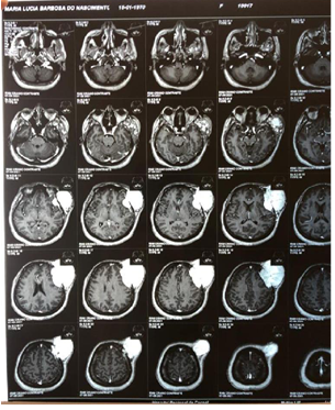
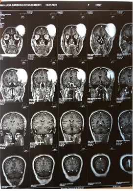
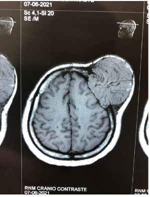
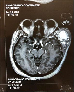
Epithelial neoplasms arise from the so-called follicular cells that have their development associated with the influence of thyroid hormones in the tissues.4 In this case report, the appearance of an expansive and compressive tumor in the extra axial region of the left forehead is observed in a female patient. A 50-year-old patient in category II of the Bethesda classification, widely used to evaluate the aspiration cytological results, found a benign nodular pattern of follicular cells in that atypical area, being indicative of an epithelial neoplasm. Therefore, it reflects on the influence of thyroid hormones that generally affect women eight times more often than men.1 With advancing age, many endocrine systems show a gradual change in production, metabolism, action and plasma concentration of hormones, therefore, it is observed that the serum levels of T3 and T4 are reduced and normal or slightly increased levels of TSH and, therefore, an increase in the prevalence of thyroid disorders among geriatric patients is observed, reaching around 2 .0% to 4.0%, while in the general population the prevalence is 0.5% to 1.0%.5,6 Around the age of 50, women experience hormonal changes with hypoestrogenemia and serum levels of follicle stimulating hormone (FSH) that directly impact the follicular cells contributing to the appearance of epithelial neoplasms.7 After the resonance, another aspect of relevance and concern in the case was evidenced, the fact that there is bone destruction in the region affected by the tumor and the patient is urgently referred to a reference unit (Figure 1A - Figure 6B).

Figure 2A Contrast-enhanced magnetic resonance imaging showing a tumor in the extraaxial region of left frontal involvement.

Figure 2B Contrast-enhanced magnetic resonance image showing a tumor in the extraaxial region of left frontal involvement.

Figure 3A Contrast-enhanced magnetic resonance image showing a tumor in the extraaxial region of left frontal involvement

Figure 3B Contrast-enhanced magnetic resonance imaging showing a tumor in the extraaxial region of left frontal involvement in coronal section.

Figure 4A Contrast-enhanced magnetic resonance image showing a tumor in the extraaxial region of left frontal involvement in sagittal section.

Figure 4B Contrast-enhanced magnetic resonance imaging showing a tumor in the extraaxial region of left frontal involvement in coronal section.

Figure 5A Contrast-enhanced magnetic resonance image showing a tumor in the extraaxial region of left frontal involvement with axial section.

Figure 5B Contrast-enhanced magnetic resonance imaging showing a tumor in the extraaxial region of left frontal involvement.
In summary, it is confirmed that the objective of the work was to characterize the case report presented as an important learning tool for medical practice and a primordial clinical experience for the development of an in-depth analysis of epithelial neoplasms and their tumor capacity, even in an unusual region as in this case in which it was observed in the extra-axial area of the frontal bone. Furthermore, the role of thyroid hormones in influencing this abnormal growth of follicular cells can be evidenced, especially in patients more prone to thyroid disorders, such as women around 50 years of age.8,9 Furthermore, it is not uncommon to find patients with presentations atypical clinics and different possible triggering factors. Therefore, further studies regarding the aspects of this type of tumor and how it manifests are necessary.
None.
The authors declare no conflicts of interest.

©2022 Soares, et al. This is an open access article distributed under the terms of the, which permits unrestricted use, distribution, and build upon your work non-commercially.