Journal of
eISSN: 2373-6410


Case Report Volume 15 Issue 4
1Department of Neurology, Mohammed V Military Teaching Hospital, Rabat, Morocco
2Faculty of Medicine, Mohammed V University, Rabat, Morocco
3Department of Neurophysiology, Mohammed V Military Teaching Hospital, Rabat, Morocco
Correspondence: Ibrahim Touré, Department of Neurology, Mohammed V Military Teaching Hospital, Rabat, Morocco, Tel 00212691190740
Received: September 10, 2025 | Published: September 25, 2025
Citation: Touré I, Benmoh Y, Béogo CI, et al. Acute ataxia and clonus revealing hashimoto’s encephalopathy (SREAT): a case report. J Neurol Stroke. 2025;15(4):98-101. DOI: 10.15406/jnsk.2025.15.00630
Introduction: Hashimoto’s encephalopathy, also known as steroid-responsive encephalopathy associated with autoimmune thyroiditis (SREAT), is a rare autoimmune condition characterized by neuropsychiatric symptoms and elevated antithyroid antibody levels. Its clinical presentation is highly variable and may include diffuse or focal central nervous system involvement, typically evolving in a subacute, relapsing–remitting pattern.
We report the case of a patient diagnosed with SREAT at our institution, whose condition was revealed by continuous segmental motor phenomena of the right hemibody upon awakening.
Case description: This is the case of a 45-year-old female patient with no known pathological history, who woke up in the morning with segmental motor symptoms on the right side and a speech arrest. She was admitted six hours after the onset of symptoms for management of functional impairment of the right hemibody, associated with a speech disorder. Neurological examination revealed right flaccid hemiparesis, predominantly affecting the right arm and face, ataxia of the right hand with the phenomenon of a wayward parietal hand, and Broca's aphasia.
A cerebral CT angiography was performed, followed by a brain MRI angiography, which revealed cortico-subcortical inflammatory-like lesions in the left fronto-parieto-temporal region, bilateral thalami, and caudate nuclei. Cerebrospinal fluid (CSF) analysis showed a mild increase in protein levels (0.7 g/L), normal cytology, absence of oligoclonal bands, and no intrathecal synthesis.
Twelve hours after the initial symptoms, the patient developed focal status epilepticus, which was treated with phenobarbital, leading to recovery of speech but persistence of motor symptoms. Thyroid antibodies were highly positive (anti-thyroid peroxidase antibodies at 771 IU/mL, anti-thyroglobulin antibodies at 615 IU/L), along with a positive antinuclear antibody (ANA) titer (1:320). Given the signs of encephalitis associated with autoimmune thyroiditis and after ruling out other differential diagnoses, a diagnosis of Hashimoto’s encephalopathy was made.
The patient received intravenous methylprednisolone (1g/day) for three days, followed by an oral dose of 60 mg/day with adjunctive therapy. After six days of corticosteroid treatment with unsatisfactory improvement (persistence of motor symptoms), intravenous immunoglobulin (IVIG) therapy was initiated at a dose of 2 g/kg over five days, without adverse effects. The clinical condition improved dramatically, with complete resolution of motor symptoms after three days. The patient developed a Cushingoid appearance on oral corticosteroids (60 mg/day) with a 5 kg weight gain, but no clinical relapse. This led to the introduction of azathioprine (2 mg/kg/day) and a gradual tapering of corticosteroids to 20 mg/day over six weeks. After 10 months of follow-up, the patient exhibited attention and concentration difficulties, but no relapse or motor sequelae.
Conclusion: Steroid-responsive encephalopathy associated with autoimmune thyroiditis (SREAT) is a rare autoimmune neuroendocrine disorder with heterogeneous clinical presentation. This condition should be suspected in any case of acute or subacute motor symptoms associated with a focal status epilepticus as the initial manifestation. Early diagnosis and appropriate treatment can prevent irreversible complications. Intravenous immunoglobulin (IVIG) therapy is a valuable alternative in cases showing poor response to glucocorticoids.
Keywords: steroid-responsive encephalopathy associated with autoimmune thyroiditis, hashimoto’s encephalopathy, anti-thyroglobulin antibodies, anti-thyroperoxidase antibodies, intravenous immunoglobulin, ischemic stroke, focal status epilepticus.
SREAT, steroid-responsive encephalopathy associated with autoimmune thyroiditis; HE, hashimoto’s encephalopathy; IVIG, intravenous immunoglobulin; IS, ischemic stroke
SREAT (Steroid-Responsive Encephalopathy Associated with Autoimmune Thyroiditis), formerly known as Hashimoto’s encephalopathy, is defined by acute or subacute encephalopathic symptoms generally associated with autoimmune thyroiditis, with responsiveness to steroids and in the absence of evidence for another cause of encephalitis. Although it is highly responsive to corticosteroids, some forms require intravenous immunoglobulin therapy. Its management poses a challenge for clinicians, due to its heterogeneous clinical presentation, the broad spectrum of differential diagnoses, and complexities in therapeutic decision-making. We report a case of SREAT revealed by right brachiofacial clonus and ipsilateral hand ataxia observed upon awakening.
A 45-year-old right-handed female patient, with no known medical history, no history of miscarriage, no toxic habits, no known exposure to tuberculosis, no history of bipolar aphthosis, and no signs of sicca syndrome. She awoke in the morning with continuous segmental motor phenomena on the right side and a sudden speech arrest, which resolved spontaneously after three hours. She was admitted six hours after symptom onset due to the sudden onset of functional impairment of the right hemibody, associated with speech arrest, without confusion, seizures, vomiting, or signs of infection.
On clinical examination: The patient was alert, followed both verbal and imitative commands, was hemodynamically and respiratory stable, and in good general condition. Neurological examination revealed right flaccid hemiparesis, predominantly affecting the brachiofacial region, graded 3/5 (MRC scale) in the right upper limb and 4/5 in the right lower limb. Deep tendon reflexes were asymmetrical, decreased on the right side. There was a mild central facial palsy. Ataxia of the right hand was noted, along with the phenomenon of a wayward parietal hand, and Broca's aphasia. The rest of the physical examination did not show an abnormality.
A brain CT angiography (Figure 1), followed by a brain MR angiography (Figure 2), was performed and revealed cortico-subcortical lesions in the left fronto-parieto-temporal region, as well as bilateral thalamic and caudate nucleus lesions, appearing inflammatory in nature, without any signs of vascular involvement. Cerebrospinal fluid (CSF) analysis showed a mild protein elevation (0.7 g/L), with normal cytology, absence of oligoclonal bands, and no intrathecal synthesis of immunoglobulins. Routine blood tests were normal, including electrolytes, renal and hepatic function, thyroid panel, calcium-phosphorus balance, complete blood count, ESR, CRP, and serologies (HCV, HBV, syphilis, and Lyme disease), all of which were negative.
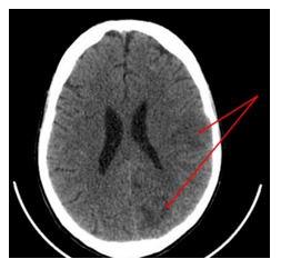
Figure 1 Cérébral CT scan showing left parieto-temporal hypodensitiy (arrow).
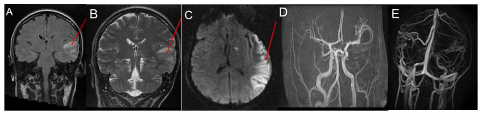
Figure 2 Brain MRI showing left parieto-temporal demyelinating lesion with cortical and sub-cortical increased signal intensity on FLAIR (A), T2 (B) and diffusion (C) weighted images (arrows). Angio MRI is normal (D + E).
Twelve hours after the initial symptom onset, the patient experienced recurrent right brachiofacial clonic seizures and continuous right versive movements, accompanied by speech arrest lasting two hours. The electroencephalogram (EEG, Figure 3) suggested a focal status epilepticus, which was treated with a loading dose of phenobarbital (20 mg/kg), followed by a maintenance dose of 200 mg every 8 hours. The speech resumed, though remained dysarthric, and motor symptoms diminished, becoming intermittent. An autoimmune encephalitis was diagnosed based on clinical and paraclinical findings. The autoimmune blood panel came back positive for anti-thyroperoxidase antibodies (771 IU/mL), anti-thyroglobulin antibodies (615 IU/mL), and antinuclear antibodies (ANA titer 1:320).
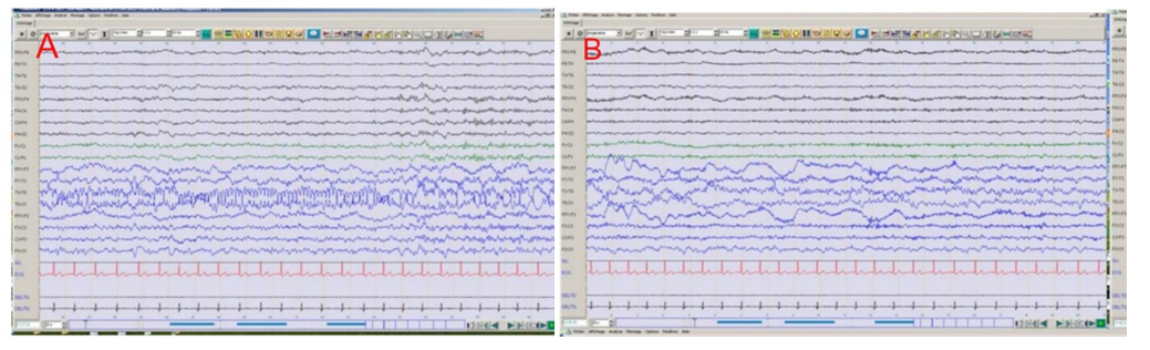
Figure 3 A) Ictal EEG: corresponding to a rhythmic activity on left temporal regions correspond to focal status epilepticus. B) Post ictal EEG: showing left temporal slow waves.
The extension workup for encephalitis was performed to exclude the main differential diagnoses and returned normal:
In the context of a clinical presentation typical of encephalitis, with markedly elevated antithyroid antibodies and a normal extension workup, the diagnosis of Hashimoto’s encephalopathy was established based on clinical, biological, and radiological findings, as well as the exclusion of major differential diagnoses. The patient was given a bolus of methylprednisolone at a dose of 1 g per day for 3 days, combined with adjunctive treatments, followed by oral prednisone at 60 mg/day. After six days of corticosteroid therapy, there was improvement in muscle strength of the right hemibody, but persistent right brachiofacial clonic seizures and ataxia of the ipsilateral upper limb remained. This inadequate response to steroids led to the initiation of intravenous immunoglobulin (IVIG) therapy at a dose of 2 g/kg over five days, without any adverse effects. The clinical condition improved dramatically, with complete resolution of motor symptoms within three days.
After six weeks of oral corticosteroid treatment (60 mg/day) and maintenance antiepileptic therapy with levetiracetam 500 mg twice daily, the patient developed a Cushingoid appearance with a 5 kg weight gain but no clinical relapse. This prompted the introduction of azathioprine (2 mg/kg/day) and gradual tapering of corticosteroids to 20 mg/day over six weeks. At 10 months of follow-up, the patient remained relapse-free without motor sequelae or seizures. However, she complained of difficulties with concentration and occasional forgetfulness.
Hashimoto’s encephalopathy is characterized by the association of neurological signs compatible with encephalitis and the presence of antithyroid antibodies.1 Its pathophysiology remains poorly understood, with one hypothesis implicating three mechanisms in its pathogenesis: cerebral vasculitis, the role of autoimmunity, and a cross-reaction between vascular inflammation and thyroid antibodies. It is a rare disorder with an estimated incidence of 2.1 per 100,000, showing a female predominance of 85%; however, severe cases have also been reported in men.2 The clinical presentation is heterogeneous, encompassing a variety of psychiatric and neurological symptoms with subacute, remitting, relapsing, or progressive evolution.
The most frequently reported neurological manifestations include cognitive dysfunction (36–100%), tremor (28–85%), altered consciousness (26–85%), transient aphasia (73–84%), seizures (52–66%), myoclonus (37–65%), ataxia (28–65%), focal deficits (27–67%), ischemic stroke episodes (18–31%), and status epilepticus (12–20%).3 These status epilepticus episodes are often resistant to anticonvulsant therapy.
Our patient’s reason for admission was sudden right hemibody heaviness associated with speech arrest, without signs of intracranial hypertension, infection, or altered consciousness, and with preserved general condition, initially suggesting a vascular mechanism (ischemic stroke). This hypothesis was promptly excluded based on neuroimaging results. Clinical examination revealed focal neurological deficit (right hemiparesis), ataxia, and cognitive involvement (aphasia), signs commonly observed in most patients with Hashimoto’s encephalopathy. Our patient developed focal status epilepticus resistant to anticonvulsants, raising suspicion of encephalitis.
Psychiatric disorders have been reported in 25–36% of patients diagnosed with Hashimoto’s encephalopathy.4 These may manifest as mood disorders, visual hallucinations, catatonia, and even acute delirium syndrome.5 Our patient exhibited no psychotic manifestations. Peripheral nervous system involvement, such as small-fiber polyneuropathy or autonomic system dysfunction associated with autoimmune thyroiditis, is rare and mostly described in adolescents, although some cases have been reported.6
Comorbidities are present in more than one-third of patients with Hashimoto’s encephalopathy and include other autoimmune diseases such as lupus, Sjögren’s syndrome, myasthenia gravis, and pernicious anemia.7
At the time of initial presentation, most patients are euthyroid; however, cases of hyperthyroidism or severe hypothyroidism have also been reported.8 These characteristics were observed in our patient, who was euthyroid and had no systemic illness.
Electroencephalogram (EEG) findings in Hashimoto’s encephalopathy are nonspecific and may show intermittent frontal delta wave activity, triphasic waves, and sometimes epileptiform abnormalities. In more than 90% of cases, intermittent slow-wave activity is described.9 The presence of high titers of anti-thyroperoxidase antibodies is considered one of the diagnostic criteria for Hashimoto’s encephalopathy, as their blood levels are elevated in 95 to 100% of cases, and in some cases, these antibodies are also detected in the cerebrospinal fluid (CSF).3,10 Anti-thyroglobulin antibodies are observed at high concentrations in the blood in 73% of cases (2) and have also been detected in the CSF.11 Some authors have reported oligoclonal band synthesis in the CSF and even lymphocytic pleocytosis.2,12 Our patient presented with clinically and EEG-confirmed focal status epilepticus and had a high blood concentration of antithyroid antibodies (anti-thyroperoxidase antibodies >6 times the normal value, anti-thyroglobulin antibodies >4 times the normal value). However, there was no oligoclonal band synthesis nor lymphocytic pleocytosis in her CSF.
The treatment of Hashimoto’s encephalopathy has been thoroughly described by other authors2,13 and can include nearly all therapeutic approaches used for other autoimmune diseases. Generally, over 90% of cases respond favorably to glucocorticoids, with a high initial dose (1 g of methylprednisolone per day in adults or 30 mg/kg in children) administered for three to seven days, followed by an average maintenance dose of 1 to 2 mg/kg with a slow, gradual taper.
Other immunosuppressants such as azathioprine, methotrexate, and rituximab are recommended by some authors for patients resistant to glucocorticoids or as maintenance therapy during glucocorticoid tapering.13 Intravenous immunoglobulins (IVIG) constitute a first-line therapeutic alternative for patients who respond poorly to glucocorticoids or in cases with associated metabolic disorders.13–15 Initially, our patient responded poorly to methylprednisolone pulses during the first three days. The intravenous immunoglobulins led to a marked improvement with complete resolution of motor symptoms.
The prognosis of Hashimoto’s encephalopathy varies but is generally good with appropriate treatment; however, spontaneous relapses occur in 12 to 40% of cases after treatment discontinuation.15 Severe cases with neuropsychological sequelae are mainly reported in patients who are diagnosed or treated late.16
None.
The authors declare that there are no conflicts of interest.

©2025 Touré, et al. This is an open access article distributed under the terms of the, which permits unrestricted use, distribution, and build upon your work non-commercially.