Journal of
eISSN: 2373-6453


Research Article Volume 10 Issue 1
1Immunology Laboratory, Ibn Rochd University Hospital Center of Casablanca, Morocco
2Laboratory of Clinical Immunology and Immuno-Allergy (LICIA), Faculty of Medicine and Pharmacy of Casablanca, Morocco
Correspondence: Loubna Mahir, Medical Biologist, Immunology Laboratory, Ibn Rochd University Hospital Center of Casablanca, Morocco
Received: March 14, 2023 | Published: March 24, 2023
Citation: Mahir L, Hsai FE, DRISSI BOURHANBOUR A, et al. Performance evaluation of immunoblot for the detection of anti-DNA antibodies compared to indirect immunofluorescence on Crithidia luciliae. J Hum Virol Retrovirol. 2023;10(1):30-32. DOI: 10.15406/jhvrv.2023.10.00261
Systemic lupus erythematosus is a severe autoimmune disease preferentially affecting the female sex. Because of the great polymorphism of the disease, the diagnosis of lupus must first be confirmed on the basis of several clinico-biological criteria. Among the biological criteria is the presence of anti-dsDNA (double stranded Deoxyribonucleic acid) antibodies. The detection of these autoantibodies requires the use of techniques with high sensitivity and specificity. We evaluated the performance of the immunoblot technique (ANA-IB) in comparison with the indirect immunofluorescence technique (CLIFT) in the detection of anti-dsDNA antibodies in 100 sera. This comparison led to the conclusion that CLIFT remains the reference method for the detection of these antibodies and cannot be replaced by ANA-IB. However, their concomitant use increases the specificity of the test.
Keywords: systemic lupus erythematosus, autoantibodies, anti-nDNA antibodies, crithidia luciliae, immunoblot
SLE, systemic lupus erythematosus; LN, lupus nephropathy; EULAR, the european league against rheumatism; nDNA, anti-native DNA; dsDNA, anti-double-stranded DNA; CL, crithidiae luciliae
Systemic lupus erythematosus (SLE) is a systemic, non-organ-specific autoimmune disease,1 severe in the absence of treatment, that predilectionally affects women during the ovulatory period (sex ratio 9 women to 1 man). Although present in all ethnic groups, it is more prevalent in non-Caucasians. While the prevalence in Europe and the United States is higher in people of African descent, SLE is uncommon in Africa.2 The condition is characterized by autoantibody production (autoAb), complement activation, and immune complex (IC) deposition. These cause damage to multiple organs: the skin, nervous system, joints, cardiovascular system, and especially the kidneys. Lupus nephropathy (LN) is a common complication of SLE, affecting one in three patients.3
The European League Against Rheumatism (EULAR) and the American College of Rheumatology (ACR) EULAR/ACR 2019 classification criteria for SLE include a positive anti-nuclear antibody at least once as obligatory entry criterion; followed by additional criteria grouped into 7 clinical criteria (constitutional, hematologic, neuropsychiatric, mucocutaneous, serous, musculoskeletal, renal) and 3 immunologic criteria (anti-phospholipid antibodies, complement proteins, SLE-specific antibodies) and scored from 2 to 10. The presence of a total greater than or equal to 10 makes it possible to affirm the existence of systemic lupus with a sensitivity of 96% and a specificity of 93%.4 Among the biological criteria, anti-native DNA (nDNA) or anti-double-stranded DNA (dsDNA) antibodies represent the most classical serological marker of SLE.5 They are present in 40-80% of lupus patients.6 Their level is proportional to the severity of NL and is rapidly decreased by treatment. An excess of anti-dsDNA antibodies precedes an exacerbation and persistent high levels indicate a relapse of NL.3
These anti-dsDNA antibodies are directed against conformations only present on native DNA. They are either complementary base bonds or secondary structures that arise during the conformation of the two DNA strands. The size of the epitope that binds to the anti-dsDNA antibody paratope is about 6 nucleotides, but 40 to 100 base pairs are required for stable antigen/antibody binding.7
Several immunological methods are used to determine anti-dsDNA antibodies. Indirect immunofluorescence on Crithidiae luciliae (CLIFT) has been considered as a first-line screening due to its sensitivity and low cost. ELISA and immunoblot techniques are also used for verification and quantification.8
In our study, we sought to compare CLIFT with Immunoblot to evaluate its performance in the detection of anti-dsDNA antibodies.
We conducted a single-center retrospective descriptive analytical study over a period of 6 months, from December 2020 to May 2021 in the immunology laboratory at the University Hospital Center (UHC) IBN ROCHD CASABLANCA in Morocco. The demographic characteristics of our study population (age and sex) were obtained from the patient records registered on the laboratory software (Kalisil version 2.10.10). Our study included all sera received from the different departments of the University Hospital Center for which a simultaneous search for anti-dsDNA antibodies by CLIFT and immunoblot was performed.
CLIFT technique: This technique is the reference technique used in our laboratory for the detection of anti-dsDNA antibodies. Crithidia luciliae (CL) is a flagellated protozoan, non-pathogenic to humans, with a modified giant mitochondria containing circular bicentennial DNA, called kinetoplast. CLIFT was performed using the IIFT: Crithidia luciliae (anti-dsDNA) kit (Euroimmun, Medizinische Labordiagnostika AG, Lübeck, Germany).9 Patient serums diluted 1:10 is incubated with this substrate. The anti-dsDNA antibodies bound to the kinetoplast DNA are then revealed by a fluorescein-labeled anti-human IgG conjugate and read under a fluorescence microscope. For all samples, the cutoff value was titer <1:10. Samples positive at a screening dilution of 1:10 or greater were titrated to an end point.
ANA IB technique: The Euroimmun Euroline ANA Profile 3 plus DFS70 (IgG) kit was used. This kit provides a qualitative in vitro determination of human IgG autoantibodies against 16 different antigens including dsDNA. Highly purified native double-stranded DNA was isolated from salmon testes. The serum was diluted 1:101 and incubated with moistened Immunoblot strips. In the case of positive samples, specific antibodies will bind to the corresponding antigenic site. To detect bound antibodies, a second incubation is performed with an enzyme-labeled anti-human IgG catalyzing a color reaction. The dried strips are then scanned with a scanner (Euroimmun AG) and evaluated with the EUROLineScan software. Depending on the signal intensity, the results are either negative (-), positive (+/++) or strongly positive (+++).10
Statistical analysis: Spearman's two-sided correlation coefficient was used to determine the relationship between the variables and results with p-value < 0.05 were considered statistically significant. The performance of ANA-IB will be measured against the CLIFT reference technique to classify our sera into positive and negative in a contingency table as follows: (Table 1)
Positive serum |
Negative serum |
|
Positive test |
True positive (VP) |
False positive (FP) |
Negative test |
False negative (FN) |
True negative(VN) |
Table 1 Contingency Table
This performance will be calculated through the following two parameters:
Sensitivity (Se): The percentage of positive individuals that the test effectively detects. In other words, sensitivity is a measure of the performance of the test when applied to positive individuals.
Specificity (Spe): The proportion of negative individuals that the test effectively detects. In other words, specificity is a measure of the performance of the test when applied to negative individuals.11
Epidemiology: One hundred and one anti-DNA examinations were performed at the immunology laboratory of CHU IBN ROCHD in Casablanca between December 2020 and May 2021 by two techniques CLIFT and ANA-IB. Our study population contains 98 patients, median age 43 years (7-74 years) with a male/female sex ratio of 0.32.
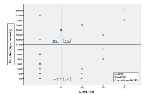
Concordance of results: The simultaneous use of both methods showed 87 concordant results including 5 positive and 82 negative and therefore a concordance of 86.14%. However, 14 discordant results were found (13.86%):
ANA-IB |
||||
Positive |
Negative |
Questionnable |
||
CLIFT |
Positive |
5 |
5 |
1 |
Negative |
3 |
82 |
1 |
|
Questionnable |
2 |
2 |
0 |
|
Table 2 Results with the two techniques CLIFT and ANA-IB

Figure 1 Correlation and agreement between CL immunofluorescence technique (CLIFT) (Threshold≥1/10) and ANA immunoblot (EUROLINE) (Threshold ≥11). Some points represent more than one patient. The value 0 is replaced by 1 for CLIFT.
Specificity and sensitivity: Questionable results were excluded during the measurement of sensitivity and specificity of the immunoblot technique. Of the 85 sera negative to the immunofluorescence technique, 82 sera also had negative ANA-IB results. Therefore, our technique to be evaluated has a specificity of 96%. Among the 10 sera tested positive to the reference method, 5 negative results were found by the immunoblot technique. Therefore, this assay method has a sensitivity of 50%.
Anti-dsDNA antibodies are usually detected and quantified by commercially available kits for enzyme-linked immunosorbent assay (ELISA, also automated versions), Crithidia luciliae immunofluorescence test (CLIFT) and radioimmunoassay methods developed according to Farr technique (FARR-RIA).12 However, CLIFT has been adopted in our laboratory as the technique of choice for the detection of anti-dsDNA antibodies.
The study by Enocsson et al. involved 178 SLE patients whose sera were analyzed by four methods including immunofluorescence on Crithidia luciliae (ImmunoConcepts) and immunoblot (EUROLINE; Euroimmun). This study showed a concordance between these two techniques of 81% with a Spearman correlation coefficient (rho) of 0.558. Nevertheless, this Swedish study found 34 discordant results or 19.10% (12 negative and 22 positive).13 Their concordance result is close to that of our study, but the discordance between the two techniques in the study by Enocsson et al was higher. However, other studies have shown lower concordance rates than our study.
In another study by Tunakan et al, sera from 46 CLIFT-positive patients were analyzed with ANA-IB. The positivity of anti-dsDNA antibodies was found in 30 patients, i.e. a concordance of 65% between the results of the two techniques. To exclude SLE, the Turkish team of "Tunakan et al" analyzed 21 CLIFT-negative sera of which 20 came back ANA-IB negative (i.e. 95%).Therefore, the results of CLIFT and ANA-IB had a high correlation coefficient (p=0.002, r=0.90).8
The Korean team of "Yang JY et al." evaluated the diagnostic performance of 6 commercial kits for anti-dsDNA detection including CLIFT and Immunoblot IB on 142 sera. The results of these two techniques were concordant in 53.8% of cases.14
The nature of the antigen and its binding, as well as other experimental details such as buffers and incubation times, are examples of factors that may affect the detection of anti-dsDNA autoantibodies and thus the lack of concordance of results in some cases.13
The limitation of our study is the lack of clinical information on the test vouchers to know the diagnosis of patients with negative results by both techniques (CLIFT and ANA-IB).
Our evaluation of the performance of immunoblot versus indirect immunofluorescence on Crithidia Luciliae in the detection of anti-dsDNA antibodies supports that CLIFT remains the reference method for the detection of these antibodies and cannot be replaced by ANA-IB. However, their concomitant use increases the specificity of the test.
Common sense therefore suggests optimizing the choice of test by following the recommendations of the French National Authority for Health (HAS) for the detection of anti-dsDNA antibodies by different techniques (in order of decreasing specificity):
In order to meet the criterion of positivity of anti-dsDNA antibodies in the context of SLE diagnosis, it is imperative that their levels are higher than the laboratory standard (> 2 times the reference dilution if ELISA test).15
None.
None.
The authors have declared that no competing interests exist.

©2023 Mahir, et al. This is an open access article distributed under the terms of the, which permits unrestricted use, distribution, and build upon your work non-commercially.