Journal of
eISSN: 2373-6453


Short Communication Volume 8 Issue 2
1Internal Medicine and Pneumology. MD. Servicio de Medicina Interna. Hospital General Guasmo Sur. Guayaquil, Ecuador
2Infectious disease. M.D. Servicio de Infectología, Hospital General Guasmo Sur, Ecuador
3GP Advisor. MD. MSc. Malongo Clinic. Angola
Correspondence: Mariolga Bravo-Acosta. Internal Medicine and Pneumology. M.D. Servicio de Medicina Interna. Hospital General Guasmo Sur. Guayaquil, Ecuador
Received: May 20, 2020 | Published: June 5, 2020
Citation: Bravo-Acosta M, Vélez-Solorzano P, Martínez-Méndez D. Clinical characteristics of Covid-19 cases in Guayaquil, Ecuador. J Hum Virol Retrovirolog. 2020;8(2):50-54. DOI: 10.15406/jhvrv.2020.08.00221
Introduction: Until April 20 in Ecuador there were 10,122 confirmed cases of Covid-19 with 507 deaths. We described the clinical characteristics of 115 confirmed Covid-19 cases.
Methods: Nasopharyngeal swab or tracheal aspirate samples were collected to perform a confirmatory test for Covid-19. Clinical, laboratory and chest radiography data, invasive mechanical ventilation, days of hospitalization and number of deaths were recorded.
Results: The mean was 54.1 years with 59% male. 96.5% had Dyspnea being the most frequent symptom and 20% had diarrhea. Hypertension and Diabetes Mellitus were the main comorbidities. The mean until death was 15.3 days with 9.2 days of hospitalization. 40.9% required invasive mechanical ventilation. 48.7% recovered, 9.6% remain hospitalized and 41.7% died. X-ray showed bilateral opacity. 46.9% had leukocytosis and 85.5% of the deceased presented lymphopenia versus 53.7% of the survivors (p<0.001). 77.4% with prolonged prothrombin time and 82.6% elevated lactic dehydrogenase.
Discussion: Respiratory symptoms are the most frequent. However, the presence of diarrhea was greater than previously reported suggesting the importance of investigating gastrointestinal disorder as the primary symptom. The fatality rate was 41.7%, like critically ill patients. The age of the deceased was older than the survivors, being 62.5% male and 52.1% with some comorbidity, both considered risk factors for severe forms of Covid-19. Lymphopenia is a critical factor associated with severity and mortality.
Keywords: SARS-Cov-2, lymphopenia, invasive mechanical ventilation, Diarrhea, mortality rate, Comorbidity, Covid-19
WHO, World Health Organization; SARSCoV-2, severe acute respiratory syndrome – coronavirus – 2; RTPCR, reverse-transcriptase polymerase chain reaction; Covid-19, coronavirus disease 2019; INSPI, Dr. Leopoldo Izquieta Pérez National Institute of Public Health Research; HT, hypertension; DM, diabetes mellitus; INR, international normalized ratio; LDH, lactic dehydrogenase; GI, gastrointestinal; ACE2, angiotensin-converting enzyme 2
In December 31, 2019, the World Health Organization (WHO) was informed about cases of pneumonia of unknown etiology in Wuhan City, Hubei Province, China. A month later, in January 2020, a new coronavirus (SARS-CoV-2) was shown to be the cause of the outbreak of severe acute respiratory syndrome officially called Covid-19.1,2 On March 11, the WHO declared Covid-19 as a pandemic.3 Until April 20, 2020, a total of 2,422,525 confirmed cases and 166,235 deaths4 have been documented worldwide and in Ecuador 10,122 confirmed cases with 507 deaths.5 In this study we describe the clinical characteristics of 115 confirmed Covid-19 cases in the city of Guayaquil, Ecuador.
Patients and data collection: 115 patients with a Covid-19 confirmed infection from February 29 to April 20, 2020 were included. The data obtained from the electronic medical record were: clinical signs and symptoms, laboratory findings and chest radiography. The number of patients who required oxygen, invasive mechanical ventilation, the time elapsed from the onset of symptoms until admission, and until intubation, as well as the mean days of hospitalization and the number of deaths were recorded.
Specimen collection and testing: Nasopharyngeal swab or tracheal aspirates samples were collected according to the Centers for Disease Control and Prevention guidelines.6 The reverse-transcriptase polymerase chain reaction (RT-PCR) was used as a Covid-19 confirmatory test7. All the tests were carried out at the National Institute of Public Health Research Dr. Leopoldo Izquieta Pérez (INSPI), Guayaquil. X-ray and Laboratory tests (complete blood count, chemical blood tests, coagulation tests and electrolytes) were performed.
Ethical Statement: The study had the approval of Guasmo Sur General Hospital board. Anonymity was preserved in accordance with the guidelines of the World Bioethics Congress and the amended Declaration of Helsinki. Statistical analysis: Categorical variables were summarized as the counts and percentages in each category. Chi-square and Fisher’s exact tests were used for categorical variables as appropriate. Differences with p values <0.05 were considered significant. All analyses were conducted with GraphPad Prism 8 statistical software.
Table 1 describes clinical characteristics detailed of all individuals in the study comparatively between each group: discharged patients, still hospitalized and dead. In all patients, the age ranged from 26 to 83 years with a mean of 54.1 years. 59% (n = 68) were male. The most frequent symptoms were dyspnea 96.5% (n=111), cough 84.4% (n=97), fever 78.3% (n=90), diarrhea 20% (n=23) and odynophagia 17.4% (n=20). Hypertension (HT) was present in 20% (n=23) of the cases, being the main comorbidity, followed by Diabetes Mellitus (DM) in 10.4% (n=12) and 7.8% (n=9) presented both. The mean number of days from symptom onset to hospital admission was 6.9; until intubation at 8 days and death at 15.3 days, with a mean number of days of hospitalization until discharge of 9.15. All the patients required oxygen support and 40.9% (n=47) required invasive mechanical ventilation. To date, 48.7% (n=56) have been discharged in good general condition, 3.5% (n=4) remain hospitalized with non-invasive oxygen support and 6.1% (n=7) under invasive mechanical ventilation. 41.7% (n=48) died.
|
Characteristics |
All Patients N=115 (100) |
Recovered patients n=56 (48.7) |
Hospitalized patients n=11 (9.6) |
Death patients n=48 (41.7) |
|
Range age (mean) - years |
26-83 (56) |
26-77 (51.5) |
30-67 (56) |
26-83 (59.4)* |
|
Sex – n (%) |
||||
|
Female |
47 (41) |
24 (42.9) |
5 (45.5) |
18 (37.5) |
|
Male |
68 (59) |
32 (57.1) |
6 (54.5) |
30 (62.5) |
|
Symptoms – n (%) |
||||
|
Disnea |
111 (96.5) |
52 (92.9) |
11 (100) |
48 (100) |
|
Cough |
97 (84.3) |
48 (85.7) |
9 (81.8) |
40 (83.3) |
|
Fever |
90 (78.3) |
44 (78.6) |
10 (90.9) |
36 (75) |
|
Diarrhea |
23 (20) |
15 (26.8) |
4 (36.4) |
4 (8.3) |
|
Odynophagia |
20 (17.4) |
16 (28.6) |
1 (9.1) |
3 (6.3) |
|
Chest pain |
12 (10.4) |
9 (16.1) |
0 |
3 (6.3) |
|
Co-morbidity – n (%) |
45 (39.1) |
15 (28.8) |
5 (45.5) |
25 (52.1)^ |
|
Hypertension |
23 (20) |
9 (16.1) |
1 (9.1) |
13 (27.1) |
|
Diabetes Mellitus |
12 (10.4) |
5 (8.9) |
2 (18.2) |
5 (10.4) |
|
Hypertension and Diabetes Mellitus |
9 (7.8) |
1 (1.8) |
2 (18.2) |
6 (12.5) |
|
Tuberculosis |
1 (0.9) |
- |
- |
1 (2.1) |
|
Without co-morbidity – n (%) |
70 (51.9) |
41 (73.2) |
6 (54.5) |
23 (47.9) |
|
Mean since the beginning of the symptoms to admission - days |
6.9 |
8 |
8.5 |
8 |
|
Mean since the beginning of the symptoms to intubation - days
|
8 |
4.5 |
8.5 |
9.5 |
|
Mean since the beginning of the symptoms to dead - days |
15.3 |
- |
- |
20 |
|
Mean of hospitalization days - days |
9.15 |
13 |
- |
- |
|
Support measures – n (%) |
||||
|
Oxigen |
115 (100) |
56 (100) |
11 (100) |
48 (100) |
|
Invasive mechanical ventilation |
47 (40.9) |
4 (7.1) |
7 (63.6) |
47 (98) |
Table 1 Comparative clinical and epidemiological characteristics of Covid-19 patients. Guayaquil, 2020
* In deceased patients 77.1%> 50 years of age vs. 53.73 of survivors (p=0.01). ^ 52.1% with comorbidity vs 29.9% of survivors (p=0.02)
In deceased patients, the mean age was 54.5 years, 77.1% being over 50 years old compared to 53.73% of survivors (p=0.01). 62.5% were male. All presented dyspnea, 83.3% (n=40) cough and 75% (n=36) fever. 52.1% (n=25) had associated chronic diseases: 27.1% (n=13) Hypertension; 10.4% (n=5) Diabetes Mellitus; 12.5% (n=6) both and 2.1% (n=1) tuberculosis. Only 29.9% (n=20) of the survivors presented comorbidities (p=0.02). The mean days from the onset of symptoms to hospitalization was 8, until intubation 9.5 and 20 until death. 98% (n=43) required invasive mechanical ventilation. Table 1.
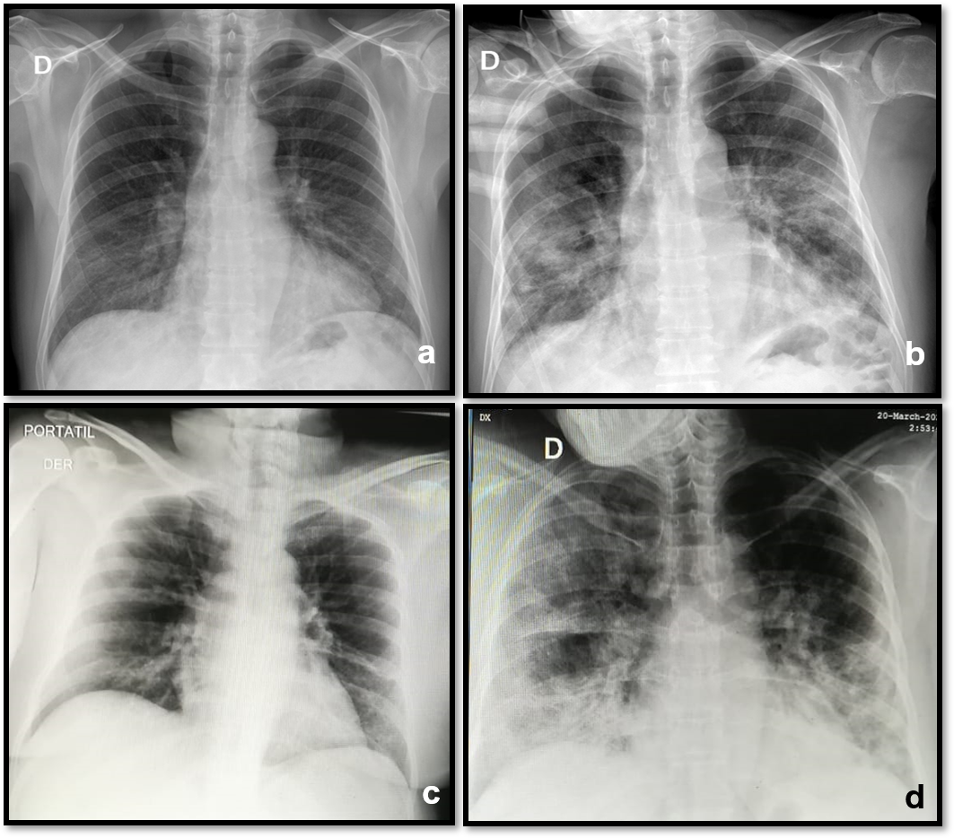
Chest x-rays were performed on 86 patients during the admission, all with bilateral opacity. None presented pleural effusion (Figure 1).

Figure 1 Chest x-ray with the evolution of two patients. a).- Male. 59 years old. March 25. At admission: Interstitial lung pattern in lower zones with right predominance. b).- March 31. in ICU: Right alveolar opacities in mid and lower zones Left hazy opacity in mid and lower zones. Dyspnea got worse and required invasive mechanical ventilation. Dies 14 days later. c).- Female. 59 years old. March 18. At admission: Lower vague hazy densities. d).- March 20. in ICU: Bilateral diffuse interstitial pattern with ground glass opacities in the right upper and lower areas. Greater lower right opacity due to peribronchovascular thickening. Dyspnea got worse and required invasive mechanical ventilation. She had a full recovery and was discharged 23 days later.
The laboratory findings at the time of admission are described in detail in Table 2. 46.9% (n=54) presented leukocytosis; 81.7% (n = 94) neutrophilia and 66.9% (n=77) lymphopenia. Only 5.2% (n=6) presented plateletpenia and 17.4% (n=20) had anemia. 10.4% (n=12) presented a prolonged activated partial thromboplastin time and 77.4% (n=89) prolongate prothrombin time, with an increased International Normalized Ratio (INR) in 37.4% (n=43). Serum creatinine was high on 13.1% (n=15) and random glucose values were high in 13.1% (n=13). 28.7% (n=33) presented hyperchloremia and 8.7% (n=10) hypokalemia, 82.6% (n=95) already had high lactic dehydrogenase (LDH).
|
Reference values |
All Patients N=115 (100) |
Recovered patients n=56 (48.7) |
Hospitalized patients n=11 (9.6) |
Death patients n=48 (41.7) |
|
|
Blood routine - n (%) |
|||||
|
Leukocytosis |
4000 – 10000mm3 |
54 (46.9) |
15 (26.8) |
5 (45.5) |
30 (62.5) |
|
Neutrophilia |
55 – 70 % |
94 (81.7) |
56 (100) |
9 (81.8) |
37 (77.1) |
|
Lymphocytopenia |
17 - 45% |
77 (66.9) |
27 (48.2) |
9 (81.8) |
41 (85.5)* |
|
Plateletpenia |
150 – 450 mm3 |
6 (5.2) |
3 (5.4) |
0 |
3 (6.3) |
|
Anemia |
11.5 - 16 g/dL |
20 (17.4) |
9 (16.1) |
1 (9.1) |
6 (12.5) |
|
Coagulation function - n (%) |
|||||
|
Prolongate Activated partial thromboplastin time |
25 - 40 seg |
12 (10.4) |
5 (8.9) |
1 (9.1) |
7 (14.6) |
|
Prolongate Prothrombin time |
9 - 12 seg |
89 (77.4) |
44 (78.6) |
9 (81.8) |
31 (64.6) |
|
International normalized ratio (INR) Increased |
0.9 - 1.1 |
43 (37.4) |
21 (37.5) |
5 (45.5) |
15 (31.3) |
|
Blood biochemistry - n (%) |
|||||
|
Serum creatinine Increased |
0.6 - 1.2 |
15 (13.1) |
2 (3.6) |
1 (9.1) |
9 (18.8) |
|
Random Glucose level Increased |
(>200 mg/dL) |
13 (11.3) |
4 (7.1) |
2 (18.2) |
6 (12.5) |
|
Lactate dehydrogenase (LDH) Increased |
81-234 U/L |
95 (82.6) |
46 (82.1) |
9 (88.1) |
41 (85.4) |
|
Electrolytes - n (%) |
|||||
|
Hyponatremia |
135 - 145 mEq/L |
5 (4.3) |
3 (5.4) |
1 (9.1) |
6 (12.5) |
|
Hypokalemia |
3.5 – 5.1 mEq/L |
9 (7.8) |
3 (5.4) |
3 (27.3) |
4 (8.3) |
|
Hyperchloremia |
96 - 106 mEq/L |
33 (28.7) |
15 (26.8) |
4 (36.4) |
13 (27.1) |
Table 2 Comparative Laboratory results of patients with Covid-19. Guayaquil, 2020
*85.5% lymphocytopenia vs 53.7% of survivors (p<0.001)
In the deceased patients, 62.5% (n=30) presented leukocytosis; 77.1% (n=47) neutrophilia. 85.5% (n=41) presented lymphopenia in contrast to the 53.7 (n=36) of the survivors (p<0.001). 14.6% (n=7) with prolonged activated partial thromboplastin time and 64.6% (n=31) a prolongated prothrombin time with an increased INR in 31.3% (n=15). Serum creatinine was increased in 18.8% (n=9) and random glucose values were elevated in 12.5% (n=6). 27.1% (n=13) presented hyperchloremia and 12.5% (n=6) hyponatremia. 85.4% (n=41) had elevated LDH levels.
In Ecuador, the first case of Covid-19 was confirmed on February 29, 2020, with a total of 10,122 confirmed by April 20, 2020.5 In this study, we describe the clinical findings of 115 confirmed Covid-19 patients from Guayaquil, Guayas province, epicenter of the cases described in the country.
Most of the patients presented dyspnea and needed oxygen support, until now where dyspnea is the most frequent symptom, followed by cough and fever.8–10 However, in contrast to other studies reporting low frequency of gastrointestinal symptoms, in our study, 20% of cases presented diarrhea, higher than previously reported.9,11,12 The gastrointestinal (GI) system has been proposed as a potential route of Covid-19 invasion and transmission, probably because of the interaction of SARS-CoV-2 and Angiotensin-Converting Enzyme 2 (ACE2) receptors could cause diarrhea.13,14 Our findings may contribute to understanding of Covid-19 transmission and the importance of GI disorders as one of the primary symptoms of the disease.11,13–15
The case fatality rate in this series was 41.7%, as had been reported in critically patients in Seattle and China9,16. The age of dead patients was significantly higher than that of the survivors, with a mean of 59.4 years being 62.5% male and with some comorbidity in 52.1% of the cases. This is similar to those reported in other studies where the age, male sex and cardiovascular and metabolic chronic diseases are risk factors for severe forms of Covid-19, probably related to secondary immunodeficiency.8,9,16–20
Neutrophilia with lymphopenia has been reported as a common feature in most patients, suggesting that Covid-19, as already described for SARS coronavirus, harms lymphocytes during the replication process in the respiratory mucosa, probably caused by the translocation of peripheral blood lymphocytes to the lungs21 which induces the of cytokines storm, the development of the systemic inflammatory response syndrome that rapidly progresses of multiple organs failure and the alterations evidenced in coagulation factors and detection of elevated LDH values could be early indicators.9,10,22 In our study, 85.5% of the deceased had lymphopenia, so lymphopenia in patients with Covid-19 could be a critical factor associated with disease severity and mortality.
Our study had limitations, first the lack of uniformity in the collection of clinical and epidemiological data on admission. Therefore, many symptoms that are now included were probably not registered. Secondly, laboratory tests were not available on admission for all patients.
In conclusion, the clinical and laboratory characteristics between the group of deceased and the group of survivors, age over 50 years, the presence of comorbidities and lymphopenia, were related to mortality.
We thank all the patients involved in the study, the health personnel responsible for medical care and Edna Correa, (MSc), for review the manuscript.
The authors declare have no conflict of interest.
Authors declare no funding was received for this study.

©2020 Bravo-Acosta, et al. This is an open access article distributed under the terms of the, which permits unrestricted use, distribution, and build upon your work non-commercially.