Journal of
eISSN: 2373-6453


Research Article Volume 3 Issue 6
1Clinic for Infectious Disease, Medical faculty University of Montenegro, Montenegro
2Institute for Health of Montenegro - Department of Microbiology, Montenegro
Correspondence: Bogdanka Andric, Clinic for Infectious Diseases, Podgorica, Medical Faculty, University of Montenegro, Montenegro
Received: August 19, 2016 | Published: October 20, 2016
Citation: Andric B, Mijovic G, Andric A (2016) Characteristics of Hand Foot and Mouth Disease. J Hum Virol Retrovirol 3(6): 00116. DOI: 10.15406/jhvrv.2016.03.00116
The Hand, Foot and Mouth Disease (HFMD) are an acute, highly contagious viral disease. Initial symptoms are fever, poor appetite, and fatigue. The fever occurs 1-2 days after onset of oral ulceration. Then on the distal parts of the extremities occurs rash, usually lasts for 7-10 days, and spontaneously resolved. The disease is most often had classified as benign infection, which generally does not require therapeutic treatment.
Recent findings have been warning that the number of maladies cases of HFMD in the world is growing. Especially in the last four years, serious cases with many severe complications, (aseptic meningitis, pneumonia, prolonged febrile state, long-term fatigue, muscle and joint pain, recurrent pain, expressed dehydration) increasing. Examination of the major epidemics in the world has shown that the most common causes of diseases are two types of enter viruses (Ev): Coxsakievirus (Cox) of the group A, subtype A16 (CoxA-16), and enter virus 71 (Ev-71). ECHO and other enteroviruses may also be associated with HFMD.
In September 2014, in Montenegro were registered epidemic of HFMD with 29 diagnostic cases. In the total sample of maladies, child population of 4-12 years accounted for 20 (71%), and adults for 9 (29%) cases. The first cases were registering in Pljevlja. In short time, disease has assumed epidemic proportions with 25 registered cases. In the same period in Podgorica were registered four cases. We analyzed the epidemiological, clinical characteristics of the disease, with special emphasis on diagnostic difficulties and prognosis of disease.
Keywords: HFMD, Epidemiological, Clinical characteristics, Diagnosis, Prognosis
HFMD, Hand Foot and Mouth Disease; Ev-71, Enterovirus 71; PCR, Polymerase Chain Reaction; CIR, Cellular Immune Response; CNS, Central Nervous System; Ev, Enter viruses; Cox A16, Coxsackievirus
Hand Foot and Mouth Disease (HFMD) is a highly contagious systemic infection caused by enteroviruses.1 It is most common in children, but also in adults. With disturbance of the general condition of various degrees, the disease vas characterized with occurrence of typical vesicles rash most frequently seen on the palms of the hands, soles of the feet and inside the mouth. Mucosal vesicles on the oropharyngs quickly sprayed leaving ulcerations.1,2
Investigations of major epidemics of HFMD in the world, showed that the most frequent causers of disease are the members of entero virus genus, most commonly, Coxsackievirus (Cox A16), (CoxA6) and Enterovirus 71 (Ev-71). In addition, sporadic cases with coxsackievirus subtype A4 - A7, A9, A10, B1 - B3 and B5, and ECHO viruses has been reported. Recent years have shown that increasing number of HFMD, caused predominantly by Ev-71 and Cox- A-16. The high incidence of mixed infections with Ev-71 and Cox-A16 was also observed.1-3
Enteroviruses represent a large family of RNA viruses in terms of the number of members, but on basis of the small size of virion are categorizing as least (picornaviridae). Some viruses cause certain clinical symptoms, but the virus itself may be responsible for a number of different clinical syndromes.4-6 They are contains a single-member linear RNA and glycoprotein s, characterized by high variability.7,8 This is one of the reasons for the difficulty in diagnosis and variations in clinical presentation. In 60% of infected persons, infection develops asymptomatically. HFMD is clinically manifest infection. The disease is characterizing by fever, often the first sign of infection, followed by other general symptoms. Painful sores in the mouth, hands / feet develop after the start of fever9,10 In the greatest number of HFMD cases mild and self-limiting illness last about 10 days with spontaneous regression. The most frequent complications are dehydration and fussiness. Rare and sometime difficult forms of disease with serious complications present neurological: meningitis, encephalitis, cardiovascular and pulmonary complications. Numerous investigations in the world identified the factors that influence of severity of the disease and complications. The high risk factors are the primarily characteristics of causal enteroviruses and characteristics of the hosts. There was a significant difference in age, total duration of fever, rate of respiratory and heart disturbances, shake of limbs, white blood cell count, level of glikemia and keratin kinasis (CKMB). Severe symptoms that last for a long time ware registered in immune-deficient patients (different dysfunction of immune system, in patients with diabetes mellitus, organ transplant recipients, and patients receiving chemotherapy, the malignant diseases and HIV / AIDS infection).
In some patients with HFMD respiratory tract disturbances are mostly benign usually followed by fever, sore throat and rhinitis. A difficult respiratory manifestation goes with interstitial pneumonia, mixed viral-bacterial pneumonia, rarely can progress to ARDS11-15 and even fatal diseases.16-18
HFMD infections are widespread in the world. In areas with a temperate climate, the disease has seasonal characteristics. They occur mostly in the summer and in early fall. In tropical and subtropical regions, it occurs throughout the year. Due to fecal-oral transmission, disease acquired title as "disease of dirty hands". Rarely, the infection can be transmitting via droplets and from mother to fetus. Investigations have shown that in 40- 70% of cases secondary infection at home where registered, and depend on the type of virus, overpopulation, hygienic living conditions. After 10 day, in most patients, symptoms withdraw, without consequences.7-19 In recent years, increasing number of patients in the world shows a busy registration of severe forms of the disease, including fatal.7,17,19
Specific therapy for HFMD does not exist. There is only symptomatic (analgesics, antipyretics, solution for mouth rinses). Fluid intake is an important measure to prevent dehydration, especially in younger patients. Severe infections with bacterial complications require hospitalization, the application of antibiotics and other appropriate therapy.19-21
This research was approving by the Clinic for Infectious Disease in Podgorica and Institute for Health of Montenegro - Department of microbiology in Podgorica, Montenegro. We have investigated the frequency and characteristics of HFMD in 29 patients in September 2014 in Montenegro, with standard epidemiological, clinical and laboratory methods.
Clinical diagnosis including frequency and characteristics of HFMD on based on clinical symptoms: fever, exanthema (papule-vesicular rash) with typical distribution (hand, foot and mouth), Severe cases is defined as having HFMD symptoms, in our investigated group with dominant respiratory disorder. The laboratory assay may show an increase in leucocytes in the peripheral blood, increased blood glucose, chest X-ray.
Etiological confirmation of diagnosis was establishing on the following tests: Polymerase Chain Reaction (PCR) for common amplification of nucleic acid of enteroviruses. PCR assay detect all human entero virus strains including coxsackievirus, Echovirus and poliovirus and is the test of choice for detection of acute disease in multiple specimen sources (CFS, plasma, body fluids, ocular swabs, dermal swabs and others Serological ELISA method (Antibody-capture enzyme linked immunosorbent assay) was using for diagnosis coxsackieviruses based for detection of immunoglobulin G (IgG), IgM and IgA antibodies and showed broad specificity to entero virus (e.g. Coxsackievirus A and B and other enteroviruses). ELISA method was using of the detection the other potential co-participants in infection (Chlamydia pneumoniae, Micoplasma pneumoniae, Heamophilus influenza) in patients with (respiratory) complications. Antibody response would not be reliable detectable during acute disease. Furthermore, there is a high prevalence of entero virus antibodies in the general population from previous exposure and thus serologic testing is not useful in nearly all clinical situations. Non-specific reactivity did occur however in cases of Infectious Mononucleosis and in Micoplasma pneumonia infection.
In September 2014 in Montenegro were investigated 29 cases with HFMD. The diagnosis of the disease based on epidemiological, clinical, laboratory, microbiological (PCR and serological ELISA methods). Milder forms of the disease did not require special diagnostic procedures. For severe forms of the disease, quick diagnosis has been important, for therapy and prognosis.
In our sample of 29 cases, children s population was present with 20 (68, 96%) cases, and adult population in 9 (31, 03%) cases. The epidemic began in Pljevlja with total registered 25 (86, 20%) cases with HFMD. Indicative diagnosis based on clinical recognition of the disease, and confirmed by microbiological methods. Epidemiological investigation, established the fecal-oral route of transmission of infection. The largest number of registered cases were children aged 4-12 years 20 (62, 7%), from collective centers: kinder garden7 (33, 33%) cases, and schoolchildren’s in 13 (66, 67%) cases. An adult has been representing with 9 (28%) cases.
In the same period in Podgorica, were registered 4 (13, 79%) cases with HFMD, of which 2 (50%) cases are schoolchildren’s and 2 (50%) adult cases where the infection came from family contacts.
Diagram 1
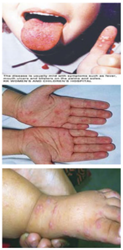
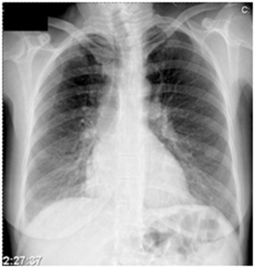
Clinically, all patients had symptoms of common infectious syndromes with fever 37.5 - 39.0C, malaise, headache, sore throat, pain in muscles and joints, and characteristic rashin the skin and mucous membranes of the oral cavity (Figure 1 A, B&C). Inflammation of the upper respiratory tract was founded all patients. Syndrome of angina, aphthous stomatitis and tonsilopharyngitis has been registering in all patients. Pneumonia is registered with 17 (58, 62%) patients, of which 14 (47%) cases were of primary interstitial pneumonia. Serious respiratory complications for mixed viral-bacterial infection (with participating Micoplasma pneumoniae, Chlamydia pneumoniae and Heamophilus influenza) in HFMD patients, is diagnosed in 3 (10, 34%) cases (Figure 2).

Figure 1 After the appearance of vesicles on the mucous membranes of the oral cavity, which can quickly turn into ulcers (A), occurs exanthema mixed type (macula, papules, vesicles) on the skin of the distal parts of the upper extremity B, lower C).

Figure 2 Primary interstitial pneumonia in our patients, were registered in 14cases, and serious respiratory complications for mixed viral-bacterial infection in 3 cases with HFMD.
To estimate the severity of pneumonia, we used clinical and laboratory parameters (age, presence of accompanying chronic infections, respiratory frequency, mental status, blood pressure, blood oxygen saturation, blood count and differential blood count), and microbiological findings. Investigations have shown that in 3 cases, the weight of pulmonary complications included mixed bacterial - viral pneumonia, with serological detection of co-infective participation of Chlamydia pneumonia, Micoplasma pneumoniae and Heamophilus influenza. Laboratory tests have shown that this severe cases are associated with total leukocyte count (>10-16 x 109), hyperglycemia, and raised level of CK-MB. These patients had a prolonged and difficult course of disease. Are treating in hospital and request antibiotic treatment. In the sample of 29 cases, with PCR method, it was confirmed participation of enteroviruses in 21 (72%) cases and in 8 (28 %) cases; we detected coxsackiaeviruses by ELISA method. Precise definition of viral serotypes was not possible due to the technical limitation of microbiological laboratory (Figure 2). Co-infective participation of bacterial agents in pulmonary complication was detecting by ELISA method.
Diagram 2
Modern epidemiological data indicate that the incidence of HFMD is continuously growing in the world from year to year.7,11,14 In some parts of the world is up to 90% in children aged less than 5 years. The incidence of diseases in older age groups increasing in the last 4 years and amount to approximately 85-95%.17 Recent studies also show that the increase the number of cases is accompanying by increase severity of disease, so it s classified in the "Dangerous diseases".14,19 Numerous studies have shown that high risk factors for severity of HFMD were representing of features the triggering viruses. Patients infected with EV-71 had significantly more frequent and harder cardiac and neurological complications. It can be expected that mixed infection between enteroviruses, for example CoxA-16 and Ev-71 with even more difficult, as well as a mix of enteroviral agents with the other infectious agents (Chlamydia, Micoplasma, Heamophilus influenza), which was confirmed during in our investigations.
Furthermore weight the disease also are significant difference in age, total duration of fever, past diseases and conditions of the immune system, rate of respiratory and heart shake of limbs, white blood cell count, blood sugar, CK-MB. Since 1980 s, multiple severe HFMD outbreaks have been documented and disease remains a significant public health problem especially in the Asia and Pacific region. In study from China 2011 showed that neutralizing antibody response was not correlating with disease severity, suggesting that Cellular Immune Response (CIR) besides neutralizing antibodies could play a critical role in controlling the outcome of Ev-71human’s infection. The recognition and correct interpretation of dermal signs of diseases that goes’ with cardiopulmonary system may do the clinical diagnosis and assessment of prognosis.
Our investigations showed the participation of the children population (of school and pre-school ages) is dominant 20 cases. Adults participated with 9 cases. In total sample of cases with HFMD, the upper respiratory disturbances have registered in all patients. The primary interstitial pneumonia, was found in 14 (47%) of patients. Serious respiratory complications for mixed viral-bacterial infection has registered in 3 (10, 34%) of HFMD patients. In our cases, the most frequent are respiratory tract complications. Identify the clinical features of disease, and the application of rapid and appropriate symptomatic therapy and antibiotic therapy in mixed viral-bacterial pneumonia, are the reason why we did not even more difficult complications. Except the transient tachycardia associated with high fever, in our series were not registered weight cardiovascular and central nervous system (CNS) complications.
The results of numerous studies around the world have shown that Enteroviruses had always baffled doctors. Studies have shown that the symptoms of the disease are often misdiagnosed. Wide range and variability of the clinical manifestations of disease include almost all organs and systems. Even in cases of well-defined clinical syndrome, what represents HFMD, respiratory, cardiovascular, gastrointestinal and other complications, represent a significant diagnostic and differential diagnostic problem.12,14,18 In one large study from USA in the total number of respondents, 39% of children with different diagnoses proved that it was an enteroviral infection.11 For the etiological diagnosis of HFMD, there are three methods (virus isolation, serological testing, PCR).11,14,15 In our series, the results have confirmed the participation of enteroviral agents in 68%, and coxsackieviruses in 22% with the use of PCR and ELISA methods. Precise definition of serotypes of causative viruses is not working out because of the technical limitations of laboratory.
Characteristics of enteroviruses are important and responsible for the difficulties of diagnosis and differential diagnosis of HFMD. The virus genome contains a single-member linear RNA. Proteins are composed of four major polypeptides. Surface protein VP1 and VP3 are the major binding site of antibodies. Domestic VP4 protein is bound to the viral RNA; replication takes place in the cytoplasm.21 After ingestion, the viruses have adsorbed to specific receptor sites on the cell surface, which corresponding to the types of enteroviruses. Viral replication occurs in the epithelial cells of the lymphoid organs of the upper respiratory and intestinal tract. Further infection spreads to the surrounding lymphatic tissue. Clinical signs of disease do not accompany this stage of primary viral multiplication. For most of the infected, the disease ends in this phase, and the infected person acquires immunity to a particular type of enteroviruses.
If after the primary replication, viruses reach the systemic circulation, a secondary viremia and scatter in different tissues and organs. The potential "target" organs depend on the type of virus and its tropism. This is the respiratory tract, CNS, vascular endothelium of the heart and blood vessels, liver, pancreas, reproductive organs, skeletal muscle, synovial tissue, skin and mucous membranes.7,9 With the end of viral replication appears circulating interferon neutralizing antibodies, mononuclear cell infiltration. Early humoral response was following by the appearance of specific IgM antibodies that appear to 7-12 day, after which occur specific IgG antibodies.
Persistent infection in the central nervous system (CNS) has been reporting at persons with hypo- and a gamma globulinemia, indicating an important role of B - cells in their elimination.10,11 Enteroviral infections are widespread in the world.2 The disease is spreads rapidly and in a relatively short time can reach pandemic proportions.7,20,21 Investigation reports around the world, especially in the last four years are reporting that frequency and severity of the enteroviral infections growing from year to year. Based on these findings, the new features, classified HFMD in the group of “threatening infections" which should provide enough research space in our midst.3,5,6 Additionally, skin rash and mucosal manifestations represent an important criterion for the clinical recognition of the disease, which proved significant during in our investigations.
In the broad spectrum of manifest Coxsackiaeviral infections, respiratory tract manifestation is frequent: colds, sore throats, bronchitis, and bronchiolytis, interstitial pneumonia, which are registries in the course of our investigations. Respiratory events in the spectrum of HFMD syndrome may be occur as serious complications, especially in immunodeficient patients as during our tests in patients with HIV/AIDS have not been proven. Our investigation has shown frequency of respiratory disorders in immune competent patients in 47% of cases. In addition, in 2% of cases, developed difficult mixed viral / bacterial pneumonia, this required additional diagnostic procedures, hospitalization and antibiotic treatment.
In our investigations, the results of etiological diagnosis of HFMD in favor of the dominant participation undefined enteroviruses with coxsackieviruses. Since interstitial pneumonia belong to the difficult manifestations of the disease, does not exclude the possibility of co-infective participation of more enteroviruses in the etiologic basis of HFMD in our cases, and on this basis the concatenation of secondary bacterial infections in 3 cases. In our investigated group, the complications as ARDS and acute respiratory failure ware not registered. However, numerous investigation reports around the world, especially in the last four years are reporting that frequency and severity of the enteroviral infections growing from year to year. Based on these findings, the new features, classified HFMD in the group of “threatening infections" which should provide enough research space in our midst.3,5,6 Additionally, skin rash and mucosal manifestations represent an important criterion for the clinical recognition of the disease, and further diagnostics of HFMD.
The hand, foot and mouth disease (HFMD) is highly contagious systemic infection caused by enteroviral agents, usually CoxA16, Cox A6 and Ev-71, or other enteroviruses. Most of these infections are asymptomatic or mild. When manifest, infection goes in addition to specific symptoms: aphthous stomatitis and the appearance of exanthema in the distal parts of the extremities (hands, feet), among the most frequent symptoms, are one of the respiratory system, as well as other disorders. Interstitial pneumonia, varying degrees of severity, was registered in immune-competent, and in immune deficient patients. It is often associated with a number of enteroviral, bacterial agents in mixed co-infections. The etiological diagnosis is complex and unreliable, in addition to being available for various serological methods and PCR. In cases with bacterial complications (mixed infections), in addition to the usual symptomatic therapy, it is necessary to include antibiotic therapy. Our investigated sample was small; it could not bring significant conclusions about this severe systemic infection, but it gave us enough to alert to the presence, reminding that it classifies as dangerous infectious diseases.
None.
None.

©2016 Andric, et al. This is an open access article distributed under the terms of the, which permits unrestricted use, distribution, and build upon your work non-commercially.