Journal of
eISSN: 2573-2897


Research Article Volume 8 Issue 1
1Conservation Department, Faculty of Archaeology, Cairo University, Egypt
2Grand Egyptian Museum, Conservation Center (GEM.CC), Giza, Egypt
Correspondence: Omar Abdel-Kareem, Conservation Department, Faculty of Archaeology, Cairo University, Egypt
Received: February 21, 2023 | Published: March 9, 2023
Citation: Abdel-Kareem O, Mahmoud R, Mertah E, et al. Documentation, investigation and analysis of a rare archaeological cartonnage object from the Egyptian Museum in Cairo, Egypt, using non-invasive methods. J His Arch & Anthropol Sci. 2023;8(1):14-20 DOI: 10.15406/jhaas.2023.08.00268
Technical and analytical investigations were carried out on an overhead mask of woman cartonnage from the Egyptian Museum in Cairo (catalogue 33279), which dated back to the Greco-Roman Period. Various non-invasive techniques were used in this study such as: AutoCAD program, multispectral imaging with Ultraviolet and Infrared Rays, optical microscope, X-ray fluorescence spectrometry (XRF), and Fourier transform infrared spectroscopy (FTIR). The structure of the cartonnage was unique and consisted of three distinctive layers, from the bottom to the top; a double layer from linen, a calcite-based plaster layer, and finally a polychrome paint layer. The study of the paint layer revealed the presence of blue, red, white, yellow, orange, brown and black pigments. Yellow color was identified as orpiment (As2S3) and yellow ochre (goethite α-FeO.OH + clay minerals) and blue as Egyptian blue (CaCuSi4O10). Two red shades were also detected of which the lighter is red lead (Pb3O4) and the darker is a mixture hematite (α-Fe2O3), red lead (Pb₃O₄) and calcite (CaCO3). Orange was identified as a mixture of orpiment (As2S3) and red lead (Pb3O4), white as white lead (PbCO3)2•Pb(OH)2,and black as magnetite (Fe3O4). The brown pigment, made up of red hematite, red lead, and black manganese, was detected for the first time in the pigment palette of ancient Egyptian cartonnage. The binding medium in linen layer was identified as Arabic gum. The study showed that cartonnage dated back to the Graeco-Roman Period because of the appearance of red lead, orpiment, and Egyptian blue. Moreover, the presence of lead in the components of Egyptian blue is considered evidence that ovens contain lead resulting in changing the manufacturing techniques of Egyptian blue.
Keywords: cartonnage, greco-roman, egyptian blue, red lead, multispectral imaging
As a result of the ancient Egyptian beliefs in resurrection and immortality (another life), they used to preserve the body of the deceased and prevent its decomposition after death, which is what we define as “embalming”. These beliefs led to the appearance of cartonnage whose main function was to protect, support the soul to return to the body, and decorate the mummy with personal funerary decorations.1,2 The cartonnage consists of multiple layers of linen and sometimes of papyrus formed in a mold and then covered with a mixture of gum or animal glue and lime or gypsum to be suitable for coloring and gilding. After drying, it is decorated with inscriptions and colors and occasionally painted in part with gold.3,5 This method has been used since the beginning of the Middle Kingdom (twelfth dynasty) and spread during the Greco-Roman period.6 The Roman era witnessed the flourishing of Cartonnage where it received great interest and became a mixture of Hellenistic art and Egyptian art owing to the faith of Greeks and Romans in the Egyptian beliefs. However, the materials used to produce cartonnage changed over time. It was common to use plastered linen in the middle kingdom, linen and stucco during the third intermediate period, old papyrus scrolls during the Ptolemaic period, and thicker fibrous materials during the Roman period.
Many studies have been previously conducted on different cartonnage pieces. A recent study about Graeco-Roman Egyptian cartonnage object showed the unique layered structure of the cartonnage as it comprises a polychrome layer, a calcite-based plaster layer, a mixed mud and sawdust support layer, and a finishing calcite plaster layer.7 Pigment colors employed were orpiment, goethite, Egyptian blue, madder, red ochre, orange as an admixture of orpiment and hematite, malachite, atacamite, gypsum, calcite, magnetite, gilding layer, and the binding medium identified as animal glue.7 Another research about Roman Cartonnage from Lisht confirmed that linen and calcium carbonate acted as support and preparation layers, respectively while the pigment layer consisted of Egyptian blue, Egyptian green, and red ocher. The Arabic gum was used as an organic media.1
One more study was about two cartonnages of two female mummies displayed in the Ismailia Museum and dated back to first century AD (Greco-Roman). Examination of these two cartonnages showed that they consist of the following: color layer, gesso layer, and linen, since 2019.3 In 2021 Mahmoud et al., studied a polychrome cartonnage mummy case that was discovered at the archaeological site of El-Lahun at Middle Egypt. The results implied linen fibers as the base support and thin calcareous layer mixed with gum Arabic as preparatory layer ‘gesso’. The pigments contained were Egyptian blue, orpiment, yellow ochre, red ochre and green pigment was created through mixing together Egyptian blue and yellow ochre.8
Moreover, in 2020 Hussein et al., studied a foot case Cartonnage excavated from Saqqara and dated back to the Late Period (712-332 B.C.) The structure of the cartonnage was found to consist of five distinctive layers, from the bottom up; two calcite-based plaster layers which are separated by double linen layers and covered with a polychrome paint layer. The study of the paint layer revealed the presence of cinnabar, red ochre, Egyptian blue, Orpiment, yellow ochre, pararealgar and the binding medium identified as animal glue. The pigment palette used was quite complex and typical of those encountered in the late and Graeco-Roman periods.9 Another example of a conducted study was in 2011 when Afifi et al., studied some cartonnage fragments belonging to the Greek‐Roman period from Hawara, Fayoum. The coarse ground layer was composed of calcite and traces of quartz as the fine ground layer use calcite only. Pigment colors were Carbon black, goethite, Egyptian blue, red lead, and animal glue was used in the four pigments as a colored medium.10 Also, in 2020 Sakr et al.,11 studied six mummies and their cartonnages found at Tell Al Sawa, Eastern Delta, Egypt. Analysis showed linen supports over a calcite preparatory layer mixed with organic binding medium of animal glue.11
Last but not least, in 2014 Abd El Aal et al.,12 studied an ancient Egyptian gilded Cartonnage with polychrome decoration found in Saqqara, Egypt and dating back to late - Greek- Roman period. The gilded Cartonnage was made of a double layer of plain weave linen soaked in gum. The first Coarse ground layer was made up of calcite and humtite. The finer second layer was white calcite. The pigment colors employed were hematite, orpiment, Gilded layer, and the binding medium identified as Arabic gum.12,13
Material investigation is a necessary step in documenting the properties of the component materials of a Cartonnage object, estimating its condition, and considering appropriate conservation treatment. The nature of a cartonnage exerts a fundamental influence on what can best be done to preserve it. Therefore, it is necessary to make a very thorough examination before any decision is made regarding the ways and means. The examination of a cartonnage as a preliminary to conservation gives not only an idea about what it is and what it is made from but also an idea about the degree of handling it will stand which is a vital factor in subsequent decision making. Also, the study of a cartonnage object may help the archaeologists in detecting more information about the cartonnage in ancient Egypt. Methods used to investigate the cartonnage must be none destructive methods as the aim of any study carried out on these cartonnage objects is to preserve them not to destruct them. In fact, there are many different useful techniques and methods that have been published for investigating the chemicals and physical properties of cartonnage dated back to various Egyptian periods.
This study aims at using non-invasive methods for examining and investigating an overhead mask of woman cartonnage from the Egyptian Museum in Cairo, which dated back to the Greco-Roman Period to understand the nature of this mask. This study will help the conservators in developing methods for conservation of these cartonnage objects that are many in the Egyptian Museums.
Historical background and description of the studied object
The studied object is an overhead mask of woman cartonnage displayed in storage area in the Egyptian Museum in Cairo (catalogue 33279) and dated back to the Greco-Roman Period (Figure 1). The overhead mask of cartonnage under study was a part of several separate pieces, consisting of mask overheads, collars, pectorals, aprons, shin pads, foot cases and ankles.9 (Figure 1a) shows detailed views of the cartonnage under study. The piece of cartonnage is approximately 30cm high and 34cm wide with a crown and jewelries that include two earrings and necklace.
The cartonnage was painted mainly with red color. The face was portrayed with big painted eyes and thick black brows. The backside of the cartonnage consists of two linens with no ornaments (Figure 1b).

Figure 1 (a) shows frontal detailed view of the cartonnage under study, (b) the backside of the cartonnage.
In general, the cartonnage suffers from weakness, fragility, dirt accumulation, stains, paint flake loss, fading, cracks, human damage in the form of the piece number written in ink, and buckling in the linen layer because of either placing it in unequipped places such as basements or fluctuations in humidity and temperature levels (Figure 2).
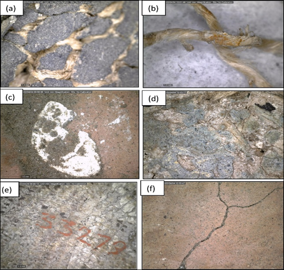
Figure 2 (a)(b) show fragility of the layers of cartonnage under study, (c) dirt accumulation and stains, (d) fading blue color, (e) human damage in the form of the piece number written in ink, (f) cracks.
Documentation using AutoCAD
AutoCAD program was used to draw cartonnage parts by shooting with a digital camera and setting a drawing scale inside the image to adjust the measurements while drawing the image. During the shooting process, the angles of the image were adjusted and then placed inside the AutoCAD program. This method is effective to document the decorations and damage of each piece separately.
Multispectral imaging
Ultraviolet rays
SYLVANIA, BLACKLIGHT-BLUE, F4W/BLB-T5, CH -, MOD 808-M, Vac 230(+/-10%), Hz 50/60, VA 40, Lamp T5 2X4W, G5, UV-A BLB IR Rays
Infrared rays
IR Illuminator, Model: S8100-30-B/C-I, Voltage: AC24V/DC12V, Current: 600mA, Wavelength: 940nm, and Serial No.: 20150625-6
Optical microscopy
The topography of the cartonnage has been studied using a Dino Capture 2.0 Version 1.5.12 microscope whereas the cross-section was examined utilizing Keyence Digital microscope type VHX 2000. The magnification varies from 50 x to 250 x.
X-Ray fluorescence (XRF)
Portable XRF device (ELIO XRF spectrometer) from XGLAB (X and Gamma-Ray Electronics) was used. It includes a set of pinhole collimators ranging from 2 mm down to 200μm, a Peltier-cooled silicon drift detector (with an energy resolution of 135eV at the Mn Kα excitation line) and a 50-kV Rh excitation tube. The operational parameters for tube voltage and anode current throughout the measurements have been fixed to 50keV and 0.7mA, and the acquisition real time was 60s. The diameter of the beam was adjusted to 600μm.
Fourier-transform infrared spectroscopy (FTIR)
To examine the textile media, FTIR spectra were obtained using a Bruker FTIR spectrometer, model VERTEX 70 fitted out with ATR crystal. The infrared spectra were acquired using an aperture of 20–100 μm, in the spectral region 600 to 4000cm−1. The resolution of 4cm−1 was used with 64 number of co-added scans for each spectrum.
AutoCAD
This method is effective to document the decorations and damage of the piece of cartonnage. According to documentation by AutoCAD program, the results showed that the object was too dirty and suffered from the damage, cracks, paint flake loss, and buckling in the linen layer (Figure 3).14
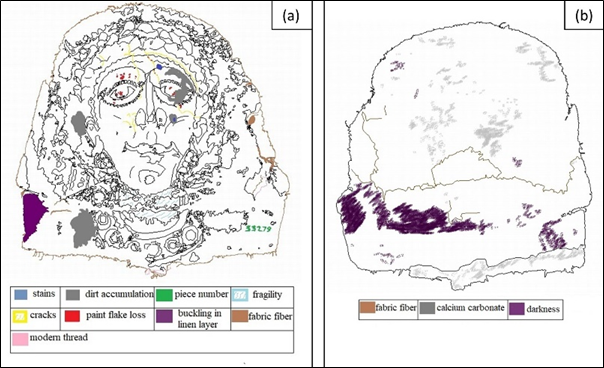
Figure 3 (a) Documentation by drawing the frontal cartonnage by AutoCAD program and showing the apparent damage on it, (b) the backside of the cartonnage.
Multispectral imaging
Ultraviolet Rays
UV Rays were used by conservators as a non-destructive analytical technique to aid in the examination of objects and identification of materials.15 The results of UVL image showed the following (Figure 4):
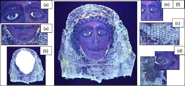
Figure 4 Documentation of the cartonnage by Ultraviolet Rays (a) shows presence of bright white emission in painted eyes and two earrings suggesting the use of white lead, (b) presence of white to yellowish emission suggesting the use of Arabic gum, (c) Textural composition 1:1, (d) dirt accumulation, (e)(f) cracks and stains.
Infrared rays
The results of VIL image showed the following (Figure 5): Egyptian blue areas in crown and necklace appeared as bright white, while all other materials appeared dark.19–21 A black color tone has been applied above the main color at the mouth and face areas.
The structure of the cartonnage
The cartonnage was unique and consisted of three distinctive layers, from the bottom to the top; one or a double layer from linen, a calcite-based plaster layer, and finally a polychrome paint layer (Figure 6).

Figure 6 a) b) Cross sectional image of the cartonnage by optical microscope by 50x showing its layered structure, c) The schematic structure of the foot case cartonnage.
Identification of textile fibers
Topography of the textile layer was studied using digital microscope. The results reveal that the fabric layer is one or double of linen layers taken 'S'-spun and plain weave (Textural composition 1:1) (Figure 7a) with the total thickness of cartonnage measuring about 0.8 mm (Figure 7b).22
The examination showed discoloration of some parts especially the external ones which turned to brown color as if being burned (Figure 7c and 7d). According to,23 the main reason for the discoloration of archaeological textiles could be the darkness of binder which is Arabic gum that was identified by Fourier-transform infrared spectroscopy.23

Figure 7 a) Digital microscopic image of textile layer: textural composition 1:1 by 50x b) thickness of cartonnage measures about 0.8 mm, c) d) discoloration fiber to brown color.
FTIR spectrum of the textile layer compared to references of Arabic gum and linen textile. According to,24 band of Arabic gum were shown at, O-H stretching band appeared at 3336 and 3279cm-1, C-H stretching bands appeared at 2898 and 2868cm-1, C-H bending bands appeared at 1451-1426-1365-1334-1313cm-1, C-O stretching band appeared at 1279,1161, 1107, 1054, 1030, 1001and 984cm-1 in the textile layer only which referred to lignin. In the last O-H bending band was appeared in textile layer only at 1623cm-1 (Figure 8).24
The plaster layer
The digital microscopic examination of the plaster layer paint showed shiny particles (Figure 9a). By using XRF for analyzing the ground layer, we proved the existence of elements (Ca: 95%) as components of calcium carbonate caco3 and small percentage of iron (Fe: 4.7%) as an impurity (Figure 9b). (Ali, 2017). Also, it had been noticed by digital microscope by 50x that the calcite-based plaster layer was sometimes covered with yellow orpiment (Figure 10a and 10b).
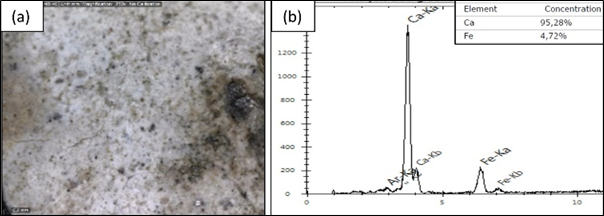
Figure 9 a) digital microscopic image of plaster layer by 50x b) XRF for plaster layer that showed Ca: 95% as component of calcium carbonate caco3 and (Fe: 4.7%) as an impurity.

Figure 10 Digital microscopic image showing that a calcite-based plaster layer was found sometimes covered with yellow orpiment.
The paint layer
Pigments are considered the most attractive targets for scientific study because their colors are yardsticks of a sense of beauty, and they provide a means for estimating ancient technologies’ ability to prepare pigments artificially.25
The colors had magical significance: red=life (blood), white=the good (light), and black=the evil (darkness). 26
Yellow pigments
Microscopic examination of the yellow paint area showed shiny and dark particles. In the morphological analysis of cartonnage, bright crystals were clearly observed under dark particles of yellow (Figure 11a).27 XRF analysis of these crystals showed pronounced atomic concentrations of arsenic (As: 2.18%) and Sulphur (S:9.66%), which propose an arsenic-based pigment (Orpiment, As2S3) in addition to presence of iron (Fe:30.51%) that presented goethite (α-FeO.OH + clay minerals) (Figure 11b).1
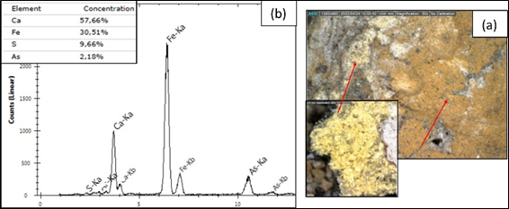
Figure 11 a) digital microscopic image of plaster layer by 50x. b) XRF for Yellow pigments; As: 2.18%, S: 9.66% and Fe: 30.51% as components of Orpiment and goethite.
Blue pigment
The microscopic image obtained on the blue paint layer demonstrated bright blue particles scattered on the calcite-based plaster layer (Figure 12a). (Figure 12b) shows the atomic concentrations of the constituting elements: calcium (Ca, 34.1%), silicon (Si, 59.56%), and copper (Cu, 4.29%), connected to the cuprorivaite mineral that presented Egyptian blue (CaCuSi4O10).13 Also, small percentage of iron (Fe: 4.7%) was found as an impurity. The presence of lead (Pb, 1.09%) in the components of Egyptian blue is considered evidence that ovens contain lead resulting in changing the manufacturing techniques of Egyptian blue.
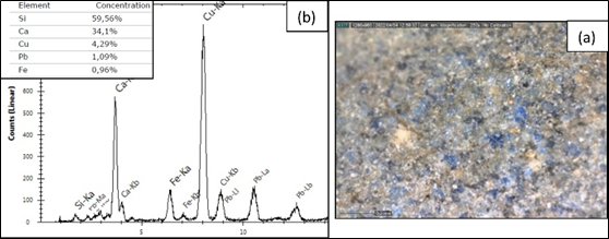
Figure 12 a) digital microscopic image of plaster layer by 250x. b) XRF for blue pigment consisting of Ca: 34.1%, Si: 59.56% and Cu: 4.29% (CaCuSi4O10).
Egyptian blue was the principal blue pigment used in Egypt from as early as the Fourth Dynasty to the Roman period and spreading all around the Mediterranean basin until the 7th century AD.28
Black pigment
The black paint taken from the eyebrow (Figure 13a), the surface morphology shows that it suffers from cracks. The XRF spectra of the black paint exhibit iron (11.15%) suggesting the presence of the iron black pigment, magnetite (Fe3O4). The presence of Ca: 61.07% referred to the ground layer, while that of Cl: 27.78% was considered an impurity that resulted in dirt accumulation (Figure 13b).26
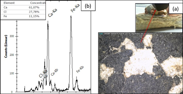
Figure 13 a) digital microscopic image of plaster layer by 50x. b) XRF for black pigment; Fe: 11.15% as component of magnetite (Fe3O4), Ca: 61.07% as ground layer, Cl: 27.78% as an impurity that resulted in dirt accumulation.
White pigment
Based on the XRF analysis of the whites of the eye, white lead (PbCO3)2•Pb(OH)2 was proved to be used as a white pigment. The spectrum revealed the presence of lead) 73.25% (in addition to traces of iron (1.18%) and calcite (25.57%) that refer to plaster ground layer (Figure 14).27
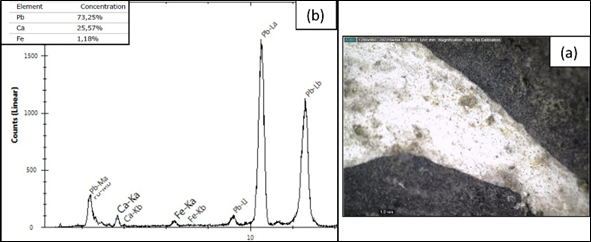
Figure 14 a) digital microscopic image of plaster layer by 50x. b) XRF for white pigment; pb: 73.25% as component of white lead (PbCO3)2Pb (OH)2, Ca: 25.57% as ground layer, and small percentage of iron (Fe: 1.18%) as an impurity.
Red pigments
Two red shades were also detected of which the lighter is red lead (Pb3O4) while the darker is a mixture hematite (α-Fe2O3), red lead (Pb3O4), and calcite (CaCO3) (Figure 15).
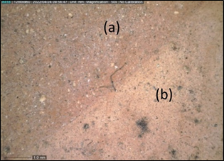
Figure 15 a) digital microscopic image of dark red paint by 50x. b) digital microscopic image of pale red paint by 50x.
Most red pigments used in ancient Egypt were earthen based colors containing iron oxide. Hematite (αFe2O3) was very common.30 This Fe based colors are longer lasting and light faster than others and are sometimes of astonishing brilliance. Two further red pigments have been imported to Egypt by the Romans: Red lead (Pb3O4) and Vermillion (HgS), so, red lead started to be employed only in the Greece–Roman periods (332 BCE to 400 CE). The introduction and production of these lead-based colorants in Egypt were likely influenced by Hellenistic and Roman painting techniques.29
The XRF chart for dark red measured a variety of elements, namely iron (Fe, 6.1%), lead (pb, 1.23%), and calcium (Ca, 92.66%), which asserted the presence of a mixture hematite (α-Fe2O3), red lead (Pb₃O₄), and calcite (CaCO3) (Figure 16).
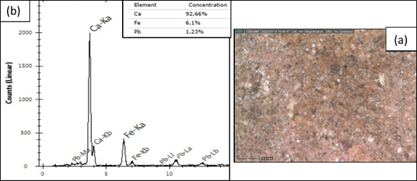
Figure 16 a) digital microscopic image of dark red pigment by 250x. b) XRF for dark red pigment showing pb: 1.23%, Fe, 6.1%, and Ca: 92.66%, as components of a mixture hematite (α-Fe2O3), red lead (Pb₃O₄), and calcite (CaCO3).
The XRF chart for pale red pigment showed the presence of pb: 7.9% as component of red lead (Pb₃O₄) and Ca: 92.8% associated with components of the gesso layer (calcite CaCO3) (Figure 17).
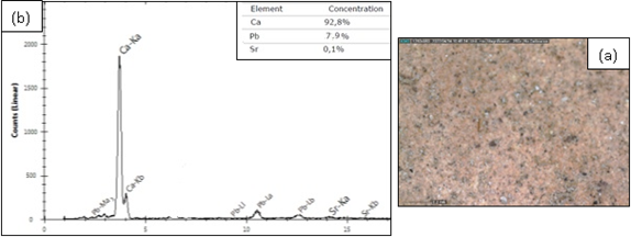
Figure 17 a) digital microscopic image of dark red pigment by 250x. b) XRF for pale red pigment showing pb: 7.9%, as component of red lead (Pb₃O₄) and Ca: 92.8% associated with components of the gesso layer (calcite CaCO3).
Orange pigment
Red lead (Pb₃O₄) was also recognized in the orange paint sample. It was admixed with (above) the yellow pigment, orpiment (As2S3) and goethite (α-FeO.OH + clay minerals) to achieve the orange shade, as revealed by the XRF analysis. In the orange paint, a noticeable amount of calcite from the underlying plaster layer was also identified (Figure 18).

Figure 18 a) digital microscopic image of dark red pigment by50x b) by 250x. c) XRF for orange pigment consisting of red lead mixed with orpiment and goethite.
Brown pigment
The brown pigment, made up of red hematite, red lead, and black manganese, was detected for the first time in the pigment palette of ancient Egyptian cartonnage. By examining the brown pigment by digital microscope by 50x, it sometimes seems purple or burgundy and appeared as grains of yellow paint (Figure 19).
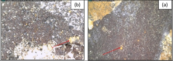
Figure 19 a) digital microscopic image of brown pigment by50x which sometimes seems purple or burgundy and appeared grains of yellow paint. b) by 250x.
The XRF chart measured a variety of elements, namely iron (Fe, 63.74%), manganese (Mn, 4.43%), lead (pb, 1.06), and calcium (Ca, 17.72%), which asserted the presence of a mixture hematite (α-Fe2O3), red lead (Pb₃O₄), and black manganese (Figure 20).
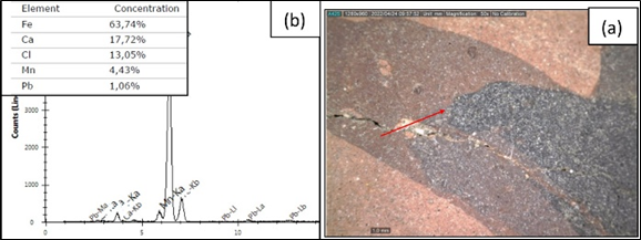
Figure 20 a) digital microscopic image of brown pigment by50x b) XRF for brown pigment: black manganese above red hematite and red lead.
Filler
Filler was found inside the crown. XRF chart revealed that it consisted of calcium carbonate caco3 admixed with orpiment (As2S3) (Figure 21).
This study revealed the materials employed in manufacturing the studied cartonnage object such as, substrate, plaster layers, paint layers, and binders. The cartonnage dated back to the Graeco-Roman Period because of the presence of red lead, white lead, orpiment, and Egyptian blue. Moreover, the presence of lead in the components of Egyptian blue is considered evidence that ovens contain lead resulting in changing in the manufacturing techniques of Egyptian blue. The obtained results in this study shed light on the techniques and material characteristics of the Graeco-Roman Period objects. The study showed that the studied object is dirty and suffers from the damage, cracks, paint flake loss, and buckling in the linen layer. Therefore, there is a necessity to conserve this object and the obtained results will help conservators to select the best appropriate conservation strategies that can be used in conservation of the current cartonnage object.
The authors would like to thank AbdelRahman Medhat Conservation Department, Faculty of Archaeology, Cairo University, Egypt for his valuable contribution with documentation, examination and analysis of cartonnage. This research did not receive any specific grant from funding agencies in the public, commercial, or not-for-profit sectors.
Author declares there are no conflicts of interests.
None.

©2023 Abdel-Kareem, et al. This is an open access article distributed under the terms of the, which permits unrestricted use, distribution, and build upon your work non-commercially.