Journal of
eISSN: 2373-4345


Short Communication Volume 4 Issue 5
Department of Orthodontics, T.M.D. and HealthCare, USA
Correspondence: Dennis J Tartakow, Department of Orthodontics, T.M.D. and HealthCare, 4712 Admiralty Way, 895, Marina Del Rey, CA, 90292, USA
Received: December 03, 2015 | Published: May 17, 2016
Citation: Tartakow DJ. How to improve our diagnostic acumen: teach it to our residents. J Dent Health Oral Disord Ther. 2016;4(5):129-130. DOI: 10.15406/jdhodt.2016.04.00124
Are orthodontists responsible for examining the occlusion, teeth and gingiva? Yes, for sure, but we also have a responsibility to use our training and understanding not just to straighten teeth, correct malocclusions or improve skeletal discrepancies of the jaws, but to ensure that any and all pathology in the head and neck is identified, documented, treated or referred for treatment. After many years of clinical practice and teaching, it occurred to me that many of our residents are missing certain aspects of their orthodontic training. Nothing is a better teacher than personal experience, however what we do and how we do it in practice often reflects upon the educators and mentors in postgraduate residency programs. The following are examples of issues and guidelines that are seldom, if ever mentioned in our teaching; they are subjects that go beyond the routine in the diagnostic process and examination.
It is the most glaring problem that is often overlooked in resident training, mostly because it is assumed that the resident knows how to write and what to write in all correspondences, diagnostic letters and patient charts, but do they? Most do not! We must prepare them to speak before a group of individuals, to address a judge and jury in the courtroom and most important, we must educate them to document correctly, writing with proper English. Speaking clearly and writing properly are THE most important aspects of documentation for communicating our thoughts, treatment plans, problems, objectives and projected outcomes. Writing clearly in a patient’s chart can make big difference years later when asked to review a patient’s record and we cannot even remember the patient’s name, let alone treating them.
Ask any medical malpractice attorney about how well dentists or orthodontists document properly in a patient’s treatment chart…you will be mortified. Most clinicians do not take the time to write adequate notes, explaining or identifying problems encountered such as compliance, oral hygiene, lack of proper appliance care, etc. besides writing so poorly that whatever is written either cannot be deciphered or makes little or no sense. Not only are many notations illegible, they are often written with shortcuts and abbreviations only known to that clinician. Most chart entries are too short, incomplete, unacceptable and inadequate. These situations occur much too often and are a poor reflection on the educators because this is our responsibility.
It can provide much more diagnostic information than measuring lines and angles by looking beyond the teeth. As a broad scan it can be used to find pathology other than dental disease. Not too long ago, a recently graduated orthodontic resident came to me beaming, stating that because of his diagnostic lectures, he spotted a carotid artery calcification on a routine cephalometric radiograph of a new 24-year old patient. Presenting with no familial or personal medical history of high cholesterol or heart disease, this calcification was never diagnosed and unbeknown to the patient. According to the vascular surgeon who removed the calcification, this pick up saved the patient’s life.
A cephalometric radiograph can help in diagnosing cervical vertebrae problems, disc disease and other spinal abnormalities. Tonsil and adenoid enlargements that contribute to airway impingement, open mouth breathing, high palatal vaults, open-bites, etc. can also be identified on a cephalometric radiograph. The list goes on, but such pick-ups can only be found if the doctor takes the time to examine the x-ray in greater detail.
It can and do show expansile lesions of the mandible whereas the panorex and cephalometric x-rays often do not. Such was the case of an 18-year old female patient who had an asymptomatic mandibular swelling and was eventually diagnosed as fibrous dysplasia. The diagnosis of fibrous dysplasia in a patient raises important questions for the orthodontist such as: (a) can a patient with fibrous dysplasia be treated with orthodontics, or (b) what are the contraindications to moving teeth in the presence of fibrous dysplasia? A rare finding indeed, but both of these views are extremely valuable tools that can facilitate early diagnosis of other pathology, especially vertebral problems caused by benign and malignant disease processes.
The SMV and PA are omnipotent in diagnosing skeletal midline discrepancies. Midline deviations are often misdiagnosed and labeled as a dental problem, when in fact there is an underlying skeletal asymmetry in the maxilla, mandible or both. Midline issues and diagnoses can easily be confirmed by using these two radiographs that beautifully demonstrate when the left and right mandibular corpi are unequal in length. How often do we blame a cephalometric radiograph with non-superimposed potion images on technique, when in fact (a) the PA view identifies the length of the mandibular rami to be unequal in length, or (b) the SMV view identifies the length of the mandibular corpi to be unequal in length. Consequences of missing this astute diagnosis can have daunting and dire treatment results. Besides, attempting to move a maxillary or mandibular dental midline may be like shoveling sand back to the ocean when the tide is coming in…a sure miscalculation that will result in relapse. These additional views can prevent misdiagnosis, poor treatment results and explain or even lead to understanding the etiology of a patient’s malocclusion: Is it skeletal, dental or both?
Nothing is a better teacher than personal experience(s) regarding what we do and how we do it in our practices. Expert training is a reflection on the educators and mentors in postgraduate residency programs. The following considerations are important subjects in the diagnostic process and examination; they are especially valuable and significant for the orthodontic resident to recognize.
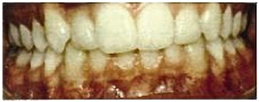
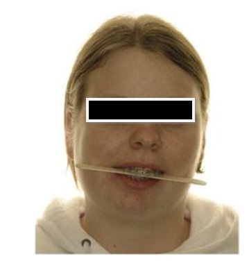
It often demonstrates dermatological diseases, tumors and other pathology of the head and neck. We can diagnose important health issues by taking the time to look. Diagnosing diseases of the skin in our patients, e.g. squamous cell carcinoma, basal cell carcinoma, melanoma, etc., is an astute part of our responsibility and demonstrates good judgment as a doctor. Because orthodontists take so many clinical photographs, very little time is required to scan for such pathology prior to examining facial structures and the dentition. Accuracy and precision are extremely important; for example, in the intraoral photo below Figure 1 is this documentation of an aberrant occlusal plane cant or just sloppy photography?

Figure 1 Intraoral photo.1
Clinical photography can identify many diseases of facial expression or appearance. Facial diseases are often related to development or physiology and can affect facial structure, facial behavior or both. Through clinical photography, we can teach the resident how to recognize various signs in the face that indicate particular diseases. Signs of facial diseases include (a) changes in appearance, (b) alterations of muscular movement, and (c) behavioral expression. Facial signs are often used to diagnose the presence of certain diseases that can be diagnosed via clinical photography. The most obvious relationships between facial signs and disease are for the genetic and congenital diseases. Specific genetic abnormalities cause such diseases as Lesch- Nyhan, Down syndrome and Cornelia De Lange syndrome, producing specific patterns of facial abnormality. Certain congenital diseases such as fetal alcohol syndrome, cretinism and hydrocephaly also produce specific facial signs and symptoms. Many infectious diseases can be diagnosed from facial signs, e.g. Lyme disease, Fifth disease, shingles and HIV infections.
These are not as popular as hand-held models and most orthodontists never consider using an articulator except for surgical cases. However, they may be extremely helpful in diagnosis, treatment planning and for medical-legal protection. When documenting patients with asymmetry such as when the cant of the occlusal plane is not level, hand-held models are often prepared inaccurately without demonstrating the exact degree of incongruity or anomaly (Figure 2). Articulated models provide excellent representation of the patient’s condition and are extremely accurate.

Figure 2 Hand-held models.1
There is much to reveal as we appraise the past and contemplate the future. Learning can be defined as useful changes in behavior resulting from reflection and experience. How can we teach our students to become better practitioners and sharper diagnosticians? Will they learn to focus on the dental problems in the context of, and in concert with a patient’s general health issues? As orthodontists, we are still responsible to diagnose pathology in the head and neck, treat or refer the patient to someone who can provide proper care. By example, we must demonstrate how to be the best orthodontist possible and the consummate expert in our field.
None.
The author declares that there was no conflict of interest.

©2016 Tartakow. This is an open access article distributed under the terms of the, which permits unrestricted use, distribution, and build upon your work non-commercially.