Journal of
eISSN: 2574-9943


Case Report Volume 8 Issue 2
1Linove private practice, Latvia
2Department of Radiology, Riga 2nd Hospital, Latvia
3Milano, Private practice, Italy
Correspondence: Alessio Redaelli MD, Milano, Private practice, Italy
Received: June 13, 2024 | Published: July 3, 2024
Citation: Linova T, Linovs V, Redaelli A. Ultrasound-guided hyaluronic acid filler injections for temporal augmentation: a case report of two patients. J Dermat Cosmetol. 2024;8(2):44-46. DOI: 10.15406/jdc.2024.08.00264
Background: The aging face often exhibits volume loss, particularly in the temporal region, where the presence of complex neurovascular structures like the superficial temporal artery increases the risk associated with rejuvenation procedures. Hyaluronic acid fillers are extensively utilized to address these concerns, albeit requiring precise techniques to avoid vascular complications.
Methods: This case study involved two Caucasian female patients, aged 41 and 37, who underwent temporal augmentation using HA fillers to correct hollowness and enhance facial contours. Ultrasound mapping of the superficial temporal artery (STA) was performed to guide the injection process. A total of 2.4 mL of HA filler was administered to each patient using a 25G, 50mm blunt cannula under real-time ultrasound guidance.
Results: The application of HA filler using the DOMINO lateral lifting technique not only restored volume but also provided a lifting effect on the medial and lower thirds of the face. Ultrasound guidance facilitated precise cannula placement within the interfascial space, ensuring the avoidance of vascular injury, with no post-procedural complications such as hematoma or intravascular filler placment observed.
Conclusions: Ultrasound-guided HA filler injections in the temporal region provide a safe and effective method for facial rejuvenation. This case highlights the importance of precise anatomical mapping and real-time procedural guidance to enhance safety and aesthetic outcomes. Future research with a larger cohort is recommended to further validate these findings.
Keywords: domino effect, hyaluronic acid fillers, temples, cannula, ultrasound mapping, superficial temporal artery
Hyaluronic acid filler injections are used to correct temporal hollowness and reduce signs of facial aging, offering significant potential for achieving pan-facial rejuvenation effects. The temporal region is recognized as a high-risk area for injections due to the complex anatomical presence of neurovascular structures, particularly the superficial temporal artery. Given the layered anatomy of the temporal region and the anatomical variations in vascular distribution, a precise technique for the placement of hyaluronic acid fillers is essential.
In this clinical case study, we performed injections in the temporal region using HA fillers through an entry point at the zygomatic arch. Prior to the procedure, we mapped the course of the superficial temporal artery using ultrasound imaging. Additionally, we utilized real-time ultrasound guidance during the injections to ensure the accurate placement of hyaluronic acid within the interfascial plane of the temporal region, thereby minimizing the risk of vascular injury.1
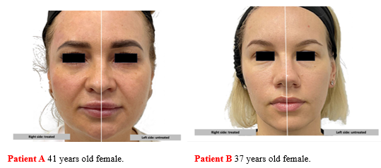
Two Caucasian female patients, aged 41 (Patient A) and 37 (Patient B), presented with volume loss in the temporal area and minimal signs of aging (Figure 1). Both patients exhibited slight upper lid ptosis and a middle face deformative aging morphotype, with no history of surgery or aesthetic injections in this area. Based on the approaches described in the study “Understanding facial aging through facial biomechanics”,2 the DOMINO lateral lifting technique was selected. This approach not only volumizes the temporal region but also provides a lifting effect on the medial and lower thirds of the face.

Figure 1 Patients A and B. The left side shows the patients before filler therapy. The right side shows the patients after filler therapy in the temporal region using 1.2 mL hyaluronic acid-based gel (25 mg/mL).
The treatment involved the use of 2.4 mL of hyaluronic acid-based gel (Art Filler Volume, 25 mg/mL) per patient (1.2 mL per side). The filler was administered using a 25G, 50 mm blunt cannula under ultrasound guidance with a Cannon I900 ultrasound machine equipped with a 24MHz probe.
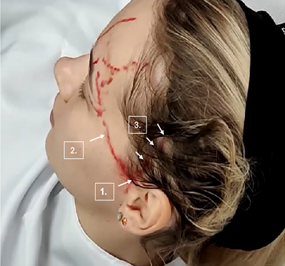
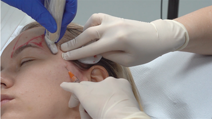
Prior to initiating HA filler therapy, the temporal artery was mapped using an ultrasound device (Figure 2). In both clinical cases, the cutaneous entry point for the cannula was selected at the midpoint of the zygomatic arch. After the insertion of the cannula and before the commencement of filler injection, a real-time ultrasound examination was conducted to verify that the cannula was correctly positioned in the layer 4 (Figure 3). After obtaining visual confirmation via ultrasound and identifying the anatomical positioning of the branches of the temporal artery (Figure 4), hyaluronic acid filler was administered using the linear retrograde technique to evenly restore the volume loss in the temporal region (Figure 5).

Figure 2 The first step, prior to initiating the invasive procedure, involves mapping the superficial temporal artery (STA) using high-resolution ultrasonography. The image shows Patient B, with the bifurcation (number 1) point of the superficial temporal artery marked in red, slightly above the zygomatic arch. The frontal branch of the STA crosses the temporal fossa (number 2), and the parietal branch of the STA (number 3) is located in the hair-bearing scalp.

Figure 3 In the second step, after the insertion of the cannula into the temporal region and before the commencement of hyaluronic acid filler injection, real-time ultrasonography was performed to confirm the position of the cannula tip (targeting layer 4).

Figure 4 Ultrasonographic image and schematic representation of the ultrasonographic structures prior to hyaluronic acid filler injection using blunt canula in the temporal region.
1. Epidermis.
2. Dermis.
3. Subcutaneous fat;
4. Superficial temporal fascia;
5. Frontal branch of Superficial Temporal Artery (STA) located in superficial temporal fascia;
6. Cannula placed in layer 4 (interfascial space);
7. Deep temporal fascia;
8. Interfascial temporal fat pad;
9. Temporal muscle fibers;
10. Deep temporal artery;
11. Intramuscular vein;
12. Ultrasound reverberation artifact from the cannula;
13. Surface of the frontal bone;
14. No ultrasound signal behind the temporal bone.

Figure 5 Ultrasonographic image and schematic representation of the ultrasonographic structures during hyaluronic acid filler injection with blunt canula in temporal region.
1. Epidermis;
2. Dermis;
3. Subcutaneous fat;
4. Superficial temporal fascia;
5. Frontal branch of Superficial Temporal Artery (STA) located in superficial temporal fascia;
6. Cannula placed in layer 4 (interfascial space);
7. Deep temporal fascia;
8. Interfascial temporal fat pad;
9. Temporalis muscle fibers;
10. Intramuscular artery (medial temporal artery branch);
11. Intramuscular vein;
12. Ultrasound reverberation artifact from the cannula;
13. Surface of the frontal bone;
14. No ultrasound signal behind the temporal bone;
15. Hyaluronic acid filler between the superficial and deep fascia layers (interfascial space).
This clinical case study demonstrates that hyaluronic acid filler injections can be effectively and safely administered in the temporal region using ultrasound guidance, and that the DOMINO technique is a winning strategy. By mapping the superficial temporal artery prior to the procedure and employing real-time ultrasound during injections, we achieved precise filler placement within the interfascial plane. This technique minimizes the risk of vascular injury, ensuring a safer approach for patients seeking correction of temporal hollowness and facial rejuvenation. Our findings suggest that integrating ultrasound guidance into the injection protocol enhances the accuracy and safety of hyaluronic acid filler treatments in anatomically complex regions. Future studies with larger sample sizes are warranted to further validate these results and refine the technique.
None.
The authors declare no conflict of interest.

©2024 Linova, et al. This is an open access article distributed under the terms of the, which permits unrestricted use, distribution, and build upon your work non-commercially.