Journal of
eISSN: 2574-9943


Mini Review Volume 4 Issue 1
1Department of Dermatology, The Federal University of Bahia (C-HUPES/UFBa), Brazil
2Department of Dermatology, Division of Pediatric Dermatology, University of São Paulo Clinics Hospital (HC- FMUSP), Brazil
Correspondence: Bruno de Oliveira Rocha MD, Rua Marechal Floriano, 420, Edf. Residencial Sidarta, Apto. 301. Canela. Salvador, Bahia, Brazil, Tel +55(71)99389-2323
Received: January 08, 2020 | Published: January 27, 2020
Citation: Rocha BDO, Oliveira ZNPD, Fernandes JD. Skin and subcutaneous cell tissue bacterial infections in newborns. J Dermat Cosmetol. 2020;4(1):1-6. DOI: 10.15406/jdc.2020.04.00139
Bacterial skin infections are especially common in children of tropical regions, varying clinically from a process superficial (such as folliculitis), to a deeper infection (such as necrotizing fasciitis). Infections of skin and subcutaneous tissue are frequent reasons for medical consultations in primary health care services and hospitalization in pediatric patients. In neonates several factors confer an increased susceptibility to bacterial infections of the skin and its complications. Herein, we review essential aspects of the main bacterial skin infections in newborns and nurselings.
Keywords: skin infection, bacteria, newborn infection, antibiotic, microbiology
The flora of the human skin is composed of several species of bacteria, especially Gram-positive cocci. Under normal conditions, the release of free fatty acids by these organisms creates an environment unfit for the installation of pathogenic bacteria. Disturbances of skin integrity, however, allow infections to occur.1–5
Bacterial infections of the skin and subcutaneous cell tissue are especially common in children and tropical regions, ranging clinically from a superficial infection (such as folliculitis) to a deeper infection (such as necrotizing fasciitis).4,5 The most common etiological agents are staphylococci and streptococci, although other Gram-positive and some Gram-negative bacteria may also be involved.5 In this context, it should be noted that methicillin-resistant staphylococci, of both nosocomial and community origin, are increasingly being imputed.6
Skin and subcutaneous tissue infections are frequent causes not only of primary care appointments but also of hospitalization in the pediatric range. Proper management of the disease should take into account the etiological diagnosis and the degree of systemic impairment of the child.3
In neonates, the immaturity of the immune system and the still limited action of the protective flora confer a greater propensity for bacterial skin infections.7 The neonate’s cutaneous flora microorganisms are illustrated in Table 1. Neonates differ from older children not only in that immune immaturity, but also by the larger body surface and by having greater transepidermal water loss.
Micrococcaceae |
Coagulase-negative staphylococci |
Diphtheroid |
Corynebacterium |
Gram-negative bacteria |
Acinetobacter |
Fungus |
Malassezia spp. |
Table 1 Colonizing flora of newborn skin
The newborn’s skin, notably more fragile and sensitive, undergoes a process of adaptation to the extrauterine environment. In the neonatal period, the cutaneous pH tends to neutrality and the epidermal lipid content is reduced. The large amount of proteoglycans also confers greater water content in the newborn's skin, facilitating transdermal absorption of toxins.8,9
These factors cause skin infections in the neonate to evolve rapidly to severe dehydration, with great risk of complications. In preterm or low birth weight newborns complications and death from skin infections are even more frequent.
Among childhood skin infections, impetigo is the most common, and may occur even before the third day of life.2,10 Hot and humid regions (diaper, armpit and neck areas) are the most frequently affected by lesions, which, when due to its superficiality, it usually does not leave scars, although it may evolve with residual hypochromia in dark-skinned patients.3,10 Regional lymphadenopathy may be present in some cases.1,11 Infection or asymptomatic colonization of caregivers considerably increases the occurrence of impetigo in neonates, increasing morbidity and mortality, prolonging hospitalization and burdening health services.7
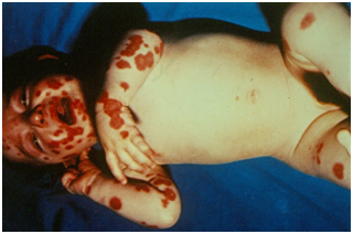
Non-bullous or contagious impetigo presents with fragile and small vesicles and pustules that rupture and dry, giving rise to tightly adhered pustular and melicic crusts (Figures 1 & 2). In neonates, there is a lower tendency to form crusts on impetigo. Lesions are more common on the face, in the perinasal region, but may also affect the extremities.3,11 Impetigo is a primary infectious process of the skin and, in cases where the infection settles in some other pre-existing dermatosis, it occurs. if the name of impetiginization.1,10,11 Complication with fungal or gram-negative infection may occur.3,12

Figure 1 Bullous lesions with halo erythematosus in the anterior thorax of a patient with impetigo. On the face and upper limbs, exulcerated lesions with some melic crusts predominate.

Figure 2 Flabby and erythematous blisters and blisters on the face and extremities of a patient with impetigo.
Bullous impetigo, on the other hand, is more frequent in the diaper and face region, presenting with flaccid blisters of sero-purulent content, surrounded by an erythematous halo.2,10 After rupturing, the blister gives rise to a circinate plaque with purulent/honey-like crusts and some satellite lesions.3,10 Fever and other such constitutional symptoms may be present.
About 85% of cases of non-bullous impetigo are associated with Staphylococcus aureus infection.1,10,11 Group A streptococci can be identified in up to 30% of lesions, 11 being more common in early lesions.12 groups B, C, G and F are rarely found. Epidemic outbreaks of bullous impetigo are associated with Streptococcus pyogenes infection of serotype M.3,11
In bullous impetigo, most cases are also associated with Staphylococcus aureus (80%), inoculated by scratch or insect bite lesions.10,11 This bacterium produces epidermolytic toxins A and B (exfoliatins), which interact with desmogleina-1 to form the blister (Table 2).11,12 Bullous impetigo is theorized to be a localized form of staphylococcal scalded skin syndrome.10 It is worth noting that in bullous impetigo the Nikolsky sign is negative.1 Predisposing factors include poor hygiene and underlying skin infections (viral, fungal and bacterial), as well as conditions accompanied by pruritus and excoriation (atopic eczema).10 Restricted intrauterine growth, maternal multiparity, and low socioeconomic status are also associated.
Bacterium |
Toxins |
Group A Streptococci |
SPE-A |
Groups B, C, F and G Streptococci |
SPE-s |
Table 2 Toxins produced by streptococci and staphylococci
The systemic antibiotics of choice are erythromycin, clarithromycin or cephalosporins. If there is evolution with systemic symptoms, hospitalization with intravenous administration of antibiotics and notification of the hospital where the child was born is necessary to observe contamination of the nursery. Isolation of affected patients should be ensured in order to prevent outbreaks. Impetigo in neonates can be reduced by more than half with prenatal zinc supplementation.13
Retapamulin, a semi-synthetic pleuromutilin antibiotic, has recently been used to treat impetigo. Good results were obtained and the Food and Drugs Administration approved its use, although only in children older than nine months.1
Two to five per cent of cases may develop acute glomerulonephritis, which is more closely related to streptococcus pyogenes strains of serotype M 2, 49, 55, 57 and 60 in children aged one to three years.2,11,12 Nephrogenic strains of S. aureus induce immune complex deposition in the subendothelial region of the glomerular capillary. Subsequent inflammation causes loss of the glomerular capillary barrier and is clinically manifested by hypertension, edema, hematuria and proteinuria, triggered 18 to 21 days after the onset of the cutaneous infectious process.1,10 Although impetigo treatment does not influence the occurrence or severity of glomerulonephritis,1,10,12 rheumatic fever can be prevented by taking the right measures.10 Cellulitis and haematogenic dissemination are some of the possible complications,1,11,12 although they are quite rare events.1
The boil results from a staphylococcal folliculitis that deepens into the dermis, affecting the sebaceous gland.2 The regions most affected are those exposed to friction (armpits, buttocks, neck and face), with typical lesions being erythematous perifollicular nodules that may present necrosis or central rupture.2,10,11 Usually resolve leaving scars. The term anthrax or carbuncle refers to the confluence of two or more boils.1 In this condition, fever is more common and the practitioner should be aware of the possibility of progression to gas-containing abscesses or necrotizing fasciitis, which require immediate surgical debridement.2
Folliculitis caused by Malassezia furfur typically occurs on the dorsum, pectoral region, and proximal upper limbs due to the lipophilic character of the fungus. Perifollicular papules are more discrete and sparse pustules may occur.1 In the presence of seborrheic dermatitis and pityriasis versicolor the suspicion that the causative agent is M. furfur increases. On direct examination with KOH hyphae and spores are visualized. Although topical antifungal treatment is effective in primary resolution, recurrence is frequent.1
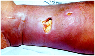
The ecthyma is a deep and painful pyogenic skin infection caused by Streptococcus pyogenes and Staphylococcus aureus, the first being the most common.1 It occurs mainly in the legs and buttocks, in trauma regions, insect bites or abrasions, especially when there is poor hygiene. or poor nutrition. Early erythematous-based pustules evolve with ulceration and adherent crust formation, making the edge of the lesion more pronounced (Figure 3).10,11 Fever and lymphadenopathy may be associated. Resolution is relatively slow and scarring.11

Figure 3 Erythema and edema, with poorly defined borders, around an ulcerated lesion, located on the medial face of the lower limb. It is a case of the ectima that complicated with cellulite.
Cellulitis is an acute infection of the dermis and subcutaneous cellular tissue that presents soft, swollen, erythematous plaques, with local heat and poorly defined edges (Figure 3), usually in sites of unhealthy skin (surgical wounds, fungal infection and abrasions or trauma).2,10,12 The agent can still penetrate through a respiratory tract infection, otitis media or sinusitis.
The most common etiological agents are Streptococcus pyogenes and Staphylococcus aureus,2,3,12 although a violet coloration of the lesion is highly suggestive of Haemophilus influenzae infection, the main agent in children under three years of age.2,10,11 Periorbital cellulitis in patients under five years old are usually caused by Streptococcus pneumoniae. In the case of newborns, the most common agents are group B streptococci. In the face of a bite report, one should think of Pasteurella multocida (cat and dog bite) or Eikenella corrodens (human bite).3,11
There are usually regional lymphadenopathy, fever, and other constitutional symptoms.2,3 Immunodeficiency and diabetes mellitus are strongly associated predisposing factors and should be investigated.11
The prognosis of cellulite is favorable if diagnosed and treated early. Otherwise, serious complications may occur, such as subcutaneous abscess, necrotizing fasciitis, osteomyelitis, bacteremia, septic arthritis, thrombophlebitis, and involvement of the eyes, heart valves, and central nervous system.3,12 Another important complication is poststreptococcal glomerulonephritis.11 rare in the first month of life, necrotizing fasciitis usually has a fatal course in 59% of patients. In this case, surgical approach and parenteral antibiotic therapy should be instituted immediately.
Like cellulite, erysipelas is an acute infection of the dermis and subcutaneous tissue, not uncommon in neonates and younger children.14 What distinguishes these two entities is that in erysipelas only the most superficial part of the subcutaneous tissue is affected, and there is intense inflammation around lymphatic vessels.3,11 The most common agent is Streptococcus pyogenes, but Staphylococcus aureus, Staphylococcus pneumoniae, and group B, C, D, and G streptococci can also be isolated. Conditions that interfere with skin integrity will allow bacteria to enter (ulcerations and insect bites), 1,11,14 however, are not necessary conditions for their occurrence.2 Venous insufficiency, lymphatic obstruction, diabetes mellitus and nephrotic syndrome should be investigated.11 In neonates, infection can originate from the umbilical stump, spreading to the abdominal wall and also to the external genitalia in those undergoing postectomy.
Unlike cellulite, erysipelas plaques are well defined and slightly hardened, maintaining the other characteristics (erythema, pain and local heat).1,11 Vesicles, blisters and petechiae may be present. Skin lesions are usually preceded by fever and malaise, similar to a viral cold.1,3,2,11,14 Lower extremities and face are the most frequent sites of involvement.
Histologically, a neutrophilic interstitial infiltrate with dermal edema and dilation of lymphatic and capillary vessels may be observed. Gram or Giemsa stains may reveal streptococci.1,14 Latex agglutination test for streptococcal antigens on biopsy specimens may be positive.11 Among the main diseases that may resemble the clinical picture of erysipelas, the following may be mentioned: hives, contact dermatitis and pharmacoderma.11
Although extremely questionable, a randomized double-blind clinical trial demonstrated a faster improvement in patients receiving anti-inflammatory drugs (steroid or otherwise) in conjunction with antibiotic therapy.14 In about 20% of cases there is recurrence and resolution often shows post inflammatory hyperpigmentation.1,11 The mortality rate is less than one percent and complications are less frequent than in cellulitis, but the professional should be aware for the possibility of poststreptococcal renal and cardiac involvement.1,3,14
Perianal streptococcal dermatitis is a superficial infection caused by Streptococcus pyogenes, predominantly affecting three to four year-old male children and is very rare in newborns. More than 50% of the cases are associated with pharyngotomidalitis and the previous occurrence of impetigo may predispose to its development.10,11 Erythematous, bright, well-demarcated and pruritic plaques around the anus are the typical lesions that may develop with perianals fissures. Pain on defecation often leads to fecal retention.11
Staphylococcal scalded skin syndrome (SSSS) or Ritter's disease is caused by Staphylococcus aureus phagotype II (types 3A, 3B, 3C, 55 and 71), which colonizes the nasopharynx, conjunctiva, skin wounds, umbilical cord and urinary tract.1,11 The epidermolytic toxins produced by Staphylococcus aureus are hematogenously disseminated.1 The serine proteinase activity of these toxins leads to skin cleavage due to their action on desmoglein-1, causing granular layer splitting and generalized detachment of the superficial epidermis.11
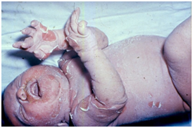
The incidence peaks are in neonates and children up to five years old.10,11 SSSS occurs more frequently in newborns due to the lack of antibodies against staphylococcal toxins and renal immaturity, slowing down the clearance of these toxins. Initially, a fever, malaise, irritability, and diffuse erythema with rapid progression to erythroderma are observed, with more pronounced erythema in the flexural and perioral regions. Subsequently, blisters form, with subsequent diffuse exfoliation, perioral, perinasal and periocular crusts, with underlying shiny erythema (Figure 4) (Figure 5).10,11 Cleft lip and fissures are common and the oral mucosa is spared.10 Fever, diarrhea and vomiting are common in neonates. Dehydration and intensification of the toxemic condition may occur in case of diagnostic and therapeutic delay.

Figure 4 Exfoliation of flexural regions, perioral crusts, and erythema underlying lesions in infant staphylococcal scalded skin syndrome.

Figure 5 Diffuse exfoliation and erythema, perioral and perinasal crust in staphylococcal scalded skin syndrome in newborn.
In addition to NET, which is the main differential diagnosis, SSSS must also be differentiated from other bullous diseases (bullous impetigo and bullous mastocytosis), scarlet fever, Kawasaki disease, and toxic shock syndrome.1
The resolution occurs in about 14 days and without scars, except in the occurrence of secondary infection. When much of the integument is affected, hydroelectrolytic disturbance and loss of thermal regulation may occur. Bacteremia, cellulitis, endocarditis, and pneumonia are some of the most common complications.10,11 The mortality rate in children is much lower than in adults (3% vs. 50%, respectively).10
Most bacterial skin and subcutaneous tissue infections are caused by staphylococci and/or streptococci species. In most cases, antibiotic therapy should be empirical, taking into account the local bacterial resistance profile (Table 3). Invading the epidermal barrier as little as possible, avoiding contact with soap and not removing the caseous vertex are simple and efficient measures to prevent these infections in newborns. In patients under three years of age, attention should be paid to the need for gram-negative coverage. Correcting environmental factors such as poor hygiene should also be part of the treatment plan.
Type of Infection |
Etiological agents |
Treatment |
Impetigo |
Staphylococcus aureus |
Mupirocin or topical fusidic acid |
Folliculitis |
Staphylococcus aureus |
Topical mupirocin, erythromycin, benzoyl peroxide or clindamycin |
Ecthyma |
Streptococcus pyogenes |
Topical Mupirocin |
Cellulitis |
Streptococcus pyogenes |
Penicillin, Clindamycin or Cefazoline |
Erysipelas |
Streptococcus pyogenes |
Penicillin, cephalexin, clindamycin or cefazolin |
Perianal Streptococcal Dermatitis |
Streptococcus pyogenes |
Penicillin or cephalexin |
Staphylococcal scalded skin syndrome |
Staphylococcus aureus fagotipo II (types 3A, 3B, 3C, 55 and 71) |
Cloxacillin and gentamicin, or clindamycin and cefotaxime |
Table 3 Etiological agents and therapeutic options in bacterial skin and subcutaneous tissue infections
The authors declare that they have no conflicts of interests.
None.
None.

©2020 Rocha, et al. This is an open access article distributed under the terms of the, which permits unrestricted use, distribution, and build upon your work non-commercially.