Journal of
eISSN: 2574-9943


Case Report Volume 6 Issue 1
Department of plastic surgery, Faculdade Ciências Médicas de Minas Gerais, Brazil
Correspondence: Luiz Eduardo Toledo Avelar, Department of plastic surgery, Faculdade Ciências Médicas de Minas Gerais, Brazil
Received: November 23, 2021 | Published: March 3, 2022
Citation: Avelar LET, Real J, Haddad A, et al. Sexual dimorphism and hyaluronic acid treatments of male patients. J Dermat Cosmetol. 2022;6(1):15-17. DOI: 10.15406/jdc.2022.06.00200
There are several differences between the male and female face. These differences are found not only in the three thirds of the face, but also in all topographic layers. When studying the skull of both genders, we can see that the male skull is bigger, heavier and has more volume than the female.
From top to bottom, when we analyze the superior third of the face, we can see the frontal region is completely different in male and female craniums. The female frontal region is more vertical and rectified with the frontal eminence well pronounced. The male frontal region is more oblique, does not have the pronounced frontal eminence superiorly, but has an important protrusion above the superior orbital rim, called the supraorbital ridge (Figure 1). This gives the frontal region an irregular surface on profile view and is an important sign of ageing since it becomes more prominent in elderly patients.
When we see a young male patient with a pronounced supraorbital ridge or even in a female patient, the injection of hyaluronic acid dermal fillers is well indicated for rejuvenation in the case of the former or femininization in case of the latter.
The structural differences between the male and the female mandible (sexual dimorphism) are easily noted. Anthropologically, the gonium is the most lateral point of the mandible body and can be well pronounced in both genders but is definitely more pronounced in men. Likewise, the mandible ramus in men is stronger and more rectified, giving male patients a wider mandible and a more square shaped face – in men, the distance between the gonium is similar to the distance between the most lateral point of the zygoma. In women, an oval shape is more common, and this should be preserved when providing aesthetic treatments, in order to avoid the masculinization of the female face (Figure 1).6,10,14

Figure 1 Male (left) and female (right) skull, both of 40-year-old individuals at time of death. Note how the gonium (red arrow) are present in both genders, but the ramus of the male mandible is wider and more rectified. The male bone structure has a square shape whereas the female skull has an oval shape.). The intergonial distance is also wider in male subjects and the chin is more angulated and rectified (yellow line). The width of the chin in defined by the distance between the canine teeth (dotted yellow line).
The mentum is also very different in male and female subjects. Women have a rounder, more oval shape, which is compatible with the other structures of the female skull, whereas in men, the chin usually is more rectified and angulated. We believe the width of the chin should not have soft tissues such as the nostrils or the corners of the mouth as its anatomical reference like it has been pointed by some authors. We understand that the soft tissues and its position on the face can differ a lot regarding individual characteristics, ethnical and the ageing process of each patient. Although the teeth positioning will also change regarding genetics and ageing, a vertical line between the canine tooth and the first premolar tooth will normally correspond to the width of the chin and can be one more anatomical reference to better define chin width when we want to treat this area, either for enhancing or defining the chin (Figure 1).
When treating male patients, a common request is to enhance masculine features of the face, which usually can be achieved by addressing the structure of the lower third of the face. Men who feel that their face looks more child-like or portrait an idea of being younger than they really are can also benefit from enhancing the lower face. This is also true when treating trans patients that request a more masculine look.
When the goal is masculinization, we start by enhancing the gonium. We palpate the gonial angle and mark a point about 1.5 to 2 cm above and medially to the angle, in a diagonal direction (Figure 2). Then, we inject this point with a needle, in the subcutaneous plane, above the SMAS, so to avoid trauma to the parotid gland.

Figure 2 To identify the injection point, we palpate the gonial angle, in the junction of the ramus and the body of the mandible (blue lines). About 1,5 to 2 cm superiorly and medially we determine the point of injection (yellow circle). We a bolus of high G prime hyaluronic acid with a needle, in the subcutaneous plane, above the SMAS.
Since our goal is to elevate the skin and create more projection of the gonium, it is important to use a hyaluronic acid with high G prime. It is important to remind that the masseter is a very strong muscle and that when we inject fillers in the supraperiosteal plane in this area, and therefore under the masseter, the product will suffer constant pressure from the muscle, and will most likely be deformed and will not have a long-lasting effect.
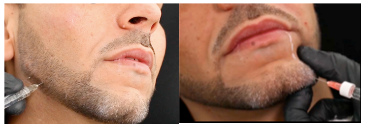
After achieving the desired projection, the patient will show a more squared structure of the face and will likely have an uneven transition between the treated area and the rest of the mandible. We then follow with the treatment of this transition with the placement of more hyaluronic acid, this time with intermediate G prime and a more flexible consistency (Figure 3). It is important to remember to treat the ramus of the mandible as well (the pre auricular area) to maximize the results of the masculinization.

Figure 3 Left: After placement of the HA bolus, we continue to define the mandible and even the transition between the treated area and the rest of the mandible, to ensure the treatment has a natural effect. Right: treating the most lateral point of the chin.
After determining the most lateral point of the chin, we inject this area with a high G prime AH in the supraperiosteal plane and, if needed, we can also inject more medially, with less product, in order to rectify the chin inferiorly. The goal of the treatment is to create projection of the skin and as a result, obtain a more masculine, faceted and strong face.
Below are three examples of outcomes achieved with this technique with before and after photos. The after photos were taken 30 days post-procedure (Figures 4–6).
None.
The author declares there is no conflict of interest.

©2022 Avelar, et al. This is an open access article distributed under the terms of the, which permits unrestricted use, distribution, and build upon your work non-commercially.