Journal of
eISSN: 2574-9943


Case Series Volume 8 Issue 4
1Dermalaser KPW. Lima, Perú
2Instituto de investigación en ciencias biomédicas – INICIB – Universidad Ricardo Palma, Lima, Perú
3Medical Doctor, Dermatologist, Expert in Exposome, Perú
4Plastic surgeon, Perú
5Master in medicine, Perú
Correspondence: Kateryn Michelle Perez Willis, Dermalaser KPW, Medical Doctor, Dermatologist, Expert in Exposome, Avenida del Pinar 152, Surco, Lima-Peru, Tel 511970514344
Received: September 30, 2024 | Published: October 15, 2024
Citation: Perez W, Kateryn M, Hernández PI. Safety and efficacy of female chin enhancement and facial contouring with a 3-point injection technique, an imaging follow up with ultrasound and magnetic resonance. J Dermat Cosmetol. 2024;8(4):97-101. DOI: 10.15406/jdc.2024.08.00275
This research aimed to assess the effectiveness of a 3-point injection technique using hyaluronic acid (25mg/ml HA reticulated Revanesse Shape G’ 203) to enhance the chin and mandibular border with hyaluronic acid 25mg/ml HA reticulated Revanesse Contour G’ 150. The technique involved injecting the filler into specific planes (supraperiostium for chin enhancement and subcutaneous for mandibular contouring) to achieve improved facial aesthetics. By combining needle and cannula, precise targeting was possible without severe complications. Thirty female participants with retrognathia and undefined mandibular border underwent the procedure and were evaluated at 21 days, and 9 months post-injection. Ultrasound technology facilitated accurate injections by visualizing facial structures and blood vessels. Additionally, one participant underwent magnetic resonance imaging with 3D reconstruction to confirm the precise placement of hyaluronic acid in the intended injection plane.
Keywords: hyaluronic acid, genioplasty, magnetic resonance imaging ultrasonography, retrognathia, (Fuente: MeSH – NLM)
Hyaluronic Acid injections are one of the top 5 non-surgical procedures all over the world,1,2 patients are seeking more angled and defined faces daily in our consultations without any surgical or downtime procedures.3 As the population ages, plastic surgeons and dermatologists must remain aware of the treatments to prevent and reverse the external signs of aging.4 In the mandibular region, Retrognathia, commonly referred to as chin retrusion, is a prevalent facial irregularity that can significantly impact an individual's self-confidence and overall facial appearance,5,6 Skin laxity and soft tissue sagging in the jawline may lead to jowling and chin ptosis along with reduced chin projection.7,8 Over the past several years, hyaluronic acid (HA) injectable dermal fillers have become the most popular agents for soft tissue contouring and volumizing. Although they are not free of complications, fillers have a good safety profile, mainly if the appropriate filler is selected for each patient and procedure.9 Many authors prefer needles because they are considered more precise, as the injector can exactly target the tip of the needle to the desired place ensuring accurate placement of the filler, other authors claim blunt tip cannulas to be safer and less traumatic than the needles.9 However, during this study, we combined both tools and we had a great outcome without any severe complication.
Design and study area: A quasi-experimental series of case studies was conducted.
Population and sample: This study features a group of 30 Latin American women, aged 30-40, who were referred due to concerns about their facial structure, specifically an undefined jawline and a short chin. The participants were selected through a non-random, convenience-based sampling method.
Study variables and instruments: We study the injection technique and safety in the area of Injection. We used Ultrasonography Claerius L20 to ensure the layers of application, magnetic resonance with a 3D reconstruction, Global Aesthetic Improvement Scale (GAIS Scale) post procedure.
Procedures: All patients were treated with hyaluronic acid 25mg/ml (Revanesse Shape G’ 203) on the chin and was placed on the supraperiostium layer in the pogonion area for the chin enhancement and with hyaluronic Acid (25mg/ml HA reticulated Revanesse Contour G’ 150) in the mandibular border on the subcutaneous layer in order to achieve a beautified facial contour. Every patient had an Ultrasonography Claerius L20 performed on the chin and mandibular area in order to guarantee the exact layer of application of the hyaluronic acid and one patient was performed a magnetic resonance with a 3D reconstruction in order to visualize the hyaluronic acid in the different layers described in this study, the 3-point injection technique was performed as described.
Step 1: We applied 0.05cc of Lidocaine with epinephrine intradermal in the 3 injection points used in the technique with a 30G needle. The anesthesia was applied in order to minimize the pain during the insertion of the 21G Needle and the 21G Cannula.
Step 2: Deep supraperiosteal bolus was injected in the chin area (Pogonion) with a 21G needle (Figure 1). The amount of the supraperiosteal bolus on the pogonion used was from 0.5 ml to 1ml depending on each patient. The amount of filler used differed between patients, as it was adjusted to accommodate individual variations in chin shape and mandibular structure.
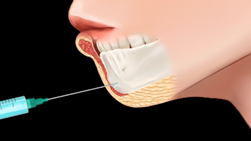
Figure 1 The landmark for this injection point with a 21G Needle is the pogonion. Reticulated HA (Revanesse Shape G’ 203).
Step 3: A linear injection on the subcutaneous tissue on the mandibular border with a 21G Cannula, the landmark for this injection was an imaginary line drawn on the border of the mandible. The amount of hyaluronic acid used was from 0.5 ml to 1 ml each side depending on the individual need (Figure 2).
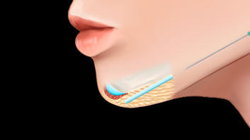
Figure 2 A linear injection of Reticulated Hyaluronic Revanesse Contour G’ 150 was applied on the subcutaneous tissue on the mandibular border with a 21G Cannula.
Step 4: A linear injection of hyaluronic acid (Revanesse Contour G’ 150) on the subcutaneous tissue above the DAO muscle with a 21G cannula in a fanning technique. The landmark for this injection was an imaginary line drawn from the border of the mandibule to the beginning of the DAO (corner or the mouth - see Figure 3) The amount of hyaluronic acid used was from 0.2 ml to 0.5 ml each side.
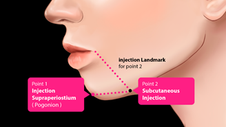
Figure 3 Illustrates the reference point for injection, serving as a precise landmark for the injection.
Global Aesthetic Improvement Scale (GAIS Scale) was used Post procedure. GAIS is a generalized aesthetic quality assessment by the observer. Before and after pictures were taken with Lifeviz, Quantificare in order to compare the results after the procedure.
All patients were treated with Revanesse shape in the chin area (Pogonion) and Revanesse contour on the subcutaneous tissue in the mandibular border and above the DAO muscle with a fanning technique on the area. All patients agreed to the study and had an inform consent explaining possible adverse reactions.
To ensure accurate placement of hyaluronic acid, 30 patients underwent ultrasound imaging (Claerius L20) of the chin and jawline. Additionally, one patient had an MRI with 3D reconstruction to visualize the filler's distribution across various tissue planes, as described in this study (Table 1).
|
Rating scale |
|
Description |
|
1 |
Very Much Improved |
Optimal Cosmetic result in this subject |
|
2 |
Much Improved |
Marked improvement in appearance from the initial condition, but not completely optimal for this subject |
|
3 |
Improved |
Obvious improvement in appearance from initial condition but a re-treatment is indicated |
|
4 |
No change |
The appearance is essentially the same as the original condition |
|
5 |
Worse |
The appearance is worse than the original condition |
Table 1 Global aesthetic improvement scale (GAIS)
Statistical analysis: A descriptive analysis of images and GAIS Scale was performed.
Ethical considerations: All patients agreed to the study and had an inform Consent explaining possible adverse reactions and all patients signed the informed consent form. The study was approved by the Clinic's Derma Laser KPW Ethic Scientific Committee. Safety was assessed through the recording of all adverse events after the injections, including physician and patient observations throughout the course of the study.
This study enrolled 30 patients (females). The range of age was from 30 to 40. The results showed that 23 patients (77%) were very much improved after the procedure, scoring 1 on the GAIS scale, 5 patients (17%) showed much improved, scoring 2 on the GAIS scale, 2 patients (7%) were improved, scoring 3 on the GAIS scale. Overall, these results suggest that the treatment was effective for most patients, improving the definition of their chin and mandibular border. During this study none vascular occlusion was reported, the reports showed minor transient adverse reactions like bruising, edema and pain on the injection site (Table 2) (Table 3).
|
Age |
GAIS score 21 days after treatment |
|
32 |
2 |
|
33 |
2 |
|
35 |
1 |
|
33 |
1 |
|
34 |
1 |
|
37 |
1 |
|
38 |
1 |
|
38 |
1 |
|
39 |
1 |
|
40 |
1 |
|
33 |
1 |
|
31 |
2 |
|
38 |
1 |
|
35 |
1 |
|
35 |
1 |
|
34 |
1 |
|
30 |
2 |
|
30 |
1 |
|
32 |
1 |
|
31 |
1 |
|
33 |
1 |
|
34 |
1 |
|
37 |
3 |
|
37 |
1 |
|
40 |
1 |
|
32 |
3 |
|
33 |
2 |
|
36 |
1 |
|
35 |
1 |
|
36 |
1 |
Table 2 GAIS Scale 21 Days after treatment
|
Age |
GAIS score 9 months after treatment |
|
32 |
2 |
|
33 |
2 |
|
35 |
1 |
|
33 |
1 |
|
34 |
1 |
|
37 |
1 |
|
38 |
1 |
|
38 |
1 |
|
39 |
1 |
|
40 |
1 |
|
33 |
1 |
|
31 |
2 |
|
38 |
1 |
|
35 |
1 |
|
35 |
1 |
|
34 |
1 |
|
30 |
2 |
|
30 |
1 |
|
32 |
1 |
|
31 |
1 |
|
33 |
1 |
|
34 |
1 |
|
37 |
3 |
|
37 |
1 |
|
40 |
1 |
|
32 |
3 |
|
33 |
2 |
|
36 |
1 |
|
35 |
1 |
|
36 |
1 |
Table 3 GAIS Scale 9 months after treatment
Safety
The safety of the hyaluronic acid injections was evaluated by monitoring and recording any adverse events or side effects reported by patients and observed by physicians throughout the study. Common side effects of hyaluronic acid injections include swelling, pain, redness, bruising, lumps, firmness, tenderness, itching, and skin discoloration.1 However, in our study, no severe adverse reactions occurred; only mild and temporary side effects were reported, which are listed in Table 4 (Figures 4–11).
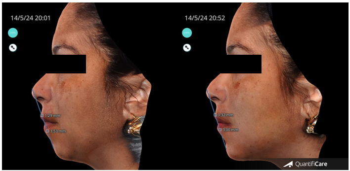
Figure 5 Before and after the 3 Point Injection Technique with the measurements from the nose, Lip and chin.
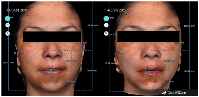
Figure 7 shows the transformative results of the 3-Point Injection Technique, as visualized using Quantificare and Lifeviz technology. The before-and-after comparison, viewed from the front, reveals a noticeable increase in facial length and a more defined, angular facial contour.
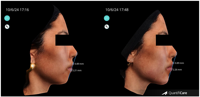
Figure 9 Before and after the 3 Point Injection Technique with the measurements from the nose, Lip and chin.
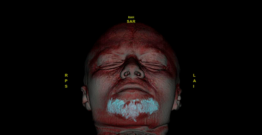
Figure 10 MRI scan shows the hyaluronic acid filler was precisely placed using the 3-Point Injection Technique.
|
Bruising |
Edema |
Pain in the injection points |
None complications reported |
|
6 |
3 |
4 |
27 |
Table 4 Transient adverse reactions described
Mandibular contouring and the chin plays a crucial role in facial structure and profile. A well-defined chin can exude confidence and balance. Fortunately, there are modern techniques with hyaluronic acid to assure a beautified contour and profile. Bo Chen et al.,10 described a technique based on Anatomical morphology, in this technique they describe important anatomical landmarks for injection like the pogonion for projection as the authors also describe in this article, the paragonion which we don’t use as a landmark for our technique.
During this technique they do not specify the concentration of Hyaluronic Acid but they mention is a high elasticity HA, they describe the use of a 27 Gauge needle for the injections and a cannula for a subcutaneous fanning to soften the prejowl sucus, and reduce the labiomandibular groove on both sides, also the median injected volume of HA was 1.85 ml. which is minimum compared to our technique. Holt S, et al.,11 describe a nonsurgical Augmentation of the Chin Using a lateral to medial approach with Hyaluronic acid,11 this technique is described as indicated for patients who present mild to moderate chin retrusion and resorption, the boundaries for this technique were indicated as the following: The superior boundary of the chin compartment was identified by outlining the mentolabial crease, and the lateral boundary was identified by outlining the labiomandibular fold, they used a 20 mg HA with lidocaine (the cohesivity or G prime of the filler is not specified) and a 27 gauge needle, the mean volume of HA injected was from 1 to 1.5 ml which is less volume than the Bo Chen et Al described and our technique, this may be due to the fact that this technique is described mainly for mild to moderate chin retrusion.
Our technique utilizes a relatively higher amount of Hyaluronic Acid, averaging 2.5-3cc, compared to similar methods. This increased volume may be attributed to the severity of retrognathia in our patient population, requiring more filler to achieve optimal results.
A randomized trial conducted by Marcus et al.,12 evaluated the effectiveness and safety of using hyaluronic acid fillers for chin augmentation and correction of chin retrusion. Their findings align with our study's conclusions, showing that using Hyaluronic acid for chin and mandibular border was safe and effective.
Alloplastic implants are best for those patients who have adequate vertical chin height but need more projection because implants can only produce significant changes in anterior projection.7,13 Surgical genioplasty is a larger procedure that can correct all deficiencies. Both these surgical approaches involve extensive dissection and thus confer higher risk of significant complications such as implant rejection or migration, scarring, infection, bone resorption, mental nerve injury, and also result in permanent alteration of the chin which may not look natural.13–15
The 3-Point Injection Technique significantly enhanced the chin and jawline contours of our patients. Notably, patient results remained consistent at 21 days and 9 months post-procedure, as measured by the GAIS Scale. A limitation of this study is that all the patients were females. The combined use of cannulas and needles proved highly effective for precise filler placement, confirmed by ultrasound and MRI. While minor temporary side effects occurred, the technique was deemed safe for all patients. We recommend this technique be performed by experienced physicians with specialized training for optimal outcomes and high patient satisfaction.
None.
None.
The authors declared that there are no conflicts of interest.

©2024 Perez, et al. This is an open access article distributed under the terms of the, which permits unrestricted use, distribution, and build upon your work non-commercially.