Journal of
eISSN: 2574-9943


Case Report Volume 3 Issue 3
1Sparsh Skin Hair & Laser Clinic, India
2Belagavi Institute of Medical Sciences, India
Correspondence: Sushruth Kamoji, Sparsh Skin Hair & Laser Clinic, Belagavi, India
Received: May 12, 2019 | Published: May 22, 2019
Citation: Kamoji S, Gurudev S, Patil MN. Rhinoscleroma – a unique presentation. J Dermat Cosmetol. 2019;3(3):75-77. DOI: 10.15406/jdc.2019.03.00117
Rhinoscleroma is caused by a gram negative bacilli – Klebsiella pneumonia rhinoscleromati. It is a chronic slowly progressive granulomatous infection predominantly affecting the upper respiratory tract and the tracheobronchial tree. Though it may be fatal, it is mildly contagious. It is usually prevalent among the unhygienic, malnourished and overcrowded with sporadic foci in Central Europe, Egypt, India, Indonesia, USA & Africa. It is known to spread through the nasal droplets.1
Clinically in 95-100% of cases, Rhinoscleroma begins primarily in the nasal cavity, at the junction between the stratified squamous epithelium of the vestibule and the respiratory epithelium. It may also involve nasopharynx (18-43%), larynx (15-40%), trachea (12%) and bronchi (2-7%).2 Involvement of the oral cavity is quite uncommon. Evincing this fact, we came across only two case reports from India.3,4
Arriving at the conclusion of Rhinoscleroma solely depends on the histopathological examination, although the presenting features and imaging techniques sum up to outline a presumptive diagnosis of the disease. Here we are reporting an unusual presentation of rhinoscleroma involving the oral cavity.
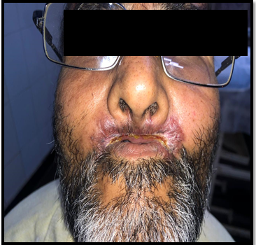
A 54year old male, who underwent tooth extraction 7years ago, presented with complaints of multiple and recurrent oral ulceration along with difficulty in opening of mouth. He had gradual loss of dentition over the last 6 years. The recurrent ulceration led to involution of oral cavity to an extent of permitting mouth opening to barely1 and a half finger (Figure 1). Medical and surgical history was unremarkable and the patient was irregularly on medications for the same from various doctors.
Examination of the patient revealed stenosed oral aperture with erythematous crusted plaques at the angles of mouth and upper lip along with fissuring. There was surrounding mild induration. Central part of the lower lip was relatively spared (Figure 2). Regional lymph nodes were palpable.

Figure 2 Erythematous crusted plaques at the angles of the mouth, philtrum, upper lip along with fissuring; Central part of the lower lip was relatively spared.
Histopathological evaluation of the excised tissue revealed numerous Mikulicz cells and Plasma cells under H & E Stain, with the former being predominant (Figure 3) (Figure 4). Patient was subjected to baseline investigations including hemogram, Liver function test, Renal function test, Serum electrolytes, serology which were found to be normal. Radiography of head and neck was normal. The patient was started on Tab Tetracycline 2gram/ day in four divided dose. Clinically, the lesion has improved with reduction of crusting and induration in the last 2months (Figure 5) (Figure 6).

Figure 3 & 4 Post treatment improvement in the crusting and induration 2months post treatment with Tetracycline.
Rhinoscleroma is known to progress through 3 overlapping stages. The first stage is Exudative/rhinitic/catarrhal/ atrophic stage which is characterised by nonspecific rhinitis, purulent rhinorrhea, crusting & nasal obstruction. This is followed by proliferative or granulomatous stage in which there can be granulomatous nodules in the nose, pharynx and larynx which may also be associated with dysphonia, anosmia, anesthesia of soft palate and epistaxis. The final stage is that of sclerosis during which the previously formed nodules are replaced by dense collagen in the form of fibrosis. In many cases all three stages can be found at the time of diagnosis.1,5
The mucopolysaccharide in the caspsule of Klebsiella rhinoscleromatis and an altered immune response along with alteration in the CD4+ and CD8+ ratio leads to ineffective macrophage production and increased bacterial replication.6 Histopathological picture is pathognomonic. A dense infiltrate is observed consisting mainly of plasma cells.
Mikulicz cells are large round vacuolated histiocytes which show the presence of gram‐positive or giemsa stained bacilli or amorphous clusters of mucopolysaccharide. Russell bodies are plasma cells with aggregates of condensed immunoglobulins.5,7 Both these cells are characteristic of this condition.
The clinical mimickers include Mucocutaneous leishmaniasis, paracoccidioidomycosis and other dimorphic fungal infections, Rhinosporidiosis, leprosy, yaws, tertiary syphilis, nasal tuberculosis, sarcoidosis, Wegener’s granulomatosis, nasal natural killer cell/T- cell lymphoma, basal cell carcinoma and squamous cell carcinoma, Extranodal Rosai–Dorfman disease.1,3,5,7
Rhinoscleroma poses a diagnostic and therapeutic challenge due to its chronic course, need for a prolonged therapy and incessant relapses. Thorough clinical evaluation of the patients, especially when the lesions are pronounced, Histopathology assessment with immunoperoxidase studies and special stains - to identify the antigenic structure of the organism; Endoscopy and imaging techniques could precisely offer the diagnosis of rhinoscleroma.
The treatment comprises of antibiotic therapy with broad spectrum Tetracycline – forms the first line of treatment, at a dose of 2g/day, given in divided doses for 6months, followed by 1 g/day for a similar period of time. Cephalexin in similar doses is preferred while recurrent.1,5
Sclerotic lesions in the fibrotic stage of the disease process responds to Ciprofloxacin, 250–500mg twice a day for 3months and a twice a day meticulous nasal lavage with saline for 4weeks. Surgical modality remains the mainstay for the scarring evident along the respiratory apparatus during healing process.1,5,8
The presence of oral involvement without that of nasal cavity or pharynx was quite unusual and hence posed a diagnostic challenge. We report this case for its unique presentation.
None.
Author declares that there is no conflict of interest.

©2019 Kamoji, et al. This is an open access article distributed under the terms of the, which permits unrestricted use, distribution, and build upon your work non-commercially.