Journal of
eISSN: 2574-9943


Case Report Volume 3 Issue 1
University Hospital Hassan II, Lot N 8, Adarissa II, Fes, Morocco
Correspondence: Mrabat Samia, University Hospital Hassan II, Lot N 8, Adarissa II, Fes, Morocco, Tel 0021 2689 2709 24
Received: December 28, 2018 | Published: February 1, 2019
Citation: Samia M, Elloudi S, Laamari K, et la. Multiple dermatofibromas in a patient with lupus erythematosus and Sjögren’s syndrome: a new case report. J Dermat Cosmetol. 2019;3(1):13-14. DOI: 10.15406/jdc.2019.03.00107
Dermatofibroma (DF) is a common benign skin tumor. It is often a solitary lesion occurring on the lower limbs. Multiple DFs is rare. This is associated with auto-immune conditions and immunosuppression. We report a new case of multiple dermatofibromas in a patient with lupus erythematosus and Sjögren’s syndrome.
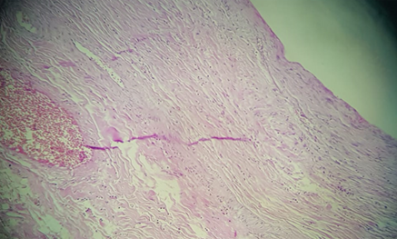
A 34–years- old female, was diagnosed with lupus erythematosus and Sjögren’s syndrome, was treated with antimalarials and steroids. She presented few months after the diagnosis, rapid onset of cutaneous lesions. At the moment of the diagnosis, she was taking 15mg a day of prednisolone. Clinical examination showed 18 well-circumscribed round fierm brown round papules, with a size of 3 to 15mm in diameter, with dimpling signs. The lesions were on the buttock, the thighs and the lower back. The papules were clinically typical of dermatofibromas. Dermoscopy found five different patterns: most lesions had a scar like central white patch surrounded by a pigmented network. Other patterns included a homogeneous pigmented pattern, a composed pattern, lentigo-like and seborrheic keratosis-like patterns. Excision biopsy specimen taken from the thigh found a proliferation of fibrohistiocytic spindle cells between the collagen fibers, making well-demarcated nodules located in the dermis, with no mitotic figures. The histological findings allowed us to retain the diagnosis of dermatofibroma.
DF is a very common benign tumour. It often presents occurs as a single which can be observed in an otherwise healthy person.1 Multiple eruptive dermatofibromas (MEDF) are rare.2 Barf and Shapiro were the first to describe multiple eruptive dermatofibromas in 1970.3 The presence of five to eight dermatofibromas (DF) appearing within a period of 4 months or at least 15 lesions defines multiple eruptive dermatofibromas.4 Many case reports have associated the appearance of multiple dermatofibromas in patients with autoimmune diseases like systemic lupus erythematosus, myasthenia and pemphigus vulgaris. They have also been reported in patients with altered immune states as HIV infection, organ transplant, acute myeloid leukaemia.5 These findings have led to think that multiple dermatofibromas have a strong link with altered immunity state and autoimmune diseases, and so should not be considered as benign neoplasm but reactionnal tumors instead.4‒6 Histologically, DFs are characterized with fibrohistiocytic proliferation made mainly of spindle-shaped cells. These spindle-shaped cells are a result of transformation of fibroblasts or endothelial cells. Recent research has led to the conclusion that DF should be considered as an immune reactive process in which dermal antigen presenting cells activate an immune response to an unknown stimulus.7 Many triggering factors have been reported such as insect bites, trauma and virus infections.1,8 Furthermore, it has been shown that the serum of patients with multiple DFs had platelet-derived growth factor and fibroblast growth factor in it, these factors stimulate fibroblast proliferation and therefore lead to the appearance of DF.9 DF lesions are characterized with increased mast cells; they have a key role in the pathogenesis of DF with by secreting certain growth factors.10 MEDF are clinically and histologically similar to the solitary one which is commonly present in immune competent patients (Figures 1‒3).11

Figure 3 Histological section showing fusiform cells arranged in short bundles, with some histiocytes. Laterally, the lesion infiltrates thick dermal collagen. There are no nuclear atypies. No atypical changes in nucleus.
In summary, multiples dermatofibromas are often associated with altered immune states. However, there exact pathogenesis is still unknown. To our knowledge, this is the second case reporting multiples eruptive dermatofibromas in a patient with lupus erythematosus and Sjögren’s syndrome.
Authors declare that there is no conflict of interest.

©2019 Samia. This is an open access article distributed under the terms of the, which permits unrestricted use, distribution, and build upon your work non-commercially.