Journal of
eISSN: 2574-9943


Case Report Volume 2 Issue 2
University of Taubaté, Brazil
Correspondence: Carolina Forte Amarante, Rodrigo Fernando Grillo Avenue, room 609; Jardim dos Manacás, Araraquara, São Paulo country, Zip code 14801-5, Brazil
Received: February 18, 2018 | Published: April 4, 2018
Citation: Amarante CF. Lymphomatoid contact dermatitis: the importance of its diagnosis, treatment and follow up. J Dermat Cosmetol. 2018;2(2):123-124. DOI: 10.15406/jdc.2018.02.00059
Contact dermatitis is a frequent, pruritic/itchy inflammatory dermatosis, caused by external agents in contact with the skin. It represents 4 to 7% of all dermatological appointments, besides being one of the main occupational diseases. The lymphomatoid contact dermatitis (LCD), described by Gomés Orbaneja in 1976, is defined as a particular form of allergic, persistent and chronic contact dermatitis, with clinical and/or histopathological characteristics similar to cutaneous lymphoma. The purpose of this article is to describe a case of LCD showing the necessity to be attentive to its diagnosis and prescribe the therapeutics to control its dermatological lesions efficiently.
Keywords: lymphomatoid contact dermatitis, patch test, pruritic, ethylenediamine, CD4‒positive cells
LCD, described by Gomés Orbaneja in 1976, is defined as a particular form of chronic and persistent allergic contact dermatitis, with clinical and/or histopathological characteristics similar to cutaneous lymphoma.1,2
This type of contact dermatitis is part of the spectrum of skin lymphomatoid hyperplasias also known as pseudolymphomas1‒3 which are formed of lymphoproliferative reactive processes of T or B cells resulting from a variety of causes.2 Several allergens such as gold, nickel, glutaraldehyde, ethylenediamine and N‒isopropyl‒N9‒phenyl‒phenylenediamine (IPPD) have been described as a trigger factor of LCD1‒7 having as its main and most accepted immunological mechanism the delayed hypersensitivity, Gell and Coombs type IV with the sensitization of T‒cells.
From the histopathological point of view, the findings between cutaneous lymphomas and pseudolymphomas can be indistinguishable.1,2 The absence of acanthosis, presence of prominent epidermotropism, deep lymphocytes monomorphic infiltrate, monoclonality in the genic rearrangement and destruction of the skin adnexals are findings that suggest a real cutaneous lymphoma of T cells. Alternatively, the findings of epidermic spongiosis or spongiotic micro‒vesicles are more associated with LCD. This being so, the definitive diagnosis is performed from the correlation of clinical history data, histopathology and immunohistochemistry, in addition to the positive contact test.1
A 66 year‒old‒man presented with erythematous to violaceous nodules and plaques on his face, anterior chest and posterior cervical region for three years (Figure 1) (Figure 2). He works as a fork lift.
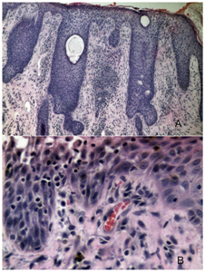
The histopathological exam from a biopsy of his face showed acanthosis with a granular layer thickened in some parts of epidermis or even absent, focal parakeratosis, moderate spongiosis, and exocytose of typical lymphocytes and focal superficial necrosis of epidermis. There was moderate lymphocytic infiltrate around superficial vessels. The diagnosis was compatible with eczematous dermatitis (Figure 3).

Figure 3 (A) BK: HE 100x. Epidermis show focuses of spongiosis, lymphocyte exocytosis, hypogranulosis and parakeratosis. There is a superficial moderate perivascular lymphomononuclear inflammatory infiltrate. (B) Image 002: HE 400x. Superficial Necrosis of the epidermis, spongiosis and exocytosis of lymphocytes.
Immunohistochemical panel of the lymphoid infiltrate did not show malignant criteria. The contact test was strongly positive for potassium dichromate and nickel sulfate, and positive for Peru Balsam, cobalt chloride, (MIX) and PPD (MIX) perfume.
The patient was advised to keep distance from suspected substances and dark clothing and was mometasone prescribed. After 2months, the patient returned showing only a residual hyperchromia (Figure 4) (Figure 5).
LCD has a chronic nature, benign at first, but its diagnosis is challenging due to the difficulty in finding the particular allergenic substance. Allergic contact dermatitis can be clinically and/or histologically similar to cutaneous lymphoma.1‒9
It manifests clinically as erythematous, violaceous or brownish plaques or nodules which can coalesce when associated to pruritus.1‒6 Psoriatic like erythroderma can occur.3 The finding of atypical CD4‒positive cells in the epidermis may suggest mycosis fungoides. However, alterations in the epidermis, such as spongiosis, are findings in favor of LCD.8 In spite of being a benign disease at first, individuals need to be vigilant towards its progression to a cutaneous lymphoma, as if the aggression persists; there is a possibility of abnormal mitosis of the T‒lymphocytes involved in the pathogenesis of the disease.9
In conclusion, we reported a case of LCD with a clinical picture that resembled lymphoma, but the histopathological exam with immunohistochemical analysis helped to exclude this and favoured a diagnosis of a spongiotic process.
None.
The author declared that there are no conflicts of interest.

©2018 Amarante. This is an open access article distributed under the terms of the, which permits unrestricted use, distribution, and build upon your work non-commercially.