Journal of
eISSN: 2574-9943


Short Communication Volume 2 Issue 6
1Department of Dermatology, University Hospital Hassan II Fez, Morocco
2Department of Anatomopatology, CHU University Hospital Hassan II Fez, Morocco
Correspondence: El Jouari Ouiame, Department of Dermatology, University Hospital Hassan II Fez, Roas sidi Hrazem Fez, Morocco, Tel 2126 4576 8798
Received: October 05, 2018 | Published: November 15, 2018
Citation: Jouari ELO, Gouzi I, Senhaji G, et al. Benign or malignant? J Dermat Cosmetol. 2018;2(6):102-103. DOI: 10.15406/jdc.2018.02.00096
Warts are one of the most frequent benign neoplasms. It is the third most common skin disease in childhood and is probably even more common in adulthood. We report a particular case of pseudotumoral presentation of warts mimicking a verrucous squamous cell carcinoma in a 60-year-old man.
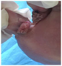
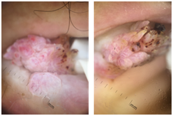
A 60-year-old man with a 3years’ history of chronic intertrigo of the space between toes presented of with painful tumors of this region that appeared months prior to seeking medical attention. The clinical examination revealed two juxtaposed, well defined, tumors of 1cm with warty surface, covered by hemorrhagic crusts, located to the last space between toes of the right foot (Figure 1). The tumors were adherent to the skin but mobile from the deep plane. Dermoscopy of the lesion showed a filiform appearance, with dotted (pinpoint) vessels and red streaks located in the center of the papillae that correspond to hemorrhages. As well as, spindle vessels surrounded by a whitish halo (Figure 2). No palpable lymphadenopathy was noted. The main diagnosis of verrucous squamous cell carcinoma was strongly being made through the history of chronic lesion, suspicious clinical appearance and the presence of spindles vessels in dermoscopy. A surgical biopsy excision of one tumor was performed. Histological examination revealed a wart (Figure 3 & 4). The patient had undergone a total surgical excision of the second tumor. At one year of decline, no recurrence was noted.
We aim to report a particular case of pseudotumoral presentation of warts. Warts are induced by over 100types of human papillomavirus (HPVs) and can affect any race. They are subdivided into genital and non-genital types.1 Plantar warts are the most common type’s subtype of non-genital warts, and several procedures and topical treatments have been used in its treatment, such as cryotherapy, salicylic acid and CO2 laser.1 The particularity of our case is the very important size of tumors, the painful character of the lesions, the occurrence on a site of chronic intertrigo and the presence of spindle vessels surrounded by a whitish halo, that are also seen in squamous cell carcinoma. The tumors were strongly suspicious. Hence, the excision biopsy was mandatory to eliminate a possible malignancy, particularly a squamous cell carcinoma. The oncogenic role of human papillomavirus (HPV) has been elucidated in numerous published reports.2 The role for HPV in development of cutaneous SCCs remains less clear. Although, verrucous squamous cell carcinoma is a relatively rare tumor, and it can be associated with high-risk, oncogenic HPV subtypes.2 These malignancies are typically caused by the compounded mutagenic effects of HPV infection and cumulative sun exposure on the skin, a 2-hit phenomenon.3 All patients should have a thorough history and physical examination to create a differential diagnosis, and there should be a low threshold for biopsy when cases are not clear cut.3 HPV-related malignancies continue to be an issue for patients, so a large representative biopsy specimen is important to judge the structure of the lesion in order to establish the diagnosis and to exclude foci of verrucous carcinoma.

Figure 1 Two 1cm tumors with warty surface, located to the last space between toes of the right foot.

Figure 2 Dermoscopy showing a filiform appearance, with dotted vessels, red streaks located in the center of the papillae and spindle vessels surrounded by a whitish halo.
None.
Authors declare that there is no conflict of interest.

©2018 Jouari, et al. This is an open access article distributed under the terms of the, which permits unrestricted use, distribution, and build upon your work non-commercially.