Journal of
eISSN: 2373-6437


Mini Review Volume 10 Issue 3
Vascular Surgery, Aberdeen Royal Infirmary, United Kingdom
Correspondence: Yen Ming Chan, Aberdeen Royal Infirmary, Scotland, United Kingdom, Tel +44 345 456 6000, Fax +44 122 455 2553
Received: January 22, 2018 | Published: June 6, 2018
Citation: Chan YM, Lazaravicuite G, Renwick B. Surgical decannulation of veno-arterial extracorporeal cardiopulmonary resuscitation (VA-ECPR): a technical note. J Anesth Crit Care Open Access. 2018;10(3):97-99. DOI: 10.15406/jaccoa.2018.10.00369
post-procedure, interventionist, VA-ECPR, ECPR, ECMO, SFA
VA-ECPR, veno-arterial extracorporeal cardiopulmonary resuscitation; ECMO, extracorporeal membrane oxygenation; CFA, common femoral artery; PFA, profunda femoral artery; SFA, superficial femoral artery
In this endovascular era, the popularity of seldinger technique or percutaneous approach coupled with advancing device technology has not only led to increase used of femoral vessels as access site but also insertion of larger bore vessel sheath (18Fr-24Fr).1 This concept is no longer confined to the use of surgeons or interventionist. Rather, it is now increasingly commonplace for critical care physicians to be performing procedures such as an emergent veno-arterial Extracorporeal Cardiopulmonary Resuscitation (VA-ECPR) or Extracorporeal Membrane Oxygenation (ECMO) by the bedside.2,3 This result in a unique surgical conundrum as the femoral vessels are cannulated under time pressure, in suboptimal environment with no immediate fluoroscopic facilities and physicians who may not have skills to deal with access site complications.1
Therefore, one of the challengesfaced by vascular surgeonstoday in addition to dealing with access site complicationis in providing expertise indecannulation of these large bore access sheath in ECPR or ECMO on successful weaning. Althoughmultiple studies exist regarding the different cannulation techniques for ECPR or ECMO.4–8 Little is available in a way of decannulation techniques.9 We describe a technical note on surgical decannulation of a percutaneously inserted ECPR in a patient with congenital variation of femoral artery and share tips for consideration in future practice.
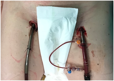
Percutaneous cannulation of 17Fr venous sheath and 24Fr arterial sheath performed by critical care physicians (Figure 1). First, bilateral groin incision is made. The venous sheath was exposed in the common femoral vein (CFV) with sloops for proximal and distal control (Figure 2). Next, the arterial sheath was exposed in the superficial femoral artery (SFA). Congenital variation of retroinguinal common femoral artery (CFA) bifurcation was subsequently confirmed with profunda femoral artery (PFA) noted alongside SFA (Figure 3). The venous sheath was removed, CFV clamped and venotomy oversewn with 5/0 prolene double breasted (Figure 4). After clamping SFA, the arterial sheath was removed and decision made to use ipsilateral long saphenous vein as patch repair. Embolectomy performed prior to patch angioplasty with 5/0 prolene (Figure 5). The distal reperfusion cannula is then retrieved from distal SFA and primarily repaired. Bilateral lower limb perfusion was acceptable post-procedure.

Figure 1 17Fr venous sheath (black arrow) and 24Fr arterial sheath (black pointer) with distal perfusion cannula in patient’s right and left groin respectively. This was inserted percutaneously by critical care physicians.

Figure 2 Venous sheath in-situ common femoral vein (black arrow) with sloops for proximal and distal control.
Since it was first discovered in 1950s, the indication and technology of ECMO has revolutionised from used only in cardiac surgery to the bedside as in the case of ECPR.10 Although more refined guidance is required and ethical issues such as public’s acceptability of ECPR has yet to be established, the use of ECPR is currently viewed as appropriate and is increasing.11 This is reflected upon the ten-fold rise in ECPR in all adult ECMO used according to the Extracorporeal Life Support Organisation (ELSO) registry.12 Richardson et al reported that survival to discharge for ECPR is 29% compared to conventional CPR (CCPR) with rates varying from 15–17%.13 ECPR is also associated with improve neurological outcome particularly during the first 3-6 months when compared to CCPR.14 Meanwhile, improvement in the design of cannulae and sheath has led to an increase used of percutaneous peripheral cannulation in ECMO in place of open approach.15 The benefit of this is faster cannulation with less bleeding on insertion but it does not come without risk.4
Vessel complications from percutaneous cannulae placement ranges from 5–10%.9 These include limb ischemia, vessel perforation andsignificant bleeding after cannulae removal, all of which require surgical revision that has significant morbidity.
Methods of decannulation described are manual compression, open approach and more recently percutaneously.8 Manual compression can only be safely performed if the sheath is less than 16Fr. Percutaneous approach is a new and promising technique but lacks robust randomised controlled studies at present.16,17 In addition, these studies show that the success of percutaneous approach to decannulation relies heavily on skilled personnel in the usage of suture-mediated closure device which are either cardiac, vascular surgeons or interventionists in both cannulation and decannulation. Banfi et al.8 suggested that if percutaneous cannulation was performed, surgical approach should be used in decannulation.8 Not only would open approach lead to less haemorrhage, it allows assessment and repair of vessels as necessary.
Our experience in surgical decannulation has highlighted a few tips for consideration in future practice. Firstly, it is crucial to consider the higher risk of bleeding in this group of patients as they are usually anticoagulated. Technically, a high index of suspicion is necessary for possibility of SFA instead of CFA cannulation as in our case. Furthermore, being mindful of anatomical variant in the groin such as high bifurcation (retroinguinal or retroperitoneal) of femoral vessels and the need for incision such as Rutherford Morrison in those setting or presence of saphena varix which require extra care during dissection. Finally, should patch angioplasty be required in the native vessels, it is prudent to avoid artificial patch such as bovine patch as indwelling sheath may be placed for duration longer than 24 hours with risk of colonisation and having an infected field. Instead, we suggest only using vein patch if reconstruction is considered with copious antibiotics.
The use of percutaneously placed large bore vessel sheath as incases like ECPR and ECMO will only be on the rising trend as technology advance medicine further. Although new approach like percutaneous closure is promising, having the skills, knowledge and experience in surgical approach to decannulation of these devices would be an invaluable art to all generations of vascular surgeons.
None.
The author declares no conflict of interest.

©2018 Chan, et al. This is an open access article distributed under the terms of the, which permits unrestricted use, distribution, and build upon your work non-commercially.