International Journal of
eISSN: 2574-8084


Mini Review Volume 3 Issue 2
Department of Breast Imaging and Women Care, Centro Diagnóstico Mon, Argentina
Correspondence: Ivelís M Sarachi, Department of Breast Imaging and Women Care, Centro Diagnóstico Mon, 7 street No. 1486, La Plata, Buenos Aires, Argentina, Tel 542215235281
Received: June 09, 2017 | Published: June 22, 2017
Citation: Sarachi IM. Vacuum assisted breast biopsy: what to do with thin breasts and superficial lesions? Int J Radiol Radiat Ther. 2017;3(2):212-213. DOI: 10.15406/ijrrt.2017.03.00057
Vacuum Assisted Breast Biopsy (VAB) is a safe and minimally invasive procedure in which a sample of breast tissue is removed for examination with a vacuum pump. It has advantages over open surgical biopsy: faster patient recovery, and lower cost and patient morbidity. With a biopsy guidance system with tomosynthesis it is possible to locate exactly the depth of target lesion. Before procedure it is necessary to choose most efficient biopsy approach. Also, the knowledge of the patient's mental and physical comorbidities, anticoagulation status, small or thin breasts, and breast implants, is important for a successful biopsy. VAB limiting factors are target less than 10mm away from the skin (with a minimum notch of 12mm), and compressed breast that reaches less than 30mm thick (part of the notch is out of the skin, not being able to perform the VAB). Rolled view is a technique that enables the clinician to perform a VAB in small or thin breasts by providing more tissue depth between the surface and the target lesion. This procedural modification adds versatility to the VAB technique, extending its potential clinical application.
Keywords: VAB, tomosynthesis, small, thin, breast
VAB, vacuum assisted breast biopsy; DBT, digital breast tomosynthesis
Before the radiologist perform breast biopsy, it is necessary to review the patient’s mammograms to determine the projection in which the target is best seen and the shortest distance from the skin to the target. To perform breast biopsy, the breast is held in compression for stabilization, digital breast tomosynthesis (DBT) is used for localization of the target, and a vacuum-assisted biopsy device. The patient could be positioned prone (with the breast pendent through a hole) or in an upright position (with the patient seated). Targets are usually calcifications, architectural distortion, and focal or developing asymmetries. VAB is a tissue sampling technique that uses devices and imaging guidance to remove samples of breast tissue through a small skin incision, under local anesthesia. Does not require stitches for closure.1‒3 The needle used in the VAB has a notch at its end. Once the needle has been positioned, the notch is open and a rotating cutting device removes the tissue sample, which is carried through a tissue collection receptacle. It takes twenty minutes to performed, and patients can usually return to normal activities soon after the procedure, without the costs and discomforts of an open surgical biopsy. Compared with open surgical biopsy, image-guided breast biopsy is equally accurate, reliable, and reproducible. More common complications include hematoma (less than 10%) and infection (less than 1%). In this article, we review strategies and procedural modifications that aid in successful completion of breast biopsy in lesions <1cm from the skin surface or <3cm depth of breast tissue from skin to chest wall (under compression).1‒4
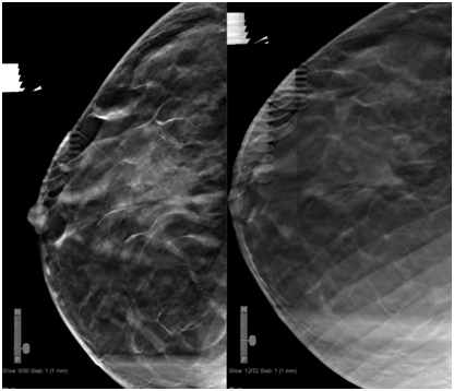
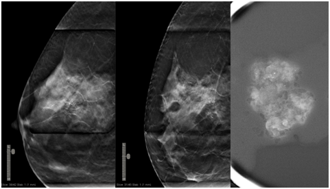
We included all patients who underwent DBT-guided VAB from November 2015 through May 2017, a total of 200 patients. A Selenia Dimensions system (Hologic) was used for all patients. The patient was placed into compression in the direction from which the needle would traverse the shortest amount of breast tissue, with a fenestrated paddle. In case of thin breast and superficial lesions we performed rolled views that allows the possibility of generate virtually more depth to the target and more thickness to the breast. Then the technologist compressed the breast. Lidocaine (1% buffered with sodium bicarbonate) was used for local anesthesia, and a larger gauge needle (ATEC 9 or 12-gauge needle) was placed at the selected target with DBT, delivering 100-300mg of tissue per sample. The notch where the sample is taken is variable (12mm or 20mm). Radiography of the specimen was routinely performed while the patient remained in the biopsy position. Finally, if we removed all microcalcifications we placed a clip marker into the biopsy site.
DBT-guided VAB allows use of the full detector size for imaging and provides lesion depth information without requiring triangulation. It facilitates target lesion re identification and sampling of even non-calcified masses.1,2 In our practice, the mean total procedure time was 15minutes. The mean time needed to identify and target the lesion was 2minutes (Figure 1 & 2).

Figure 1 First craniocaudal (CC) acquisition in which target is located at 6 mm away from the skin (left). Second CC acquisition with rolled view, where we located virtually the target deeper into the breast, which is 12 mm away from the skin (right). Lesion was an architectural distortion with anatomopathological result of a typical lobular hyperplasia.

Figure 2 First lateromedial (LM) acquisition in which target is located at 4 mm away from the skin (left). Second LM acquisition with rolled view, where we located virtually the target 14mm away from the skin (right). Lesion was microcalcifications with anatomopathological result of non-invasive ductal carcinoma.
Superficial lesions and thin breast represent a limitation to performing VAB. Rolled view is techniques that allow us relocate the target deeper generating more thickness to the breast. It wasn´t required to repeat breast biopsy, but if despite rolled view technique it is necessary to cancel breast biopsy, open surgical biopsy or short-interval follow-up imaging must be recommended because of the risk for malignancy.
None.
Author declares that there is no conflict of interest.

©2017 Sarachi. This is an open access article distributed under the terms of the, which permits unrestricted use, distribution, and build upon your work non-commercially.