International Journal of
eISSN: 2574-8084


Case Report Volume 8 Issue 1
1Faculty of Medicine, University of New South Wales, Australia
2Genesis Care, Mater Hospital, Australia
3Chatswood Dermatology Centre, Australia
4GenesisCare, Northern Cancer Institute, Australia
5Immunopathology Department, NSW Health Pathology, Westmead Hospital, Australia
6Faculty of Medicine, University of Sydney, Australia
Correspondence: Prof Gerald Fogarty, Radiation Oncology, Mater Sydney, Crows Nest, NSW Australia, Tel +61 2 9458 8050, Fax +61 2 9929 2687
Received: March 16, 2021 | Published: March 29, 2021
Citation: El-Turk N, Holt N, Gorjiara T, et al. Superficial radiotherapy and volumetric modulated Arc therapy for skin cancers within hamartomatous skin in patient with PTEN mutation: A case report. Int J Radiol Radiat Ther. 2021;8(1):26-30. DOI: 10.15406/ijrrt.2021.08.00291
Phosphatase and tensin homolog (PTEN) gene acts as a tumour suppressor gene. Mutations of this gene are a step in the development of many cancers. Sufferers can have large fields of symptomatic hamartomatous skin change especially in sun exposed areas. RT has been reported to cause increased acute toxicity in this cohort.
A 78-year-old fit male had a confirmed PTEN variant LRG_311t1 Exon 5, c353A>C. Symptomatic skin lesions of left frontal scalp and left nasal ala were confirmed on punch biopsy to be basal cell carcinoma (BCC) and he was referred for definitive radiotherapy (RT). He was treated with lesion based superficial radiotherapy to the left nasal ala to a total dose of 50 Gy in 25 fractions given at 5 fractions per week using a Xstrahl 300 machine via a 3cm circle applicator at 30cm source surface distance with a generating energy of 100 kV. The left frontal scalp was treated with a field-based volumetric modulated arc therapy technique to a planning target volume (PTV) of 74.8cm3 to 45 Gy with a simultaneous integrated boost PTV to 55 Gy of 4.1 cm3 to the BCC, all in 25 fractions. He developed the expected desquamation, erythema and mucositis within the nasal field and desquamation and erythema in the left temple. The PTEN mutation had no visible increase on the acute side effect profile compared with those without the mutation. After more than 6 months, the areas treated with RT remained clear of symptomatic hamartomatous skin change with no late toxicities.
To our knowledge this is the longest benefit received of any treatment for fields of symptomatic hamartomatous skin change associated with PTEN mutation. It is also a report of not observing increased acute toxicity of RT in the definitive treatment of skin cancer in those with proven PTEN mutation. This one case adds evidence that definitive RT to skin may be delivered safely in this cohort. More studies with multiple patients with longer follow up are needed to confirm that those suffering with PTEN mutation can be safely and successfully treated with definitive RT for skin cancer and fields of symptomatic hamartomatous skin change with no increase in late effects.
Keywords: PTEN, PTEN Hamartoma Tumour Syndrome, hamartomatous skin change, Australia, skin cancer, basal cell carcinoma, superficial radiotherapy, volumetric modulated arc therapy, toxicity
PTEN, Phosphatase and tensin homolog; RT, Radiotherapy; SXRT, superficial RT; VMAT, volumetric modulated arc therapy; SIB, technique with a simultaneous integrated boost
Phosphatase and tensin homolog (PTEN) gene acts as a tumour suppressor gene.1,2 PTEN gene mutation has an incidence of 1:200 000, although this is thought by some experts to be underestimated.3 Mutations of this gene are a step in the development of many cancers.4 Every patient with PTEN mutation develops hamartomas in different tissues, especially in the skin.5 Sufferers can have large fields of symptomatic hamartomatous skin change especially in the sun exposed areas, for which there is no recommendation for therapy.
Radiotherapy (RT) may be needed in those with PTEN mutations for cancer. RT has been reported to cause increased acute toxicity in such cohort. The clinical data is limited. There is no published data in the RT of skin cancer for those suffering from PTEN mutation. In this article, we have reported on the definitive radiation therapy for skin cancer and for fields of symptomatic hamartomatous skin change in a patient with PTEN mutation.
Patient consent was obtained for reporting this case study. A 78-year-old male with a good general condition, had a confirmed PTEN variant LRG_311t1 Exon 5, c353A>C. This was diagnosed on family review following the diagnosis of immunodeficiency and inflammatory bowel disease of a son with macrocephaly. Our patient had phenotypic issues typical of a PTEN mutation including a past history of renal cell carcinoma with micro metastases to prostate and left ureter, multiple parathyroid cancers and multiple BCCs. These were all treated with excision. He had a low B cell count (1.06% of peripheral blood lymphocytes, reference interval 6-28) and low CD8 T Cell count (84μL, reference interval 200-690 μL), typical of PTEN mutation. He also had macrocephaly and large fields of symptomatic hamartomatous skin change especially in the sun exposed areas. These areas had been treated with various topical therapies when there were symptomatic exacerbations. He had punch biopsy proven basal cell carcinoma (BCC) of left frontal scalp and left nasal ala and was referred for definitive RT.
On examination, he had macrocephaly and hamartomatous skin typical of PTEN mutation. Our patient described using topical treatments for symptomatic relief of his hamartomatous skin, providing only temporary relief. He also described having skin that burns very easily when in the sun (compared to others with similar Fitzpatrick skin type), despite the use of sun protection from early adulthood. He had BCCs with a left nasal alar lesion (Figure 1), and a scalp lesion within a field of actinic damage (Figure 2A-C). He had a RT consultation and, while the usual side effects were also described, the radiation oncologist (RO) (GBF) explained that, due to the paucity of research in the area, there was no way of knowing what sort of normal tissue reactions the radiation would induce in the context of a PTEN mutation. It was further discussed that the mutation may predispose to more serious acute and late reactions, and that there may be need to take a treatment break, or even curtail the treatment at some point to reduce the toxicity. His treatment was planned as per department protocol (Figure 3). The nasal alar was planned with superficial RT (SXRT) and the forehead was planned with a field-based volumetric modulated arc therapy (VMAT) technique with a simultaneous integrated boost (SIB).
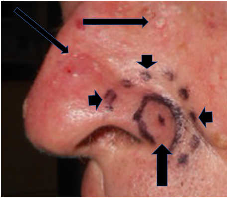
Figure 1 Nasal alar lesion at SXRT planning. Vertical thick arrow indicates BCC. Short arrows delineate SXRT area. Long thin arrows show skin hamartomatous change on the left nares.
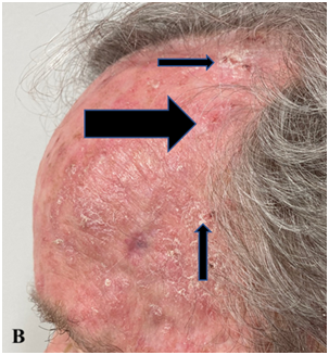
Figure 2B Left lateral projection with thick horizontal black arrow showing biopsy proven BCC. Thin black arrows show surrounding actinic change amongst hamartomatous change.
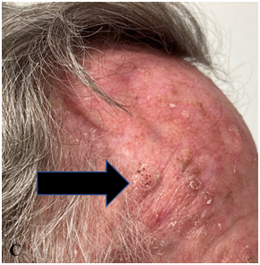
Figure 2C Right lateral projection with thick horizontal black arrow showing skin hamartomatous areas.
Figures 2A-C: Forehead at presentation showing hamartomatous change, active BCC and surrounding actinic change.
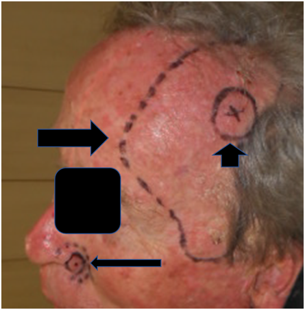
Figure 3 Planning areas. Long thin horizontal black arrow showing nasal alar SXRT field; Long thick horizontal black arrow showing left lateral VMAT field; Short thick black vertical arrow showing left lateral VMAT – SIB field.
He was treated with lesion-based SXRT to the left nasal ala with a total dose of 50 Gray (Gy) in 25 fractions given at 5 fractions per week. A Xstrahl 300 machine with energy range 50 – 300kVp (Xstrahl Group, Walsall, UK), was used for the delivery of a 100 kVp beam via a three centimetre (cm) circular applicator at 30 cm SSD (source surface distance). For each fraction the nostril was packed with wet gauze and a septal shield was employed to decrease the amount of mucositis. The left frontal scalp was treated with a 0.8 cm gel bolus field-based VMAT technique,6 using a TrueBeamTM Linear accelerator (Varian Medical Systems, Inc., CA, USA) to a planning target volume (PTV)7 of 74.8 cubic centimetres (cm3) to 45Gy (PTV45) with a SIB PTV to 55 Gy of 4.1 cm3 (PTV55-SIB) to the BCC (Figure 4). He was reviewed by the RO on a weekly basis during RT. The RT was completed in the planned time without any break.
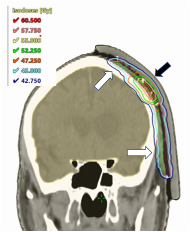
Figure 4 Coronal dosimetry of VMAT volume for left temple, planned in Eclipse V15.6 planning system (Varian Medical Systems, Inc., CA, USA). White arrows show PTV45 doses. Black arrow shows PTV55-SIB doses.
Following the completion of treatment course, the patient developed the expected desquamation, erythema and mucositis within the nasal field and desquamation and erythema in the left temple. The radiation side effects were not dissimilar to the toxicities expected in patients without a documented PTEN mutation. It appeared that the PTEN mutation had no visible increase of the acute side effect profile. He returned in 4 weeks demonstrating resolution of his cancers, field change and acute RT toxicity (Figure 5A-B). There was no unexpected acute toxicity. After more than 6 months, the areas treated with RT remained clear of hamartomas with no late toxicities (Figure 6A-C).
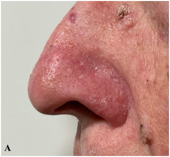
Figure 5A 4 weeks post RT showing complete response in SXRT area with almost completely resolved skin toxicity. There was total resolution of the in-field hamartomatous area. There is a suggestion of a decrease in the nearby surrounding out-of-field skin hamartomatous area. Please compare with figure 1.
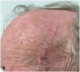
Figure 5B 4 weeks post RT showing complete response in VMAT area with almost completely resolved skin toxicity. Note resolution of the in-field skin hamartomatous area.
Figure 5A-B: 4 weeks post RT
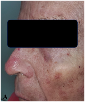
Figure 6A Six months post RT. Thin black arrow showing complete resolution in SXRT area. There was total resolution of the in-field hamartomatous area. There is a continuing suggestion of a decrease in the nearby surrounding out-of-field skin hamartomatous area. Please compare with figure 1.
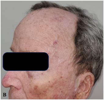
Figure 6B Six months post RT showing complete response in VMAT area of the skin hamartomatous area, especially when compared with Figure 2B prior to RT.
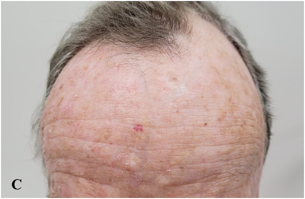
Figure 6C Six months post RT. Thin black arrow showing untreated, skin hamartomatous area on the left forehead. Thick black arrow showing treated skin with continuing in-field clearance of the skin hamartomatous area on the left forehead. There is a suggestion of a decrease in the nearby surrounding out-of-field skin hamartomatous area if this Figure is compared with Figure 3.
Figures 6A-C: Six months post RT.
The PTEN gene acts as a tumour suppressor gene through the action of its protein product, phosphatase and tensin homolog (PTEN), which negatively regulates the Protein kinase B (PKB, also known as Akt) signalling pathway. This kinase plays a key role in multiple cellular processes such as glucose metabolism, apoptosis, cell proliferation, transcription, and cell migration.1,2 Mutations of this gene are a step in the development of many cancers including thyroid cancer, renal carcinoma, glial tumours and melanoma.4 Mutations in the PTEN gene can promote the development of non-cancerous tumors called hamartomas. Almost all patients with PTEN mutation develop these hamartomas in their skin.5 Some of these may cause diagnostic difficulty and potentially be mistaken for neoplasms or for extensive skin field cancerisation.8 PTEN loss of function in skin may be enhanced by prolonged UVA exposure, most commonly from sun exposure, and may be a major source of UV induced skin cancer formation in these patients.9
Radiotherapy may be needed in those with cancer and PTEN mutations. PTEN mutations generated radiation sensitivity in cells grown in culture10 and PTEN deficient cells were unable to repair DNA double strand breaks induced by RT, compared with normal cells.11 These laboratory findings suggest that PTEN deficiency may increase clinical sensitivity to RT, including more acute toxicity in normal tissues within the RT areas or volumes.
The clinical data of PTEN deficiency on toxicity is limited to small series and case studies only. As far as oncological outcomes are concerned, Bruine de Bruin et al studied retrospectively the prognostic value of PTEN expression found on immunohistochemical staining on local control in 52 patients accrued between 1990 and 2008 with T1-2 supraglottic laryngeal squamous cell carcinoma (LSCC) treated with radiotherapy only. There was a significant association between PTEN expression and worse local control and between lymph node status and local control.12 Increased risk of malignant transformation of paediatric gliomas treated with RT was reported by Broniscer et al.13 Gits et al. in a review documented cases that had reported the development of diffuse intrinsic pontine glioma in patients with PTEN deficiencies treated with RT for medulloblastoma.14 As for increased toxicity in normal tissues, Tatebe et al reported on a breast cancer sufferer with PTEN germline mutation who had greater than expected acute and late toxicity following two separate courses of radiotherapy.15
Increased carcinogenesis from RT in those with PTEN deficiency is a risk. However, there is no data on increased skin malignancies following RT to skin in this patient population. Fortunately this will be easy to observe and we have encouraged our patient to seek lifelong surveillance from his dermatologist looking for new skin cancers especially within the skin fields that have been irradiated.
The incidental long term results of RT to hamartomatous skin was a real surprising result. At six months, the hamartomas in the treated skin had not returned and there was no evidence of long term side effects from the treatment. Gonzalez et al documented in a case study that shave removal of hamartomas resulted in recurrence in the area within 2 months.16 Yehia & Eng recommend that while interventions are available (including excision, topical agents, curettage, cryosurgery and laser ablation), treatment should be avoided unless malignancy is suspected or lesions are symptomatic because relief from symptoms is only temporary and there is rapid regrowth of hamartomas.17 Molvi et al reported that systemic acitretin (an oral retinoid) can be used to treat hamartomas with good response, however the lesions return on discontinuation of the medication.18 Our patient has demonstrated continuing complete in-field response in skin affected by PTEN associated skin hamartomatous areas. There are suggestions that with both modalities there is a decrease in the nearby surrounding out-of-field skin hamartomatous area as well. To our knowledge, resolution of lesions at six months is the longest recorded ‘cure’ of hamartomas.
To our knowledge this is the first report of definitive RT for skin cancer in PTEN sufferers. We could not find reports with RT with acute or long term toxicity in skin in the definitive treatment of skin cancer in those with PTEN deficiencies. In our case, there no rise in acute effects was observed. We have also incidentally documented a patient having received the longest benefit from any treatment of PTEN associated skin hamartomatous areas that are known off. This is an exciting and promising result, potentially introducing a new treatment of hamartomatous skin with better long term local control compared to the available treatments. More studies with multiple patients with longer follow up are needed to confirm that those suffering with PTEN mutation treated with definitive RT for skin cancer have no increase toxicity, and that they receive prolonged benefit from RT for at least in-field PTEN gene mutation related skin hamartomatous change.
This case study documents a patient treated with definitive RT for skin cancer to a standard skin cancer dose with two separate and common modalities for skin, SXRT and VMAT, without an appreciable increase in toxicity. To our knowledge, this is the first report of not observing any increased acute toxicity of RT in the definitive treatment of skin cancer in those with proven PTEN mutation. A new finding was that RT also appeared effective for durable control of in-field PTEN gene mutation related skin hamartomatous change, especially when compared to current therapies for such a problem. There was also a suggestion with both modalities of out-of-field skin hamartomatous change that warrants more investigation. More studies with multiple patients with longer follow up are needed to confirm that those suffering with PTEN mutation can be safely treated with definitive RT for skin cancer and control of areas on skin with PTEN related skin hamartomatous change.
None.
The author declares there are no conflicts of interest.

©2021 El-Turk, et al. This is an open access article distributed under the terms of the, which permits unrestricted use, distribution, and build upon your work non-commercially.