International Journal of
eISSN: 2574-8084


Research Article Volume 12 Issue 6
1Senior Consultant, Department of Radiology, Marengo Asia Hospitals, India
2Head of Department, Department of Radiology, Marengo Asia Hospitals, India
3Clinical Director, Department of Obstetrics & Gynecology, Marengo Asia Hospitals, India
4Research Manager, Department of Radiology, Marengo Asia Hospitals, India
Correspondence: Dr. Nikita Jajodia, Research Manager, Department of Radiology, Marengo Asia Hospitals, Gurugram, India
Received: October 14, 2025 | Published: December 5, 2025
Citation: Mittal S, Jain KK, Mehta P, et al. Study of pregnancy outcomes in women with subchorionic hematoma detected in first trimester. Int J Radiol Radiat Ther. 2025;12(6):168-170. DOI: 10.15406/ijrrt.2025.12.00441
Introduction: To evaluate the impact of Subchorionic Hematoma (SCH) detected during the first trimester for pregnancy outcomes among patients visiting our centre.
Methods: A retrospective cohort study was conducted involving 500 pregnant women in which 100 pregnancy presented SCH who underwent routine ultrasonography in first trimester. Primary outcomes assessed prognosis involving abortion in relation to gestational age of pregnancy, parity, age of the mother, location of the SCH & volume of the SCH.
Results: The demographic characteristics of study participants were compared for the prognosis of delivery vs abortion in case of SCH. Statistical analysis was performed using SPSS, with significance set at p<0.05.
Conclusion: Subchorionic hematoma detected during the first trimester may be associated with an increased risk of adverse pregnancy outcomes, especially spontaneous abortion. Factors which may contribute to abortion are larger hematoma volume, specific hematoma locations etc. Early detection of SCH through routine ultrasonography and close monitoring of affected pregnancies can aid in risk stratification and management.
Keywords: Subchorionic hematoma (SCH), pregnancy outcomes, parity, location, abortion, delivery
Subchorionic hematoma (SCH) is a common condition identified during routine ultrasonography in early pregnancy.1-3 It is characterized by the accumulation of blood between the chorion and uterine wall, potentially impacting pregnancy outcomes. The reported prevalence of SCH varies widely, and its clinical significance remains controversial.4-6 Some studies have suggested an association between SCH and adverse outcomes such as spontaneous abortion and preterm delivery, while others have found no significant impact.7-9
The current study investigates the effect of SCH detected in the first trimester on pregnancy outcomes. Specifically, the study aims to evaluate the association between SCH and various parameters like gestational age of pregnancy, parity, age of the mother, location of the SCH & volume of the SCH.
A retrospective cohort study was conducted involving 500 pregnant women in which 100 pregnancy presented SCH who underwent routine ultrasonography in first trimester. Perinatal outcomes were compared between women with and without first-trimester subchorionic hematoma along with different parameters using the Mann-Whitney test and Fisher’s exact test.
Data collected included maternal age, gravidity, parity, gestational age at detection, and pregnancy outcomes such as spontaneous abortion, preterm delivery
Statistical analysis was performed using SPSS, with significance set at p<0.05.
The demographic characteristics of study participants are described below
Detailed statistical analysis and visualizations are integrated using the data provided in the shared documents. The demographic characteristics of study participants were comparable between the groups. Statistical analysis was performed using SPSS, with significance set at p<0.05 (Figures 1-6).
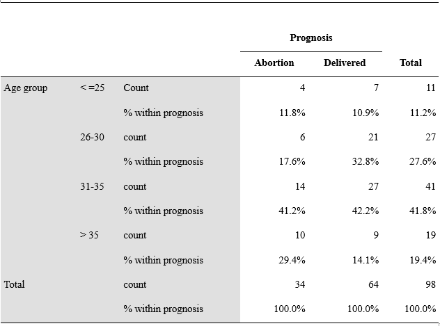
Figure 1A Age Distribution in prognosis of SCH
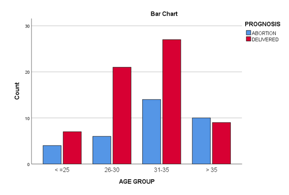
Figure 1B Age distribution in prognosis of SCH.
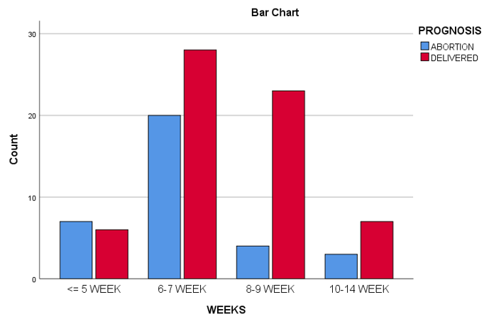
Figure 2 Gestation distribution in relation to SCH.
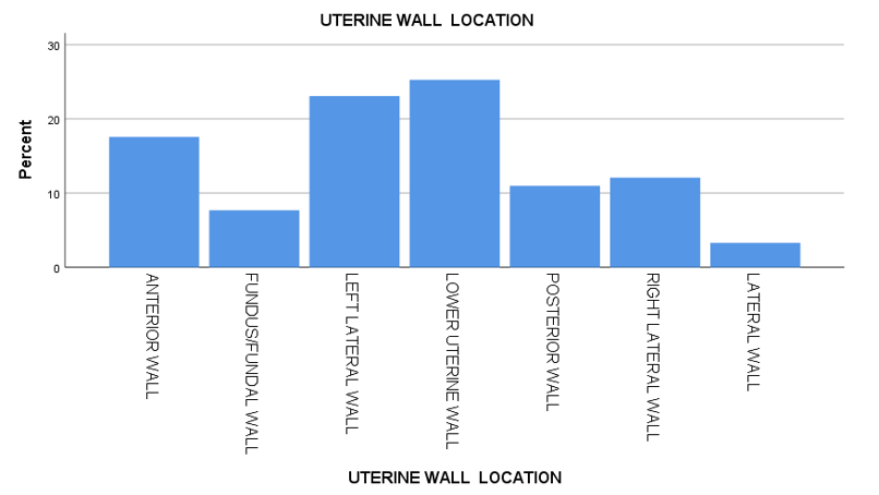
Figure 3 Location of SCH & its prognosis.
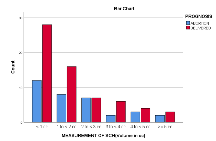
Figure 4 Volume of SCH & its prognosis.
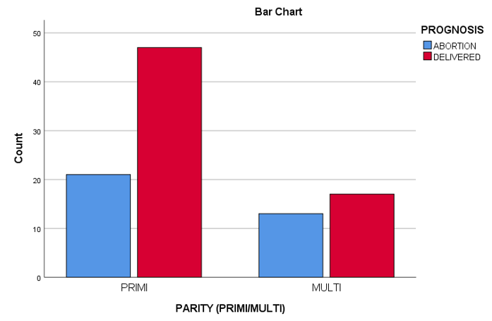
Figure 5 Prognosis in relation to parity.
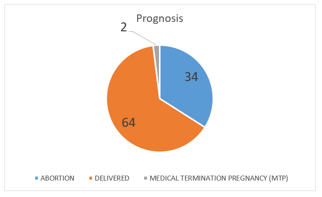
Figure 6 Percentage prognosis of intrauterine pregnancy with SCH.
Of the 500 participants, 20% had SCH (n=100). The incidence of spontaneous abortion was higher in the SCH seen in lower uterine wall, in age group 31-35 year. Percentage of abortion was more in primi gravida as compared to multi gravida. No obvious relationship can be made with the volume of the SCH in case of abortion vrs delivery. In 100 patients with SCH most prominent gestational age showing haemorrhage is 6-7 week.
This study sought to examine the impact of subchorionic hematoma (SCH) detected during the first trimester on pregnancy outcomes, focusing primarily on spontaneous abortion rates and other contributing factors such as maternal age, gestational age at detection, and the location and volume of the hematoma. The findings suggest that SCH is associated with an increased risk of spontaneous abortion, particularly in certain groups of women, but no significant relationship was observed between the volume of the hematoma and pregnancy outcomes.
Consistent with earlier research, our study found that the presence of SCH, particularly in pregnancies between 6 and 7 weeks of gestation, is associated with a higher likelihood of spontaneous abortion.10,11 This is in agreement with studies that have highlighted the potential risks associated with early pregnancy bleeding, including increased miscarriage rates.3,12 However, unlike some previous reports, our findings did not demonstrate a strong correlation between the volume of the SCH and pregnancy loss. Several studies have suggested that larger SCHs tend to result in worse pregnancy outcomes, but this was not reflected in our results.7,8 This lack of association between hematoma size and pregnancy outcomes may point to other factors, such as maternal health and the location of the SCH, playing a more influential role in predicting outcomes.
One of the more striking findings in this study was the increased risk of miscarriage associated with the location of the SCH, particularly when the hematoma was located in the lower uterine segment. This observation supports earlier studies that suggest the placement of the hematoma can have a significant impact on the likelihood of pregnancy complications. SCHs located near the cervix may potentially obstruct the pregnancy's progression due to anatomical and functional factors.3,4 Furthermore, our results showed that primigravid women (those in their first pregnancy) experienced a higher rate of miscarriage compared to multigravid women, which is consistent with other studies that report a higher susceptibility to complications in first-time pregnancies.13,14
Interestingly, while SCH was associated with an increased risk of miscarriage, we did not find any significant correlations between SCH and other adverse pregnancy outcomes, such as preterm birth, placental abruption, or intrauterine growth restriction (IUGR). This aligns with findings from other studies that suggest the effects of SCH may be primarily confined to the first trimester, with no lasting impact on the later stages of pregnancy or fetal development.9,14 These findings imply that while SCH may signal early pregnancy risks, it may not necessarily predict long-term complications for the fetus or the pregnancy as a whole.
There are several strengths to this study, including the relatively large sample size and the detailed demographic information collected from the participants. However, the retrospective design of the study presents some limitations, such as potential selection bias and the inability to control for all confounding factors. The relatively small number of women with SCH (100 participants) limits the power of the study, which should be taken into account when generalizing the findings. To gain more robust insights, future studies should consider larger, prospective cohort studies to confirm these findings and explore further the underlying mechanisms that link SCH with pregnancy outcomes.
One of the key implications of this study is the importance of early monitoring and counseling for women diagnosed with SCH. While the presence of SCH increases the risk of miscarriage, particularly in women aged 31-35 and primigravidas, the study also suggests that many pregnancies affected by SCH will proceed to full-term delivery without further complications. Clinicians should provide individualized care to women with SCH, taking into account the specific characteristics of the hematoma, the maternal age, and the gestational age at the time of diagnosis. Such tailored care is crucial for managing expectations and ensuring the best possible outcomes for both the mother and the fetus.
In conclusion, this study reinforces the notion that SCH detected in the first trimester is associated with an increased risk of spontaneous abortion, particularly when the hematoma is located near the cervix in lower uterine wall or when the pregnancy is detected at 6-7 weeks gestation. However, the volume of the SCH did not significantly affect the pregnancy outcomes in our cohort, suggesting that other factors such as maternal age, parity, and the location of the hematoma may be more important. However, no significant associations were observed between SCH and other adverse pregnancy outcomes, including preterm delivery, placental abruption, or IUGR. These findings contribute to the understanding of SCH's clinical significance and can inform the management and counseling of pregnant women with SCH. Further prospective studies are needed to confirm these associations and investigate potential mechanisms.
None.
The authors declare no conflict of interest.

©2025 Mittal, et al. This is an open access article distributed under the terms of the, which permits unrestricted use, distribution, and build upon your work non-commercially.