International Journal of
eISSN: 2574-8084


Case Report Volume 2 Issue 4
Department of Radiodiagnosis and Imaging, India
Correspondence: Sneha Karwa, Department of Radiodiagnosis and Imaging, Memorial Hospital and Medical College, Ahmednagar, Maharashtra, India, Tel 919890849900
Received: October 29, 2016 | Published: March 14, 2017
Citation: Karwa S, Patil VV. Role of magnetic resonance cholangiopancreatography in biliary disorders. Int J Radiol Radiat Ther. 2017;2(4):95-99. DOI: 10.15406/ijrrt.2017.02.00032
Magnetic resonance cholangiopancreatography or MRCP is a technique that uses a powerful magnetic field to evaluate the disorders of liver, gallbladder, bile ducts, pancreas and pancreatic duct. This technique was introduced in 1991 & has evolved rapidly over last two decades as it is non-invasive, does not require anaesthesia or contrast enhancing material and is less operator dependent. Although at present endoscopic cholangiopancreatography (ERCP) is the gold standard in evaluating the pancreaticobiliary disorders but is invasive, operator dependent and results in complications such as cholangitis, pancreatitis, haemorrhage and perforation of duodenum which have limited its routine use and has made MRCP as a routine investigation in evaluating biliary tree disorders. Furthermore MRCP is superior to ERCP in diagnosing the biliary disorders in the settings where biliary anatomy is distorted due to previous pancreatic biliary surgeries. MRCP is a technique that uses heavily weighted T2 sequences which highly enhances static or slowly flowing bile against non-enhancing surrounding soft tissue which makes the evaluation of biliary tree more accurate and easy.
Keywords:magnetic resonance imaging, magnetic resonance cholangiopancreatography, biliary ducts, gallstone, cholangiocarcinoma, liver, gallbladder, cholangitis, pancreatitis, haemorrhage, perforation, duodenum, anatomy, technique, diagnosing
ERCP, endoscopic cholangiopancreatography; MRCP, magnetic resonance cholangiopancreatography; US, ultrasonography; CT, computed tomography; PTC, percutaneous transhepatic cholangiography; haste, half fourier acquisition single-shot turbo spin echo; MRI, magnetic resonance imaging
Obstructive jaundice or biliary tract disorders are often common complaint of patients, and the majority of these patients turned out to have cholelithiasis. Biliary disorders are more common in females than males. Correct methods to detect common bile duct and pancreatic disease in patients with obstructive jaundice are important for treating surgeon to carry out appropriate treatment. For this purpose surgeons prefer the diagnostic modality which is non invasive, safe and highly sensitive in diagnosing biliary disorder as the treatment approach varies highly depending on the cause of biliary obstruction.
Evaluation of suspected biliary obstruction has traditionally involved a variety of imaging modalities including Ultrasonography (US), Computed Tomography (CT), and invasive cholangiography that includes Endoscopic Retrograde Cholangio-Pancreaticography (ERCP) and Percutaneous Transhepatic Cholangiography (PTC). Currently the non-invasive diagnosis of bile duct obstruction mainly relies on US and CT; however the accuracy of these techniques is limited because of low sensitivity for the diagnosis of stones in common bile duct. When compared with US and CT, ERCP is more accurate but is invasive and operator dependant and is associated with 1-7% morbidity and 0.2%-1% mortality.1 Also ERCP may be technically difficult and even impossible where anatomical variants are encountered or where anatomy is distorted due to previous surgical procedure.2 This fails the ERCP to examine the biliary and pancreatic ducts, upstream of obstruction making it difficult to exclude synchronous lesions and to plan appropriate therapeutic intervention. MRCP has emerged as a potent non-invasive alternative approach to evaluate the pancreatico-biliary system. The lack of need for sedation, i.v. contrast and radiation exposure and the advantage of it being non-invasive, able to delineate lesions at all levels in addition to being highly sensitive has made MRCP an important alternative to ERCP.3 MRCP plays vital role in diagnosing biliary tract disorders as it is non-invasive, operator independent with diagnostic accuracy
Study consists of fifty unselected patients of different age groups in whom there was clinical suspicion of biliary disease. This is a prospective cross sectional study carried out in department of radio diagnosis, DVVPF’S Medical College and Hospital, Ahmednagar. All cases of biliary pathology attending DVVPF’S Hospital, Ahmednagar were included in study, excluding those with cardiac pacemakers, prosthetic heart valves, cochlear implant or any metallic implant.
Technique of MRCP
The MRCP technique is performed with heavily T2-weighted turbo spin echo sequences and fast gradient echo sequences in which stationary fluid has resultant high signal intensity.4 It takes advantage of high signal intensity of body fluids on heavily T2W MRI. Static or slow moving fluid filled structures such as bile duct appears hyper intense areas, whereas background tissues some signal. This inherent difference in signal intensity enables MRCP to be carried out without contrast.5,6 MRCP is usually performed with heavily T2W sequences by using fat spin echo or SSFSE (Single Shot Fast Spin Echo) technique and both a thick collimation and thin collimation multi section technique with a torso phased array coil. The coronal plane is used to provide a cholangiographic display and the axial plane is used to evaluate CBD.7
For MRCP patients should be nil by mouth for 3-4 hours prior to the procedure. This reduces fluid content within the stomach, decrease duodenal peristalsis and promote the filling of gall bladder. MRCP is done by using 2 sequences - breath-hold and non-breath-hold sequences. The breath-hold sequence acquires a single slab of data, between 40 and 80mm thick, in 1 or 2seconds. Thin slabs (4mm thick) can also be acquired using breath-hold T2-weighted half Fourier acquisition single-shot turbo spin echo (HASTE) sequences. These are obtained in coronal or oblique coronal views. In addition, the MRCP involves acquiring multiple thin collimation slices, a non-breath-hold, respiratory-triggered 3D turbo spin-echo (TSE) T2-weighted sequence, (1.5mm) that can be post-processed on an imaging workstation. The source images from a thin collimation multislice acquisition are reviewed in addition to the reconstructed images, in order to demonstrate small stones or other intraductal pathology that may be obscured by reconstruction effects.
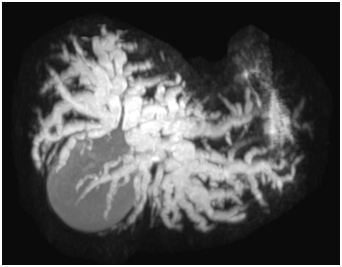
In the present study the cases of duct calculi predominated and was seen in 16 patients (32%) fallowed by congenital (choledochal cyst) in 12(24%) and gall bladder masses in 6(12%).In our study, patients of biliary pathology especially stricture and mass lesion in lower part of CBD were better evaluated by MRCP. In patient with Klatskin tumour, in which hepatic ducts were better evaluated by MRCP (Table 1‒3) (Figure 1‒7).
Sex |
No. of Cases |
Percentage% |
Males |
17 |
34% |
Females |
33 |
66% |
Total |
50 |
100 |
Table 1 Sexwise Distribution in the Biliary Diseases
Age (Years) |
No. of Patients |
Percentage (%) |
0-18 |
3 |
6 |
19-40 |
16 |
32 |
>40 |
32 |
62 |
Total |
50 |
100% |
Table 2 Age Wise Distribution in Biliary Diseases
Diagnosis |
No. of Cases |
MRCP Diagnosis Accuracy (Based on Final Diagnosis of ERCP, Histology, Operative Findings) |
A) Congenital |
12 |
|
Choledochal cyst |
12 |
100% |
B) Duct Calculi |
16 |
|
In lower end of CBD |
7 |
100% |
In mid part of CBD |
5 |
100% |
In CHD |
4 |
100% |
C) Stricture |
6 |
|
Benign |
3 |
100% |
Malignant |
3 |
100% |
D) Mass Lesion |
16 |
|
Klastkin tumour |
4 |
100% |
Periampullary carcinoma |
6 |
84% |
GB mass |
6 |
100% |
Total |
50 |
Table 3 Number of Patients Showing Various Diseases

Figure 3 Hilar Cholangiocarcinoma on MRCP.
Non Visualisation of Confluence of Right and Left Hepatic Ducts with Dilated IHBR.
ERCP, histopathological reports and post-operative findings were compared. MRCP was 98% accurate in diagnosing the diseases. False negative result in one patient was due to technical problem. In this patient MRCP diagnosis was mass lesion in 2nd part of duodenum, but on operation it was pancreatic head carcinoma.
Anatomy of biliary system
Biliary tract includes right and left hepatic ducts, joins to form common hepatic duct. Cystic duct from gall bladder joins with common hepatic duct to form common bile duct. Common bile ducts have supraduodenal, retroduodenal, pancreatic and intraduodenal segments. Sphincter of oddi typically encircles the terminal portions of biliary and pancreatic ducts and their common channel (Table 4).
Intrahepatic |
Portahepatic |
Suprapancreatic biliary obstruction |
Intrapancreatic sites |
Primary sclerosing carcinoma |
Cholangiocarcinoma |
Pancreatic carcinoma |
Pancreatic carcinoma |
Space occupying lesions and liver diseases |
Primary sclerosing carcinoma |
Metastatic diseases |
Pancreatitis |
Gall bladder carcinoma |
Pancreatitis |
Choledocholithiasis |
|
Metastatic diseases |
Strictures |
Cholangiocarcinoma |
|
Strictures |
Cholangiocarcinoma |
Ampullary carcinoma |
|
|
|
Choledocholithiasis |
Duodenal carcinoma |
Table 4 Common causes of biliary obstruction
Cholelithiasis and choledocholethiasis
Choledocholithiasis accounts for most cases of biliary obstruction. Patients with obesity, increasing age, hyperalimentation, rapid weight reduction, illeal disease are considered at high risk of choledocholethiasis. 70-80% of gall stones in western countries are cholesterol and the remainders are pigment stones. On MRCP, calculi appear as foci of no or low signal intensity irrespective of their composition. A combination of thick slab MRCP technique and thin section multislice images increases the sensitivity for detection of large as well as small (1-4mm) stones. MRCP has a sensitivity of 81% to 93% and specificity of 91% to 95% in the evaluation of common bile stones which is comparable to sensitivity and specificity of ERCP for the evaluation of common bile duct stones.
Gall stones produce three patterns of shadowing
Choledochal cyst
This is uncommon cause of obstructive jaundice. It is frequently seen in female and asian infants.
It presents with classical triad of pain jaundice and right upper quadrant mass.
Classification of alsono- lei modified by Todani et al.8
MRCP is non invasive procedure and provides the best available projection image for revealing the extent of choledochal cyst in children and adults. The development of single shot fast spin echo (SSFE) sequence made it possible to evaluate biliary tree in infants and children those who are not able to hold the breath.
Carcinoma of the gall bladder
Carcinoma of gall bladder is fifth most common malignancy of the gastrointestinal tract. Major risk factors for gall bladder carcinoma includes gall stones in 65-95% of cases history of chronic cholecystitis in 40-50% cases and an estimated 22% of patients with porcelain gall bladder will develop gall bladder carcinoma. Gall bladder carcinoma has a peak incidence in 6th decade of life.9
Gall bladder carcinomas can manifest as a polypoid mass with an intraluminal component, a bulky exophytic mass, or a mass infiltrating liver parenchyma and occupying the gall bladder lumen. Biliary obstruction may result from direct extension of the tumour to the porta hepatic or from compression of the extrahepatic bile ducts by enlarged lymph nodes.
The primary tumour and its spread beyond gall bladder appears hyper intense on T2WI and hypo intense on T1WI when compared with liver parenchyma.
Dynamic MRI with spoiled gradient pulse sequence may differentiate benign from malignant bladder lesions. Malignant lesions demonstrate early and prolonged enhancement where as benign lesion show different pattern. Dynamic MRI can also be useful for differentiation of chronic cholecystitis from carcinoma and for evaluation of weather the tumour invades beyond serosa.10
Cholangiocarcinoma
Cholangiocarcinoma is most common tumour of bile duct. Cholangiocarcinoma is classified as 1. Intrahepatic (peripheral) tumours 2. Hilar lesions occurring just past the confluence of right and left hepatic ducts commonly referred as “Klastkin” tumours; and 3. Distal ductal tumours.9
Whenever a cholangiocarcinoma is suspected, the fallowing aspects should be assessed: biliary dilatation, level of obstruction, presence of mass, focal or diffuse thickening of bile duct walls, hepatic metastasis, lymph nodes, portal vein thrombosis and cholelithiasis.11 The accuracy of MRCP in diagnosing the presence of obstruction ranges from 91-100% whereas the level of obstruction can be correctly evaluated in 85-100% while the accuracy in the differentiation of bening and malignant obstruction has varied within 30-98%. The use of combination of MRI and MRCP improves diagnostic accuracy.
Based on the results of our study we have concluded that,
None.
Author declares that there is no conflict of interest.

©2017 Karwa, et al. This is an open access article distributed under the terms of the, which permits unrestricted use, distribution, and build upon your work non-commercially.