International Journal of
eISSN: 2574-8084


Image Article Volume 9 Issue 1
University Center of Valênca (UNIFAA), Brazil
Correspondence: Prof CLAUDIO LIMA, University Center of Valênca (UNIFAA), 161, Sargento Vitor Hugo St Zip Code 27600-000 Rio de Janeiro, RJ, Brazil, No. 161, Fátima, Valença - RJ, CEP: 27600-000, Tel +5521987184089
Received: December 12, 2021 | Published: January 24, 2022
Citation: Lima C. Revisiting five classics signs of the thoracic radiology. Int J Radiol Radiat Ther. 2022;9(1):1-4. DOI: 10.15406/ijrrt.2022.09.00315
There are a few signs in radiology which are based on common objects or patterns that we come across in our routine lives. These signs are compiled with an intention to provide a comprehensive review of the scientific basis behind these signs and their importance in thoracic imaging. With a thorough understanding of these signs in conjunction with the relevant medical history, we can have a systematic approach to the problems in thoracic imaging, which can assist in a proper patient management. The objective behind the association between such common objects and the corresponding pathologies is to make the reader understand and remember the disease process. These signs do not necessarily indicate a particular disease, but are usually suggestive of a group of similar pathologies which will facilitate in the narrowing down of the differential diagnosis. Radiological signs are classical and distinctive abnormalities, characteristic of a disease or a group of similar pathologies which can be seen either on a plain radiograph or on a computed tomography scan. These signs are generally based upon and associated with common objects and patterns that we come across in our routine lives. The objective behind establishing such an association is to help the reader understand and memorize their appearance and characteristics. The familiarity and application of these signs can facilitate in narrowing down the differential diagnosis and timely management of the disease. In this essay, we describe 5 classical radiological signs used in chest imaging, which are extremely helpful in routine clinical practice not only for radiologists but also for chest physicians and cardiothoracic surgeons.
In 2008, the Fleischner Society listed imaging terms used for description of main thoracic radiological signs. This classification updated previous glossaries published. in 1984 and 1996. This glossary paper offers some radiological examples of these terms, providing a useful guide for radiologists. In addition, some terms listed —such as the atoll sign or crazy paving sign—clearly refer to symbols or naturalistic images— and these associations provide a clear and quick explanation of the associated radiological pattern.
The human brain has different types of learning mechanisms, based on personal ways of sensing, elaborating, and retrieving information from memory. In this regard, the association with symbols and photos represents a series of tips and tricks to increase learning and assimilation of concepts. Therefore, visual memory and visual-iconographic learning have been introduced into radiological language—in the majority of cases creating an association between CT and X-ray findings depicted in images and symbols of nature. Using these associations, our brain creates an unconscious emotion linked to images, which makes the memory stronger and simplifies the learning process. In addition, the knowledge of these radiological signs increases the diagnosis specificity, there being a strong association between these imaging features and certain thoracic diseases.1
Therefore, there was a need to define some medical terms. The purpose of medical terminology is to provide a standardized language for medical professionals and multidisciplinary teams. This terminology enables effective multidisciplinary medical communication, faster data sharing and discussion of clinical cases, and integration of data into patient clinical records. In addition, it should be noted that the appropriate language can help to reduce communication errors and inadequate documentation, which ensures that medical teams can analyze patient processes more quickly and accurately and, thus, speed up diagnosis and treatment. In order to critically summarize recent evidence on image descriptors, 25 experts from Brazil and 3 experts from Portugal, all members of the Brazilian Association of Thorax Image Committee, were invited to develop a consensus. An expert panel selected topics or issues related to the most significant changes in previously published concepts, including terminology used in chest radiographs, computed tomography and magnetic resonance imaging. In chest radiology, the written radiology report is the most important and often the only communication tool between the radiologist and the referring physician.1,2
An important definition was reached by this consensus, defining the term “mosaic pattern of lung attenuation”. The term “mosaic pattern of lung attenuation” is nonspecific and refers to a pattern that occurs in a variety of lung dis- eases. The terms “mosaic perfusion” and “mosaic oligemia” should be reserved for known cases of primary vascular lung disease.2
Some terms still cause controversy, as described in a letter to the editor sent by Palmucci S.3 Kitami et al pointed out that the terms galaxy sign and cluster sign could be considered misnomers. Continiung the send letter: The galaxy sign – to the best of my knowledge – was first reported in literature in 2002, when Nakatsu et al.4 described “large nodules […] surrounded by many tiny satellite nodules. These findings were considered to simulate the appearance of a galaxy”. The sarcoid galaxy was encountered in 16 (27%) of 59 patients of this mentioned study: looking at the figures of this paper, authors provide explanation of sarcoid galaxies – showing “large parenchymal nodules” surrounded by tiny nodules.
The sarcoid galaxy sign has also been reported in the paper published by Criado et al.5 where they provide same description already outlined by Nakatsu: “Fine nodular opacities are seen around the large nodules […], and small low-attenuation spots that correspond to the spaces between partially coalescent small nodules are visible peripherally.” In another article published by Herraez et al.6 the cluster sign has been described as the presence of “multiple small punctiform nodules in the peripheral regions of the lung in patients with sarcoidosis”. This appearance is again reported in the paper published by Criado.5, which indicates that the sarcoid cluster “consists of multiple micronodules distributed along the lymph vessels on high-resolution CT images.” These High resolutinon CT signs seem to refer to a different degree of profusion of granulomas within the lung: in the first model, we have a large nodule/consolidation and some tiny nodules located in the adjacent parenchyma; in the second model, we do not observe a big nodule – but a constellation of small nodular . And the author concludes: I’m not sure that we could exclude this simple association with a galaxy, to describe the profusion of nodules; in my opinion, we could still use the term “galaxy”. Clearly, for differentiating degree of profusion of nodules within the lung, a univocal correspondence is better obtained describing the open cluster and globular cluster appearances of stars.3
Radiologic signs are recognizable, characteristic patterns used to describe abnormalities visualized on imaging modalities that ultimately aid in the diagnosis and subsequent treatment of disease. When the late pulmonary radiologist Benjamin Felson was asked about his propensity for naming signs he responded that ‘‘the name saves time, helps you remember the sign, and advertises ity They say what I mean.’’1 This pictorial essay discusses 23 classic roentgenographic signs used in thoracic imaging and presents them in alphabetic order. Its purpose is to be used as an educational review for residents, whether they are beginning their training or preparing for certification exams, and serve as a refresher and a reference to the practicing radiologist.1-7
Radiological signs are characteristic and recognizable patterns in radiology and diagnostic imaging methods, whether in plain radiograph (XR), computed tomography (CT) or magnetic resonance imaging (MRI). They are based on common objects or patterns that we find in our routines and its recognition facilitates diagnostic reasoning.7-9
We present five classics thoracic radiological signs in XR or CT that cannot go unnoticed.
We don't want to exhaust the topic by showing all the existing signs. Our objective is to present five signs of thoracic radiology that over time have been less and less described, but which nevertheless represent pathologies or conditions that remain current. Therefore, the reported signs have their clinical radiological importance and every student or physician, radiologist or not, must recognize and know how to interpret.
A retrospective study of chest radiographs and computed tomography scans of the chest from the digital and physical archive of Hospital Lourenço Jorge, reference hospital in the west side of Rio de Janeiro , Brazil, between the years 2019 and 2020.
Among the approximately 1300 archived radiographs of thoracic pathologies, the cases that could be representative for the topic were separated. The same was done with the approximately 1000 chest CT scans archived. The most relevant cases with the best documentation were separated. Once the pathologies were defined, a bibliographic survey and text production were performed.
Informed consent from the patients was not obtained, since the objective of the present study was to survey archived cases, which eventually had been carried out about three years ago, making any search for patients for this purpose unfeasible. The authors were concerned to avoid any type of identification in the images presented.
Golden S sign or inverted S
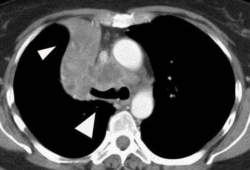
Dr. Ross Golden was the American radiologist who originally described this sign. With the collapse of the upper lobe and consequent deformation of the oblique fissure, the upper part of the S is formed and the lower part is formed by the contour of a right hilar mass or endobronchial lesion on the same side. This sign is related to obstructive bronchogenic carcinomas. Although initially used to describe signs of right upper lobe collapse, it can be applicable to atelectasis involving any lobe. Figure 11,7-11

Figure 1 Chest CT after administration of intravenous contrast in the axial plane at the level of the aortopulmonary window shows a convexity in the right hilar region creating a rounded edge with the minor fissure displaced forming the lower portion of the S (large white arrowhead). The upper portion is formed by the convexity of the collapse of the upper lobe on the same side (small white arrowhead).
Sign of 1-2-3 (Garland's Triad)
It refers to a lymph node enlargement pattern characterized by right paratracheal and right and left hilar enlargement, suggesting sarcoidosis. Leo Henry Garland (1903-1966) was an Irish-born American radiologist who published his experience on radiographs of 36 cases of sarcoidosis in an article published in 1947. A combination of bilateral hilar and right paratracheal adenopathy was found in 13 cases, 7 with pulmonary alterations and 6 without, although at the onset of the disease it was present in approximately two thirds of the cases. Figure 2.1,7-11

Figure 2 Frontal chest x-ray shows lymph node enlargement pattern (white arrowhead) characterized by right paratracheal and right and left hilar enlargement, suggesting sarcoidosis.
Double atrial contour sign
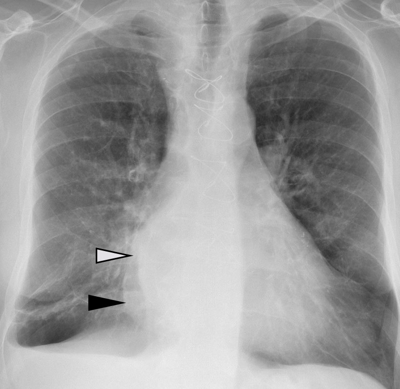
On frontal chest X-rays, this sign appears as a curvilinear density along the silhouette of the right atrium, signifying an enlargement of the left atrium. The sign can be seen in patients without heart disease, so there is a semiquantitative measurement to estimate the left atrial diameter described by Higgins et al in adult PA radiographs. If left atrial dimension is defined as the distance from the midpoint of the double density to the inferior wall of the left main bronchus, a distance greater than 7 cm was consistent with a diagnosis of left atrial enlargement, which was confirmed on echocardiography. Figure 3.1,7-11

Figure 3 Frontal chest x-ray shows curvilinear density (white arrowhead) along the silhouette of the right atrium (black arrowhead), signifying an enlargement of the left atrium.
Naclerio's "V" sign
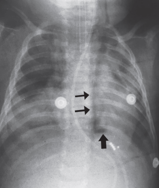
Identified on chest X-ray and depicted as two hypertransparent V-shaped lines, one along the left edge of the aorta and the other creating the continuous diaphragm sign to the left. It is produced by the presence of air between the descending aorta (vertical branch of the V) and the parietal pleura with the left diaphragm (oblique horizontal branch of the V). This sign was described in 1957 by the chest surgeon Emil A. Naclerio (1915-1985) in patients with rupture in the left posterolateral region of the esophagus, however, it is not pathognomonic, and lesions at the level of the proximal esophagus (iatrogenic or traumatic) may not present this sign. Figure 4.1,8-11

Figure 4 Naclerio's V sign. A one-year-old male, hospitalized with a diagnosis of pneumonia in the left lower lobe without satisfactory response to treatment. After the insertion of an enteral catheter, the clinical picture worsened, and a chest X-ray was requested, which showed the sign of Naclerio's V and the diagnosis of esophageal rupture with pneumoperitoneum. Vertical branch (thin arrows) and horizontal branch (thick arrow).
Continuous diaphragm signal
Continuous line of lucency extending through the midline above the diaphragm identifiable on the PA chest X-ray, representing the presence of air (pneumomediastinum) posterior to the heart and useful in differentiating a pneumothorax. Figure 5.1,8-11
Various radiological signs can be easily associated and recognized from common patterns observed in everyday life. Due to their diagnostic importance, they are fundamental for diagnostic reasoning in radiology.
None.
Authors declare no conflict of interest.

©2022 Lima. This is an open access article distributed under the terms of the, which permits unrestricted use, distribution, and build upon your work non-commercially.