International Journal of
eISSN: 2574-8084


Review Article Volume 13 Issue 2

Centro Oncológico del Note, Antofagasta, Chile
Correspondence: Dr. Alejandro Emilio Santini Blasco MD, Médico Radio-Oncólogo, Centro Oncológico del Note, Antofagasta, Chile, Tel 56 982365702
Received: January 19, 2026 | Published: March 4, 2026
Citation: Blasco AES. Review on the role of radiotherapy in the treatment of skin cancer. Int J Radiol Radiat Ther. 2026;13(1):34-37. DOI: 10.15406/ijrrt.2026.13.00450
Nonmelanoma skin cancer (NMSC) represents the most common malignancy worldwide and its incidence continues to rise, particularly in regions with intense ultraviolet (UV) exposure.1–3
Basal cell carcinoma (BCC) and cutaneous squamous cell carcinoma (cSCC) account for most cases and exhibit distinct biologic behavior and oncologic risk profiles. Although surgery remains the most frequently used curative modality, radiotherapy (RT) plays a relevant and well-established role in selected patients.4–9
In Chile, the epidemiology is variable, with the northern regions (Arica, Tarapacá, and Antofagasta) showing the highest prevalence, with a rate of 122.1 per 100,000 inhabitants, tripling the national average. This is undoubtedly due to greater sun exposure (Atacama Desert), arsenic exposure, and occupational exposure (a predominantly mining area).10
RT is especially valuable as definitive treatment in medically inoperable patients, for tumors in anatomically complex or cosmetically sensitive areas, and as adjuvant therapy in the presence of high-risk pathologic features such as positive margins, perineural invasion, or nodal involvement. This review summarizes risk stratification, contemporary indications for definitive and adjuvant RT, modern techniques with emphasis on image-guided superficial radiotherapy (IGSRT), and practical dose and fractionation strategies.4–15
Keywords: nonmelanoma skin cancer, basal cell carcinoma, squamous cell carcinoma, radiotherapy, IGSRT, hypofractionation
NMSC constitutes a growing clinical and economic burden for healthcare systems. This increase is driven by population aging, cumulative lifetime UV exposure, changes in recreational habits, and improved survival among immunocompromised patients.1–3
BCC typically behaves indolently with a very low metastatic potential, yet it can cause substantial local morbidity if inadequately treated. In contrast, cSCC carries a higher risk of local recurrence, regional lymph node involvement, and distant metastasis, particularly in the presence of adverse pathologic factors or immunosuppression.3,11-17
While surgery remains the standard approach for many patients, radiotherapy provides an effective noninvasive option with independent indications. From the radiation oncologist’s perspective, RT should be considered a primary modality in appropriately selected scenarios rather than merely a fallback option.4–9
NMSC demonstrates marked geographic variability, largely determined by latitude, altitude, and cumulative UV exposure. UVB radiation is the principal etiologic factor, inducing direct DNA damage and promoting carcinogenesis.1–3
Additional risk factors include advanced age, fair skin phototypes, male sex, prior ionizing radiation exposure, chronic cutaneous inflammation, and immunosuppression. Solid organ transplant recipients have a markedly increased incidence of cSCC and a more aggressive disease course.3,5,12
Genetic syndromes associated with impaired DNA repair or tumor suppressor pathway dysfunction (eg, Gorlin–Goltz syndrome, xeroderma pigmentosum) confer a substantially increased lifetime risk, and a prior history of NMSC strongly predicts subsequent tumors, emphasizing the importance of surveillance.3,18
Risk factors are summarized in Table 1.
|
Category |
Risk factors |
|
Demographic |
Advanced age; male sex |
|
Environmental |
Chronic ultraviolet radiation exposure (UVB > UVA) |
|
Phenotypic |
Fair skin; Fitzpatrick skin types I–II |
|
Iatrogenic |
Prior ionizing radiation; chronic corticosteroid exposure |
|
Immunologic |
Immunosuppression; solid organ transplantation |
|
Occupational |
Tar/coal/industrial chemical exposure |
|
Dermatologic |
Chronic ulcers; scars; chronic inflammation |
|
Genetic |
Gorlin–Goltz syndrome; xeroderma pigmentosum; albinism; Bloom syndrome; Fanconi anemia; Muir–Torre syndrome |
Table 1 Risk factors associated with nonmelanoma skin cancer
Note: Risk factors should be interpreted in combination; prior NMSC is a strong predictor of subsequent tumors.
BCC commonly presents as a slowly enlarging lesion with nodular, superficial, pigmented, morpheaform, or infiltrative patterns. For prognosis, precise definition of tumor borders and histopathologic growth pattern are more important than the clinical subtype.7,18
cSCC frequently appears as a keratotic plaque, cutaneous horn, or ulcerated lesion that may bleed. In situ disease is termed Bowen disease. Compared with BCC, cSCC shows a greater tendency for deep invasion, perineural spread, and lymphatic dissemination.3,11
Histologic confirmation is mandatory. Cross-sectional imaging (CT or MRI) is reserved for selected cases, such as large tumors, suspected cartilage/bone involvement, or clinical concern for perineural invasion (eg, pain, paresthesia, motor deficits).11
Risk stratification should integrate tumor diameter, depth of invasion, histologic differentiation, perineural or lymphovascular invasion, desmoplasia, high-risk location (ear, lip, periauricular region), immune status, and recurrence. Key high-risk features are summarized in Table 2.11,18
|
Feature |
Clinical relevance |
|
Tumor size |
Higher risk in lesions >2 cm (site dependent) |
|
Depth of invasion |
Greater risk with deep invasion; Clark level/dermal extension |
|
Histologic grade |
Poor differentiation associated with adverse outcomes |
|
Perineural invasion |
Increased risk of local recurrence and perineural spread |
|
Lymphovascular invasion |
Higher metastatic potential |
|
Desmoplasia |
Associated with local recurrence |
|
Anatomic location |
Ear, lip, periauricular/nasal region are higher risk |
|
Immune status |
Aggressive behavior in immunosuppressed patients |
|
Recurrence |
Higher failure rates after prior treatment |
Table 2 High-risk features for recurrence or metastasis in nonmelanoma skin cancer
Management should be individualized based on histologic subtype, tumor extent and location, risk features, patient age and comorbidities, and the anticipated functional and cosmetic consequences of treatment.3,4
Wide local excision and Mohs micrographic surgery remain the most commonly used curative approaches. Standard excision typically uses predefined clinical margins, while Mohs surgery provides comprehensive margin assessment and is particularly useful for high-risk, recurrent, or anatomically challenging tumors where tissue preservation is important.15,17
Cryotherapy, curettage with electrodessication, and topical agents (eg, 5-fluorouracil, imiquimod) may be considered for superficial or in situ lesions in selected patients, but recurrence rates are generally higher than with surgery or RT when used for invasive tumors.4,18
Systemic therapy is reserved for advanced disease not amenable to local modalities. EGFR-directed therapy and chemotherapy have been used for advanced cSCC, while Hedgehog pathway inhibitors are active in locally advanced BCC, albeit with tolerability limitations.4,17
Within this landscape, RT remains a fundamental modality as definitive or adjuvant therapy (Table 3).4–9
|
Clinical scenario |
Role of radiotherapy |
|
Medically inoperable patient |
Definitive RT |
|
Unresectable tumor |
Definitive RT |
|
Tumor in cosmetically/ functionally critical site |
Definitive RT |
|
Positive margins not amenable to re-excision |
Adjuvant RT |
|
Perineural invasion (histologic or clinical) |
Adjuvant RT ± nerve pathway |
|
Regional nodal involvement (cSCC) |
Adjuvant RT to nodal basin ± primary site |
|
Multiple recurrences |
Definitive or adjuvant RT based on context |
Table 3 Common indications for radiotherapy in nonmelanoma skin cancer
RT is noninvasive, reproducible, and generally well tolerated, and can achieve high rates of local control with acceptable cosmetic outcomes when appropriate technique and fractionation are selected.7–11
Indications for definitive radiotherapy: Definitive RT is indicated for medically inoperable patients, unresectable tumors, lesions in cosmetically sensitive or anatomically complex sites (eg, eyelids, nose, lips, ears), and selected recurrent tumors. Figure 1.4–9
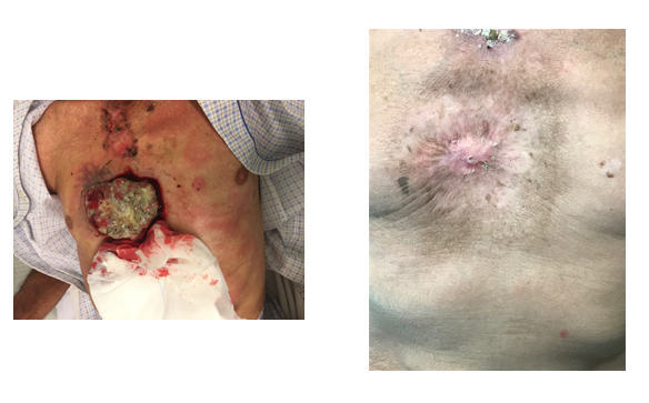
Figure 1 A 75 years man with BCC, before and after treatment with Radiotherapy.
Indications for adjuvant radiotherapy: Adjuvant RT should be considered when high-risk pathologic features are present, including positive or close margins not amenable to re-excision, perineural invasion, lymphovascular invasion, poor differentiation, deep invasion, bone or cartilage involvement, and regional nodal disease in cSCC.4–6,12
Perineural invasion is among the most clinically significant adverse prognostic factors in cSCC. In cases of clinical or radiologic perineural spread, treatment volumes should encompass the involved nerve pathway to the skull base, particularly for named cranial nerves.11
Indications are summarized in Table 3, and high-risk features in Table 2.11,18
Radiotherapy techniquesTechnique selection is central to optimizing tumor control and minimizing toxicity. The main modalities include superficial radiotherapy/orthovoltage, image-guided superficial radiotherapy (IGSRT), electron beam radiotherapy, and cutaneous brachytherapy.7–11,15
Superficial radiotherapy and orthovoltage: Superficial radiotherapy and orthovoltage (approximately 50–150 kV) are well suited for superficial, well-demarcated lesions because of rapid dose fall-off with depth, allowing sparing of deeper structures. Key practical considerations include field design with adequate clinical margins, careful immobilization, and use of applicators/cones to improve geometric reproducibility.7,8
Image-guided superficial radiotherapy (IGSRT): IGSRT integrates high-frequency ultrasound imaging into superficial RT workflows to better define tumor depth and lateral extent, enabling individualized energy selection and more accurate target coverage. This approach may reduce unnecessary irradiation of adjacent normal skin and potentially improve cosmetic outcomes, particularly for lesions in cosmetically sensitive regions.13,14
From a practical standpoint, ultrasound is typically performed at baseline to determine depth, with repeated assessments during treatment when tumor regression or uncertainty in depth is expected. Treatment margins should still account for microscopic extension and setup uncertainty; thus, IGSRT complements but does not replace standard oncologic target definition principles.13,14
Electron beam radiotherapy: Electron RT is advantageous for lesions requiring deeper penetration or larger surface coverage. Energy is selected to ensure adequate coverage of the lesion depth, and bolus is commonly used to ensure sufficient surface dose. Field shaping (custom cutouts) and attention to obliquity are essential in curved surfaces such as the scalp and extremities.8,19 Cutaneous brachytherapy: Cutaneous brachytherapy (contact applicators or custom surface molds) provides highly conformal dose distributions with rapid fall-off, and is particularly useful for small, well-demarcated lesions in anatomically complex sites. Its use requires specialized expertise and institutional resources.15Technique selection and common fractionation schedules are summarized in Table 4.
|
Technique |
Typical indications |
Common schedules (examples) |
|
Superficial RT / Orthovoltage |
Superficial, well-defined lesions |
40–50 Gy in 10–15 fractions; 50–55 Gy in 17–20 fractions |
|
IGSRT |
Superficial lesions with ultrasound guidance |
45–50 Gy in 15 fractions (institution dependent) |
|
Electron beam RT |
Deeper or larger lesions |
50–60 Gy in 20–30 fractions; 40–44 Gy in 10 fractions |
|
Cutaneous brachytherapy |
Small lesions in sensitive areas |
35–42 Gy in 7–10 fractions (HDR) |
|
Extreme hypofractionation |
Elderly/frail; logistic constraints |
36–40 Gy in 5 fractions (selected cases) |
Table 4 Radiotherapy techniques and commonly used fractionation schedules for nonmelanoma skin cancer
Note: Schedules should be adapted to tumor size, site, depth, and cosmetic considerations; BED concepts may aid regimen selection
A wide range of fractionation schedules has been used for NMSC. Contemporary practice often balances tumor control, cosmesis, logistics, and patient frailty. Guidelines support both conventional and hypofractionated regimens depending on tumor characteristics and clinical context.4–6
Conventional definitive schedules commonly range from 60–70 Gy in 30–35 fractions, whereas moderately hypofractionated regimens such as 50–55 Gy in 17–20 fractions or 40–44 Gy in 10 fractions are frequently used. Extreme hypofractionation (eg, 36–40 Gy in 5 fractions) may be appropriate for carefully selected patients, particularly older or frail individuals, with attention to cosmetic risks in large fields or thin skin areas.9,10,18
Biologically effective dose (BED) calculations (often using an α/β ratio of 3 for late skin effects) facilitate comparison across regimens. Retrospective analyses suggest that a BED₃ near 100 Gy is associated with optimal local control and acceptable cosmesis.9,19
In the adjuvant setting, 60–66 Gy in conventional fractionation (or biologically equivalent hypofractionation) is commonly employed when treating high-risk features such as perineural invasion or positive margins, and dose escalation or comprehensive pathway coverage may be considered in extensive perineural spread.4–6,12
A representative compilation of commonly reported schedules is shown in Figure 2.7–11
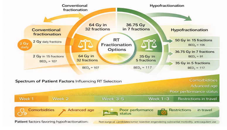
Figure 2 Representative published fractionation schedules for nonmelanoma skin cancer.
Large retrospective series have reported high local control rates with definitive RT, commonly around 90–95% for BCC and 85–90% for cSCC when appropriate technique and dose are used. Tumor size, depth, and adverse pathologic features remain key determinants of outcome.7–10,11
Acute toxicity typically includes erythema and dry or moist desquamation and is usually self-limited. Late effects may include hypopigmentation, telangiectasia, skin atrophy, and fibrosis, influenced by total dose, fractionation, field size, and technique. Cosmetic outcomes are frequently rated good or excellent when biologically appropriate fractionation is selected.8–10
Role of the radiation oncologist in multidisciplinary managementThe radiation oncologist contributes to patient selection, risk stratification, and technique and fractionation choice, and should be involved early when high-risk features or complex anatomic sites are present. Collaboration with dermatology, surgery, pathology, and medical oncology is essential to optimize oncologic control and preserve function and cosmesis.4–6,12
Radiotherapy is a central and effective modality for NMSC. When guided by appropriate risk stratification and delivered using modern techniques—including IGSRT where available—RT provides high rates of local control with acceptable toxicity and favorable cosmetic outcomes. RT should be considered a primary treatment option with well-defined indications rather than a secondary alternative.4–14
Author declare that there is no conflicts of interest.
None.

©2026 Blasco. This is an open access article distributed under the terms of the, which permits unrestricted use, distribution, and build upon your work non-commercially.