International Journal of
eISSN: 2574-8084


Research Article Volume 9 Issue 4
GenesisCare/Varian Medical System, USA
Correspondence: Kaile Li, PhD DABR, GenesisCare/Varian Medical System, 2000 Foundation Way, Suite 1100 Martinsburg, WV, 25401, Tel 3042628202
Received: August 15, 2022 | Published: September 8, 2022
Citation: Kaile L. Physical characteristics in cone beam computed Tomography (CBCT) imaging for small motion target in different directions. Int J Radiol Radiat Ther. 2022;9(4):116-119. DOI: 10.15406/ijrrt.2022.09.00333
Purpose: Fiducial marker has been an effective and intuitive way to localize motion target for lung Stereotactic Body Radiotherapy (SBRT). However, due to the complexity for motion target imaging, the optimal target localization strategy still need to be developed to improve the efficiency and effectiveness of the clinical procedure. In this study, the golden marker moving in different directions was characterized in CBCT images for optimal localization application.
Method and Materials: A Visicoil linear fiducial marker was selected for this study. The length and diameter of the marker were 5mm and 1mm. The motion was generated by Real Time Position Management (RPM) phantom from Varian Medical System. The motion was simulated to be 2.3 second breathing period and about 1cm amplitude. The motion was at three directions along the anterior-posterior (AP), left-right (LR), and In-Out (IO) of the couch. Their CBCT images were taken, and targets were done by auto-contouring with HU range from minus 900 to positive 4000. The targets were post-processed with keeping the largest part and converted to high resolution segment. Their characteristics were described by shape, volume, center of the volume and volume pixel information, which were attained by MIM Software.
Results: In this CBCT study, the volume of motion visicoil gold maker were 0.283cc, 0.348cc, and 0.271cc as moving along AP, IO and LR direction. They were corresponding to 143%, 163% and 236% of the static volume. The max HUs for each moving target were 3395, 343 and 3097, which were 49 %, 13% and 44% of those of the static visicoil. The center distances comparing to those of the static targets were 0.63cm with standard deviation at 0.02cm.
Conclusions: When the gold makers were applied for localization of treatment target in Lung SBRT, the geometric distance can be reflected in accurate level; however, the direction of motions could generate large HU difference, which could be one of factors resulted in local failure due to undefined boundary of tumor. And dosimetric characteristic in region of interest should be further investigated to understand the potential effect of local failure case even when marker tracking or soft tissue technique for localization is used.
Keywords: stereotactic body radiotherapy, motion, CBCT, fiducial marker
SBRT, lung Stereotactic Body Radiotherapy; RPM, real time position management; AP, anterior-posterior; LR, left-right; IO, In-Out; OBI, On-board imager
Quality and efficiency are the requirement for complex procedure in radiotherapy, which includes imaging simulation, target definition, dosimetry planning, target localization and dose delivery.1-4 Target motion description happens in imaging simulation, localization, and dose delivery. Fiducial marker has been an effective and intuitive way to localize motion target for lung Stereotactic Body Radiotherapy (SBRT). The effectiveness shows that markers can be used to determine the target easily in aligning to the simulation image and the accuracy can be predicted. And the intuitiveness shows that align the target to the fiducial marker, the job can be done with less depending on physician and clinical judgement. These two features make most of current clinical requirement being satisfied. However, due to the complexity for motion target imaging, the optimal target localization strategy still need to be developed to improve the efficiency and effectiveness of the clinical procedure. There are different kind of fiducial markers were available, for examples, carbon tissue marker and gold markers.5 In selection of golden marker, there are several advantages, the first is high HU range between marker and tissue, and this feature make it easily distinguish in different tissue even buried in boney structures, the second is that it magnified the variation due to motion during the imaging process, In other words, a minor change of the HUs in soft tissue can be seen in large HU difference in high z fiducial marker, which overcome the barrier of the 40HU quantitative requirement for the machine QA criteria. In this study, the viscoil golden marker6 moving in different directions was characterized in CBCT images for optimal localization application.
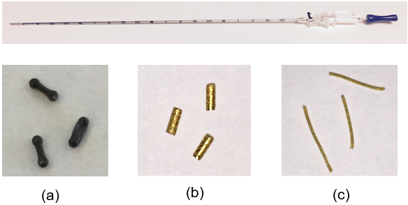
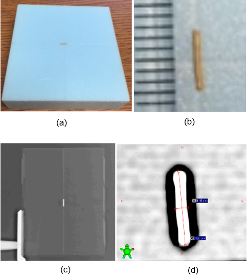
A Visicoil linear fiducial marker was selected for this study. A visicoil linear fiducial marker was show in figure 1. The length and diameter of the marker were 5mm and 1mm. The visicoil manufacture design is based on different applications.

Figure 1 Markers and Needle from different manufactures. (a) Carbon Tissue Markers, (b) Gold Tissue Markers, and (c) Gold Visicoil Linear Fiducial Makers, provided by Carbon Medical Technologies (a, b) and IBA (c).
The motion was generated by Real Time Position Management (RPM) phantom from Varian Medical System. Figure 2 shows the RPM phantom, and they were placed at different directions and form the results.

Figure 2 Gold Visicoil Linear Fiducial Maker used for this study. (a) Marker on support foam, (b) Marker magnified view with 1mm legend, (c) KV image with marker in support, (d) Magnified view of marker on KV image.
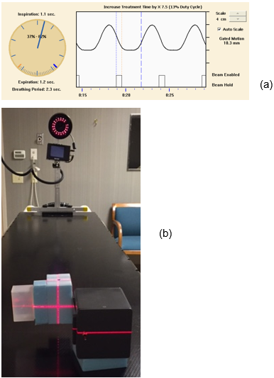
A Varian 4D camera was used to catch the signal. Figure 3 shows the camera and the motion phased signal. The motion was simulated to be 2.3 second breathing period and about 1cm amplitude.

Figure 3 (a) Phase signal generated in Left-Right (LR) direction and (b) Set-up to monitor the motion of the Visicoil.
The motion was at three directions along the anterior-posterior (AP), left-right (LR), and In-Out (IO) of the couch.
Similarly, the phantom was placed on the LINAC machine couch, figure 3 shows the positions of the phantom to generated different directions of motion. The On-board imager (OBI) of Truebeam LINAC from Varian Medical System. Their CBCT images were taken, and targets were done by auto-contouring with HU range from minus 900 to positive 4000. The targets were post-processed with keeping the largest part and converted to high resolution segment. Their characteristics were described by shape, volume, center of the volume and volume pixel information, which were attained by MIM Software (Figure 4 & 5).
In this CBCT study, the volumes of the motion visicoil gold maker were 0.283cc, 0.348cc, and 0.271cc as moving along AP, IO and LR direction. They were corresponding to 143%, 163% and 236% of the static volume. The max HUs for each moving target were 3395, 343 and 3097, which were 49 %, 13% and 44% of those of the static visicoil. The center distances comparing to those of the static targets were 0.63cm with standard deviation at 0.02cm (Table 1 & 2).
|
Marker Motion Direction |
Motion Status |
Volume (cc) |
Maximum (HU) |
Ratio between Marker volume at motion and static |
Ratio of Maximum HU of Marker at motion and static |
|
AP |
s |
0.198 |
7000 |
|
|
|
AP |
m |
0.283 |
3395 |
143% |
49% |
|
IO |
s |
0.214 |
2599 |
||
|
IO |
m |
0.348 |
343 |
163% |
13% |
|
LR |
s |
0.115 |
7000 |
||
|
LR |
m |
0.271 |
3097 |
236% |
44% |
Table 1 The volume and Maximum HU of the marker in static (s) and motion (m) status
|
Marker Motion Direction and Status |
Center X (cm) |
Center Y (cm) |
Center Z (cm) |
X |
Y |
Z |
Maximum along motion direction (cm) |
|
AP_S |
0.04 |
0.01 |
0.18 |
||||
|
AP_M |
0.08 |
0.63 |
0 |
0.04 |
0.62 |
-0.18 |
0.62 |
|
IO_S |
-0.27 |
2.55 |
-0.12 |
||||
|
IO_M |
0.07 |
2.58 |
0.53 |
0.34 |
0.03 |
0.65 |
0.65 |
|
LR_S |
-0.06 |
-0.18 |
0.21 |
||||
|
LR_M |
0.57 |
-0.21 |
0.1 |
0.63 |
-0.03 |
-0.11 |
0.63 |
Table 2 The data to simulate this process for this study
When the gold makers were applied for localization of treatment target in Lung SBRT, the geometric distance can be reflected in accurate level; however, the direction of motions could generate large HU difference, and some localization variation, which require some attention of the clinician. In other words, it also suggested that the required of fast imaging technique to lower this HU variation effect.
In diagnostic point of view, the known physical characteristics of makers could be used as a reference of different imaging modes for cross-comparison of imaging taken in different temporal points, different machines which could be in different institutions. And this feature could improve effectiveness in the application of artificial intelligent imaging analysis.
And dosimetric characteristic in region of interest should be further investigated to understand the potential effect of local failure case even when marker tracking or soft tissue technique for localization is used. And the region abutment to gold fiducial marker could possibly give information about the dose enhancement effect on gold nanoparticle characteristics and the potential of implant multiple gold markers around the tumor region to avoid the local failure due to insufficient dose due to motion target. Moreover, the microscopic structure of dosimetric described could lead to the reinvestigation of cancer control mechanism.
A recent study7 showed that the potential resonance effect in atomic level, and to activate such effect required extreme high intensity radiation beam, and the most develop high dose rate delivery technique such as ultra-high dose-rate (FLASH) technique could be a potential to realize this effect in near future. In current practice, energy specific radiation treatment is done by through directly implanting the radioactive seeds in brachytherapy or infusion of radioactive liquid into blood in nuclear medicine and both procedures involve complexity of radiation safety management procedure. Through the implantation of marker seeds in different physical materials to the treatment targets, and when these marker seeds are irradiated with high dose rate external radiation beams, the atomic resonance response of the specific energy radiation could be excited locally. The procedure could provide surgeons or clinicians a methodology involving radiation oncology care without the need of cost of radiation safety management.
In conclusion, the application of fiducial markers could lower the risk of unintended deviation in target localization even with intuitive judgement of clinicians and the application of fiducial markers could potential facilitate for computer-assistive target localization in radiotherapy procedure for given physical characteristics of fiducial makers.
None.
None.

©2022 Kaile. This is an open access article distributed under the terms of the, which permits unrestricted use, distribution, and build upon your work non-commercially.