International Journal of
eISSN: 2574-8084


Case Report Volume 2 Issue 3
Department of radiodiagnosis, Dr. DY Patil medical college and research centre, India
Correspondence: Sanjay M Khaladkar, Department of radiodiagnosis, Dr. DY Patil medical college and research centre, Pimpri, Pune- 411018, India
Received: October 31, 2016 | Published: March 3, 2017
Citation: Khaladkar SM, Chauhan RC, Chauhan S, et al. Peyronie’s disease affecting inter corporal septum – ultrasound diagnosis. Int J Radiol Radiat Ther. 2017;2(3):76-78. DOI: 10.15406/ijrrt.2017.02.00027
Peyronie’s disease also known as penile fibromatosis and plastic induration of the penis is a cause of painful erection. It is rare with a prevalence rate of 0.4-9%. It may be associated with Dupuytren’s contracture. Though its etiopathogenesis is uncertain, presence of fibrin plaque and usual dorsal location is suggestive of aberrant healing response to minor penile trauma from shear stress. Ultrasound is useful in quantifying the fibrotic involvement in Peyronie’s disease, determining the precise location of the lesion in the penis (dorsal/ventral/circumferential/septal), length, width and thickness of the plaque. It can detect fibrotic changes which have not yet calcified. We report two different cases of Peyronie’s disease with calcification in intercorporeal septum alone and another case with calcified plaque in intercorporeal septum extending along the ventral aspect of both corpora cavernosa.
Keywords: peyronie’s disease, penile fibromatosis, intercorporeal septum, tunica albugenia, plaque, ultrasound, diagnosis, sonography, cardiomyopathy, patients
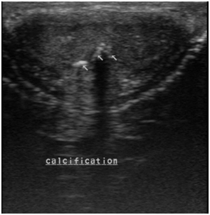
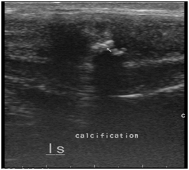
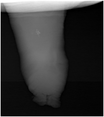
A 48year old male patient , a known diabetic presented with a palpable hard nodular lesion in the middle third of penis since 6months. Local USG of penis showed linear echoreflective calcified plaque in intercorporeal septum extending along the ventral aspect of bilateral corpora cavernosa causing posterior acoustic shadowing (Figure 1A & Figure 1B). A diagnosis of Peyronie’s disease was made. Plain radiograph of penis done with soft tissue exposure confirmed calcification (Figure 2).

Figure 1A Ultrasound mid penile shaft tranverse section shows echoreflective calcified plaques in intercorporeal septum with extension along ventral aspect of bilateral corpus cavernosa (right >left).

Figure 1B Ultrasound penile shaft longitudinal section showing multiple echoreflective calcified plaques in intercorporeal septum with extension along ventral aspect of bilateral corpus cavernosa.

Figure 2 Radiograph of penis with soft tissue exposure showing multiple calcific plaques in mid penile shaft.
Another 45year old male patient presented with painful erection and palpable nodular lesion in the middle and distal third of penis since last 8-10months to skin and veneral disease department. He was referred to the ultrasound department for sonography of the penis. Local ultrasound of penis done with linear 9-11 MHz probe revealed an echoreflective calcified plaque in intercorporeal septum measuring approximately 8mm in length and 2mm in thickness causing posterior acoustic shadowing (Figure 3).
Peyronie’s disease was first described by Francois de La Peyronie in 1743. It is also known as penile fibromatosis and plastic induration of the penis. Erectile dysfunction precedes the disease. It causes post traumatic, painful erection.1 It is seen in 1% of white men with predominance in the ages of 45-60years. Associated Dupuytren's contracture is seen in 30% of cases.1
Epidemiological data on Peyronie's disease is limited. The prevalence rate is 0.4-9% with higher prevalence in patients with diabetes and erectile dysfunction.2 Commonly associated risk factors are diabetes, hypertension, smoking, excessive alcohol consumption, lipid abnormalities and ischemic cardiomyopathy. 4% of patients with Dupuytren's contracture are reported to have Peyronie's disease.3
Though its cause is uncertain, usual dorsal location and presence of fibrin plaques is suggestive of an aberrant healing response to minor penile trauma from shear stress.4
It goes through two phases:
Repeated trauma or microvascular injury to tunica albuginea is the most widely accepted hypothesis for the etiology of the disease. This results in a prolonged inflammatory response, causing remodeling of the connective tissue and formation of fibrotic plaques with a resultant abnormal penile curvature. These fibrotic plaques can be calcified. Penile curvature worsens in 30-50% of patients, stabilizes in 47-67% of patients while spontaneous improvement is reported in 3-13% of patients.5 Plaque location can be dorsal, ventral, lateral, circumferential or septal.6 Rarely, it can also affect the corpus spongiosum.7
Tunica albuginea is a fibroelastic sheath of corpora cavernosa. It becomes less elastic with age and hence, less likely to stretch when the penis is bent or hit. This results in greater likelihood of injury with advancing age. 30% of patients with Peyronie’s disease develop fibrosis in other parts of the body, especially hands and feet.8
Penile plaques in Peyronie's disease are formed on the dorsal surface of the penis in 77% of the cases, but can also occur in the ventral and lateral surfaces and in the intercorporeal septum - 47% in the distal segment of penile axis, 36% in middle segment and 17% in proximal portion.2 Tunica albuginea has two layers, both having different structure and function. The outer layer is positioned longitudinally and is composed of collagen fibres. The inner layer has circumferential fibers surrounding and separating the corpora cavernosa. The inner layers are in close proximity to each other along the sagittal plane of the penis and forms the septa which is contiguous at the proximal portion of the penis. The septum is rich in fenestrations at distal and middle segments of the penis.2
Nodules and plaques in Peyronie’s disease develop as an inflammatory infiltrate in Smith’s space (situated between spongy corpora cavernosa and tunica albuginea). There are perivascular lymphocytic and plasmacytic infiltrates which progress to dense fibrous connective tissue. These plaques later become sclerotic and calcified. Extension to intercorporeal septum can also occur. In late stages, these lesions can occasionally undergo ossification.2
Ultrasound is the primary screening modality in the detection of Peyronie’s disease. However, noncalcified plaques without acoustic shadowing can be easily detected by endoluminal (intraurethral) Ultrasound as it provides a higher spatial resolution than conventional Ultrasound.7 It occurs most frequently where septal fibers attach to undersurface of tunica albuginea – dorsal midline and along the septum, dorsal midline with septal extension, or may occur along the ventral midline. Plaques may extend from dorsal midline circumferentially around the corpora cavernosum. Rarely a ventral plaque may form between the corpora cavernosa and spongiosum, connecting through the septum.2
Ultrasound is useful in quantifying the fibrotic involvement in Peyronie’s disease, determining the precise location of the lesion in the penis (dorsal/ventral/circumferential/septal), length, width and thickness of the plaque. It can detect fibrotic changes which have not yet calcified. Normal tunica albuginea is seen as a thin hyperechoic line covering the corpora cavernosa. Fibrotic plaques are seen as focal hypoechoic thickening of the tunica albuginea. The plaque can be diffuse or nodular. It is usually hyperechoic, but can sometimes can be isoechoic to hypoechoic. Calcified plaques appear echoreflective with posterior acoustic shadowing. Less common lesion variants are focal nodular echogenic thickening of septa without posterior acoustic enhancement or attenuation of acoustic beam.
Rare form is an isoechoic lesion (not detected on Ultrasound). These are picked up due to posterior attenuation.
Hypoechoic penile plaques are seen in acute stage when the fibrosis is least and interstitial edema is predominant. It is seen as focal thickening of peri-cavernous tissue without acoustic attenuation or enhancement.2 Periurethral fibrosis of unknown origin and fibrosis caused by urethral manipulation (Kelami syndrome) are differentials of Peyronie’s disease.7
It can be treated with oral medication, topically administered medication, shock wave lithotripsy, laser radiation therapy or surgery.2,7
None.
Author declares that there is no conflict of interest.

©2017 Khaladkar, et al. This is an open access article distributed under the terms of the, which permits unrestricted use, distribution, and build upon your work non-commercially.