International Journal of
eISSN: 2574-8084


Case Report Volume 6 Issue 4
1GenesisCare, Department of Radiation Oncology, St Vincent’s Hospital, Australia
2Melanoma Institute of Australia, Australia
3St Vincent’s Private Hospital, Australia
Correspondence: Gerald B Fogarty, Director, Department of Radiation Oncology, St. Vincents Hospital, 438 Victoria St, Darlinghurst NSW 2010, Australia, Tel 61 2 83025400
Received: August 06, 2019 | Published: August 29, 2019
Citation: Fogarty GB, Ziebell A, Nicholls S, et al. Lessons learnt from an apparent lack of clinical response following superficial radiation therapy of cutaneous squamous cell carcinoma. Int J Radiol Radiat Ther. 2019;6(4):150-151. DOI: 10.15406/ijrrt.2019.06.00237
Cutaneous squamous cell carcinoma (cSCC) is the second most common form of skin cancer and arises from abnormal growth in the squamous cells of the skin’s outermost layer due to cumulative exposure to ultraviolet radiation. Without treatment, cSCC can be life-threatening and even fatal. Radiation therapy is one of the oldest available treatments for skin malignancy and modern techniques enable the definitive treatment of non-melanoma skin cancers. Superficial radiation therapy (SRT) uses X-rays, or photons, to stop mitosis in rapidly dividing cells. As the radiation only penetrates to just below the skin’s surface, SRT is ideal for selected cutaneous malignancies, particularly when surgery is not feasible. We present two cases of cSCC in two medically unfit patients treated with SRT. Despite a lack of obvious clinical response, SRT resulted in sterilization of the lesions. Clinicians need to be aware of this phenomenon in order to avoid unnecessary salvage surgery.
Keywords: immuno-suppression, radiation, radiotherapy, skin cancer, squamous cell carcinoma, superficial X-ray
BCC, basal cell carcinoma; cm, centimetre(s); CSCC, cutaneous squamous cell carcinoma; CTV, clinical target volume; Gy, gray; mm, millimetre; RT, radiotherapy/radiation therapy; SRT, superficial radiation therapy
Cutaneous squamous cell carcinoma (cSCC) is the second most common form of skin cancer after basal cell carcinoma (BCC) and arises in squamous cells of the epidermis. Lesions are due to cumulative ultraviolet radiation and are usually found on areas exposed to the sun such as the face, ear, scalp, neck and limbs. cSCC often presents as scaly sores that may crust or bleed and not heal. Without treatment, cSCC can be life-threatening and even fatal.1,2 Radiation therapy has been used to treat skin cancer for nearly a century.3 Modern techniques have continuously developed enabling radiation therapy to be used in the definitive treatment of non-melanoma skin malignancies.4,5 The control rate of most skin cancers is at least 90 percent.3 Superficial radiation therapy (SRT) uses X-rays, or photons, to stop mitosis in rapidly dividing cells. Superficial X-ray treatment machines target the skin and spare deeper structures, and are ideal for selected cutaneous malignancies, particularly when surgery is not feasible.6 We present one case each of cSCC in two medically unfit patients treated with superficial x-rays using an Xstrahl® machine (Xstrahl Ltd, Camberley, Surrey, UK). Despite a lack of obvious clinical response, SRT resulted in complete histological sterilization of the lesions. Clinicians need to be aware of this phenomenon in order to avoid unnecessary salvage surgery.
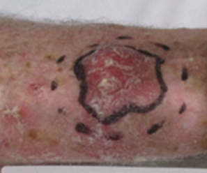
A 79-year-old male had a poor performance status and multiple comorbidities that precluded surgery. He had been treated with various therapies for multiple cSCC over the years, but radiation therapy (RT) had been his main treatment modality. The patient had also undergone an orbital exenteration due to this disease. After having had multiple lesions metachronously treated with RT with success, it was felt that subjecting the patient to a biopsy for each lesion was burdensome, and so it was no longer routinely performed. Over time, the previously treated lesions had achieved a complete response at four weeks post treatment with lesser and lesser doses of RT, meaning that the lesions were radiosensitive. The patient then presented with a one-year history of a skin lesion measuring three centimetres (cm) in diameter on the right forearm (Figure 1). Definitive superficial X-ray treatment with an Xstrahl® (Xstrahl Ltd, Camberley, Surrey, UK) machine was planned. The initial prescription was 36 Gray (Gy) in six fractions at two per week dosed to the surface using a 150KvP beam at 30 cm source to surface distance with a custom made five cm lead cut out placed on skin. The lesion did not regress during RT, nor afterwards as had been the case with his other lesion, and so a further 12 Gy in two fractions were given. The lesion only regressed from three to two cm four weeks after completion of all RT and a decision was made for surgical salvage. Excisional biopsy histopathology showed a completely excised 16 millimetre (mm) crusted lesion that on microscopy was granulation tissue only with no evidence of malignancy. There was no sign of regrowth after a year.

Figure 1 Clinical cSCC of the right forearm. Solid line is gross tumour volume. Dotted line is field edge. The tissue between is a planned margin of normal tissue.
A 67-year-old male had a poor performance status and multiple comorbidities due to long-standing renal transplantation. He had been treated with multiple therapies, including RT, for numerous cSCC over the years. His lesions were also usually radiosensitive; they did not require the recommended dose for complete response at four weeks. The patient re-presented with a three-month history of a lesion on the right forearm measuring three cm in diameter (Figure 2). The lesion was clinically determined to be a cSCC. It was treated with 36 Gray (Gy) in six fractions at two per week dosed to the surface using a 100KvP beam at 30 cm source to surface distance with a custom made five cm lead cut out placed on skin. The lesion showed no clinical regression during review four weeks after RT. Surgical salvage histopathology revealed a mass of granulation tissue measuring 24 mm in diameter with no evidence of malignancy. There was no sign of re-growth six months after surgery.
Radiation therapy and surgery are often used in the definitive treatment of cSCC.7 While surgery removes the cancer usually with a planned margin, RT treats the cancer or gross tumour volume7 with a margin of normal tissue to field edge, killing the cancer cells but not removing the resultant necrotic tissue. The latter decreases over time as normal homeostatic mechanisms remove the dead tissue and replace it with new tissue through the granulation process. Our departmental policy is to review patients at four weeks to assess response. If this process has not started, we salvage with surgery. In light of these cases, this approach may be premature. While somewhat unusual, these two cases illustrate that the medically unfit can suffer from defective homeostatic mechanisms resulting in necrotic debris that is slow to resolve post treatment. From these cases, we have learnt that more time may be needed in the medically unfit for the lesion to shrink. The histopathology of the excised biopsies in each case showed that superficial x-ray therapy delivered by the Xstrahl® machine had indeed worked. In these two patients, treatment failure was perhaps called too soon, resulting in non-essential salvage therapy with surgery. Clinicians supervising definitive radiation therapy for skin lesions need to appreciate that more time is perhaps required for lesion resolution in the medically unfit.8
These two cases of cutaneous squamous cell carcinoma in medically unfit patients demonstrate that sterilization of the treated area can occur despite a lack of clinically apparent response. Clinicians need to be aware of this phenomenon in order to avoid unnecessary salvage surgery. We suggest that resolving lesions be carefully observed, with salvage treatment reserved if this resolution stops.
None.
The authors wish to thank Aileen Eiszele of A&L Medical Communications for editorial review and for overseeing the journal submission and acceptance process.
Author declares that there is no conflict of interest.

©2019 Fogarty, et al. This is an open access article distributed under the terms of the, which permits unrestricted use, distribution, and build upon your work non-commercially.