International Journal of
eISSN: 2574-8084


Case Report Volume 6 Issue 3
1Biomedical Scientist, Radiology Department, Sírio Libanês Hospital, Brazil
1Biomedical Scientist, Radiology Department, Sírio Libanês Hospital, Brazil
2Radiologist, Siemens Healthineers, Brazil
2Radiologist, Siemens Healthineers, Brazil
Correspondence: Bertolazzi P, Biomedical Scientist, Radiology Department, Sírio Libanês Hospital, Brazil, Tel +55(11)99424-7878
Received: March 19, 2019 | Published: May 28, 2019
Citation: Bertolazzi P, Silva CFG, Mendes REV, et al. Iodine map, monoenergetic 40-kev and cinematic rendering in the diagnosis of pancreas disease: a case report. Int J Radiol Radiat Ther. 2019;6(3):84-85. DOI: 10.15406/ijrrt.2019.06.00223
Pancreatic cancer is currently one of the deadliest of the solid malignancies. Contrast enhanced multidetector-row computer tomography (MDCT) is the current modality of choice or the detection of distant metastasis in pancreatic cancer as a part of pre-operative workup, which helps decide on resectability. Therefore, to improve the early diagnostic rate of pancreatic cancer, early detection, and differential diagnosis of pancreatic masses are critical. The phase of peak pancreatic parenchymal enhancement with dual-energy imaging through the use of low–kiloelectron-volt monochromatic images or iodine MDI and how those techniques compare with conventional MDCT imaging during the same phase.
67 years-old male patient, 66 kilograms, presenting lack of appetite and weight loss in last 2 months and have no previous cancer. Patient with suspected pancreatic lesions was submitted to Computed Tomography Dual Energy (CTDE) and the iodine map, monoenergetic 40keV and cinematic rendering post-processing were performed.
Multiphasic pancreatic1−3 protocol CT scans composed of pre-contrast, early arterial and portal phase images of upper abdomen were routinely done using the SOMATOM Definition Flash (dual-source CT scanner), as default protocol of Institute. CT scans were obtained with the patient in a supine position during full inspiration. The scan range of the all phases images was from the lung base to lower margin of the iliac. The acquisition duration was approximately 13 s, and dual energy acquisition was obtained using the following parameters; 145/67 eff mAs (miliamperes), 80/Sn140 kV (kilovoltage), rotation time: 0.5 s, pitch: 0.6, resulting in 191 mGy (miligray) of DLP (Dose Length Product) or effective dose: 2.8 mSv (milisievert). 120 ml of Iodine contrast (350 mg/ml) was injected before 50 ml of saline with flow rate of 5 ml/s. The reconstruction was performed with slice thickness: 3 mm and increment: 2.4 mm, using D30f as kernel and an Abdome window.
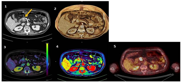
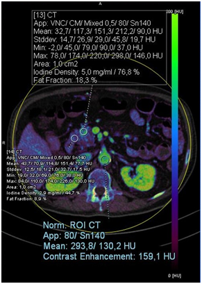
Pancreas head injury was also visualized on PET-CT and confirmed as squamous cell carcinoma with biopsy. In the iodine map, we can observe with greater definition the lesion with the attenuation of the amount of iodine, which is even more evident in monoenergetic 40 keV and we visualize it texturally by cinematic rendering, as showed in Figure 1. Quantitative analysis revealed increased Iodine enhancement (carcinoma: 5.0mg/ml; 76.8% versus normal tissue: 2.9 mg/ml; 44.7%), as well as Fat Fraction (carcinoma: 18.3% versus normal tissue: 8.9%) and greater standard deviation, what may represent greater heterogeneity of analyzed tissue when compared to the normal tissue (Figure 2).

Figure 1 Mixed image, axial slices of the upper abdomen, demonstrating squamous cells carcinoma lesion in the head of the pancreas, where 1 = computed tomography; 2 = cinematic rendering; 3 = iodine map; 4 = 40 keV monoenergetic; 5 = PET-CT to compare.

Figure 2 Quantitative analysis of the lesion in iodine map: with a normalized ROI in Descendent Aorta and two ROIs for evaluation of carcinoma lesion and normal tissue of Pancreas, showing increased Iodine enhancement as well as Fat Fraction and greater standard deviation, when compared to the normal tissue. Show an irregular, heterogeneously enhanced lesion (arrows) in the head of the pancreas, with an iodine uptake of 5.0 mg/mL, which is almost double that in the adjacent normal pancreatic tissues (2.9 mg/mL).
The use of CTDE, that is the emission of two different incidence of kilovoltage, enables post-processing techniques,4−6 among them the 40-keV monoenergetic and iodine map, which allows the evaluation of contrast enhancement with maps colored, aiding in the differential diagnosis of hypo and hypervascular pancreas lesions.5,6 CTDE venous acquisition was selected and Liver Virtual-non-contrast (VNC–syngo.via, SIEMENS, Erlangen) was used as workflow description for iodine map with ASIST perfusion as colour LUT overlay. Monoenergetic plus was used to perform 40-keV iodine map. Then, the contrast was normalized with a Region of Interest (ROI) in Descendent Aorta and two DE ROI circles were set to compare the lesion in head of Pancreas with normal tissue.4−6 Cinematic rendering is a relatively new technique of 3D visualization, which works with computer algorithms of random sampling and uses different light maps to generate a realistic representation of medical data, helping in a better visualization of the texture of the lesions, in this case we use to diagnosis of pancreatic tumors.7 Additional techniques to abdominal CTDE may help in the visualization of lesions difficult to diagnose, adding qualitative and quantitative information, leading to a differential diagnosis of the lesions.4−6
None.
Author declares that there is no conflict of interest.

©2019 Bertolazzi, et al. This is an open access article distributed under the terms of the, which permits unrestricted use, distribution, and build upon your work non-commercially.