International Journal of
eISSN: 2574-8084


Case Report Volume 12 Issue 6
 Alejandro Weber Sánchez2
1General Physician. Ángeles Lomas Hospital. Mexico
2Department of General Surgery, Angeles Lomas Hospital, Mexico
Correspondence: Pablo Weber Álvarez MD, Vialidad de la Barranca s/n cons. 410, Valle de las Palmas, Huixquilucan, State of Mexico, Mexico. CP 52763, Tel 52469527
Received: November 24, 2025 | Published: December 12, 2025
Citation: Alvarez PB, Sánchez AW. Intraparenchymal cerebral hemorrhage in a young patient secondary to de-novo hypertension and arteriovenous malformation. Case report and literature review. Int J Radiol Radiat Ther. 2025;12(6):172-174. DOI: 10.15406/ijrrt.2025.12.00442
Cerebrovascular event is defined as an extravasation of blood into the brain parenchyma, is a serious problem that affects world's population, with high mortality, which can cause permanent sequelaes. Intraparenchymal hemorrhage (IPH), defined as the extravasation of blood within the parenchyma, is a disease with high morbidity and mortality, uncommon in young patients, with higher prevalence in men. Its etiology varies depending on the age group; arterial hypertension is most common in adults and in young patients arteriovenous malformations (AVM). We present the case of a 23-year-old female patient who was admitted to the emergency department due to a de-novo hypertensive crisis that contributed to the development of an intraparenchymal hemorrhage, due to the rupture of a hemangioma. This is an uncommon presentation of this disease in young patients. Early diagnosis and a multidisciplinary approach in young patients with de novo hypertension and neurological symptoms, as in this case, is important to establish appropriate management and prevent irreversible or fatal complications.
Keywords: Intracerebral hemorrhage, cerebrovascular event, young adult, parenchymal hemorrhage, intracranial hemorrhage, arterial hypertension
Cerebrovascular event is a serious health problem, is the second cause of death in general population (9.7%).1,2 Its incidence is between 6 and 20 cases per 100,000 inhabitants per year. It is uncommon in patients aged 15 to 45 years; estimated to occur in 15% of this age group.3 The etiology of stroke in young patients is different from older patients, aneurysm or AVM rupture is the most common presentation of hemorrhagic stroke in young patients. We present the case of a 23-year-old female patient who was admitted to the emergency department due to a de-novo hypertensive crisis that contributed to the development of IPH due to the rupture of a hemangioma, which is an uncommon presentation in young patients.
A 23-year-old female patient arrived to the emergency department by ambulance. Paramedics attended her because she presented hypertensive crisis with encephalopathy manifested by headache, hearing loss, and nausea. Her blood pressure was 200/110 mmHg. Three milligrams of sublingual nifedipine was administered apparently improving her blood pressure. However, she subsequently experienced a generalized tonic-clonic seizure lasting 15 minutes, with no loss of sphincter control.
Her medical history included a genetic predisposition for diabetes mellitus from both family lines and high blood pressure from his maternal grandfather, no alcoholism, and has been using oral contraceptives (cyproterone acetate and ethinyl estradiol) since she was 19 years old.
Upon admission to the emergency room, she was conscious, oriented, with blood pressure of 130/90 mmHg, pulse of 111 bpm, respiration of 20 rpm, adequate skin color and hydration, head without alterations, neck stiffness, isochoric normoreflexic pupils, cardiopulmonary and abdomen without abnormal clinical data, the rest of the physical examination were within normal parameters.
Laboratory and imaging studies showed: leukocytosis of 14.3, neutrophils of 87%, PT and PTT within normal range. Contrast-enhanced computed tomography (CT) angiography reported IPH in the right temporal lobe secondary to a hemangioma (Figure 1).
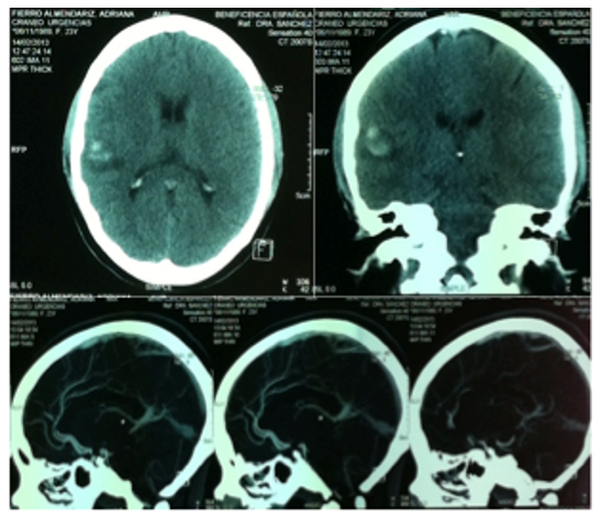
Figure 1 CT angiography with evidence of parenchymal hemorrhage.
No apparent cause of the hypertensive crisis was found, so the diagnosis of de-novo hypertensive crisis and IPH was given. She was admitted to the intensive care unit for stabilization of her blood pressure with diuretics (Lasix), and medical treatment with anticonvulsants (Diphenylhydantoin), corticosteroids, and antibiotics. With medical management, her blood pressure levels improved without further seizures. She was discharged from the intensive care unit four days later, and remained hospitalized for two more days. She was discharged from the hospital in good general condition and without sequelaes. At her follow-up appointment a week later, the patient had returned to her normal activities.
Cerebrovascular event represents the 10% to 15% of all strokes, its presentation in patients under 30 years of age is unusual, representing a challenge for diagnosis and therapeutic management.4,5 In most reports of hemorrhagic stroke in young populations, subarachnoid hemorrhage appears as the predominant form, usually associated with AVM or aneurysms.6 In contrast, IPH appears less frequently in medical literature, especially when it occurs in young patients without medical history of trauma or obvious risk factors.4,5 IPH in young patients is rare, although it is clinically important due to the functional and social sequelaes it can cause.7 It has a marked male predominance, exceeding 70% of cases in the reported series.8
Its clinical presentation varies depending on the age, location and severity of the bleeding.9 The most frequent symptoms associated with IPH in young patients include focal neurological alterations, intense headache of sudden onset, altered state of consciousness, nausea, vomiting and in some cases seizures.
Regarding etiology, the causes of IPH show an age-related distribution: cerebral vascular malformations, such as cavernomas or AVM, are more prevalent in patients under 30 years of age; while arterial hypertension is more common in the subgroup of young and older adults. However, in a considerable percentage of cases up to 32% in some series, no defined cause is found.5 Other contributing factors in this population include alcohol abuse and a high body mass index, both of which are more prevalent in young patients with IPH compared to older patients.10 The patient had a BMI of 26.4 kg/m2 and her alcohol consumption was apparently not significant, making these unlikely causes of her disease. On the other hand, the contraceptives she was taking are associated with an increased risk of ischemic stroke due to the risk of thrombosis; however, this risk is low in young women without additional cardiovascular risk factors.11,12 A meta-analysis revealed that the use of oral contraceptives does not statistically significantly increase the risk of intracerebral hemorrhage (Odds ratio: 0.92). However, a clearer association with subarachnoid hemorrhage has been observed in women with other factors such as smoking, high blood pressure, or migraine.13-15 In this context, the possibility that it was a contributing factor to the IPH in this patient is unlikely.
Cerebral AVM are among the leading causes of cerebral hemorrhage in young patients. Approximately 20% of these malformations are detected during childhood or adolescence through imaging studies requested for other pathologies, especially Magnetic Resonance Imaging (MRI). However, in most cases, their identification occurs after the cerebral hemorrhage and not incidentally.16 These alterations are detected in approximately 2 cases per 1,000 studies, and only 0.2% correspond to clinically relevant malformations such as cavernomas.16,17
De-novo hypertension, on the other hand, is more widely recognized as a relevant entity in the pathogenesis of IPH in young patients, despite the fact that it is frequently underdiagnosed. Reports do not always clearly distinguish between established and newly detected hypertension, but there is consensus that unrecognized hypertension is common in this group of patients. A significant percentage of young patients with IPH had no previous diagnosis of hypertension, which emphasize the need to establish more effective cardiovascular screening strategies, even in apparently healthy patients without evident risk factors.4,5,18
The relation between AVM and arterial hypertension in the genesis of IPH has been the subject of various studies. A recent study by Lasica et al.19 revealed that arterial hypertension is an independent risk factor for AVM rupture, even in young individuals, with an odds ratio of 3.37. This suggests that a sudden increase in blood pressure, even if recent, can precipitate AVM rupture, acting as a destabilizing hemodynamic factor on a vulnerable vascular structure.
In many cases of AVM bleeding in young patients, no clear triggering factors are identified, nor is there the presence of known arterial hypertension, which implies that the architecture of the AVM itself (such as the deep venous drainage pattern, the presence of aneurysms or high blood flow) may be sufficient to cause the hemorrhagic event.20,23 Thus, both the AVM and de-novo hypertension can operate as isolated factors, or act together amplifying the risk of hemorrhage.
For diagnosis, non-contrast head CT is the first-line study in the acute phase of IPH, due to its high sensitivity in confirming the diagnosis, locating the hematoma, quantifying its volume, and detecting complications such as intraventricular hemorrhage or hydrocephalus. MRI is also a sensitivity diagnostic method, but its initial use is limited by its availability and cost. In young patients with spontaneous hemorrhage, especially in atypical locations or in the absence of known hypertension, CT angiography is recommended to rule out underlying structural lesions such as AVM or aneurysms.22,24-26
Treatment of IPH is based on clinical stabilization measures, intracranial pressure control, management of systemic and neurological complications, and in selected cases, neurosurgical intervention depending on the location and extension of the hematoma and the underlying etiology.9 If an AVM such as an aneurysm is found, strict blood pressure control is essential: reducing systolic pressure to <140 mmHg is recommended in patients with elevated levels, using parenteral antihypertensives such as nicardipine or clevidipine, although evidence of functional benefit is limited, and renal function should be monitored.9,21 Rapid reversal of anticoagulation, if present, should be done with vitamin K and prothrombin, as recommended by the American Heart Association/American Stroke Association.9
The importance of this case is to document an episode of IPH in a patient under 30 years of age, in whom apparently de-novo hypertension and the rupture of an unknown AVM, diagnosed through imaging studies, were identified. It also illustrates how the combination of these two entities associated with cerebral hemorrhages that occur in different groups of patients, both of which occurred in this patient, acting synergistically, and substantially increasing the risk of hemorrhage into the brain parenchyma.
IPH in young patients is rare, and has a different etiology than in older adults. However, causes such as high blood pressure can also occur in young patients. In this case, the combination of an AVM and de-novo hypertension could have been the triggering factors for the hemorrhage. Although both conditions can cause IPH separately, their coexistence significantly increases the risk of vascular rupture, as in this case we report. Prompt diagnosis and early treatment are key to improving the prognosis and avoiding serious complications.
None.
No sponsor of any kind was received for the conduct of this study.
The authors declare that they have no conflicts of interest.

©2025 Alvarez, et al. This is an open access article distributed under the terms of the, which permits unrestricted use, distribution, and build upon your work non-commercially.