International Journal of
eISSN: 2574-8084


Research Article Volume 10 Issue 6
Department of maxillo-facial Surgery, School of Medicine, University of Marrakech, Morocco
Correspondence: Zahira Benzenzoum, Department of maxillo-facial Surgery, School of Medicine, University of Marrakech, Morocco
Received: December 15, 2023 | Published: December 29, 2023
Citation: Benzenzoum Z. Interest of the tongue flap in the repair of endo buccal substance loss. Int J Radiol Radiat Ther. 2023;10(6):156-159. DOI: 10.15406/ijrrt.2023.10.00373
Introduction: The tongue flapis a simple, reliable, but little-used technique, presenting an excellent alternative for reconstruction of oral cavity substance loss.
Materials, methods: We carried out a retrospective study over a 3-year period, involving 10 cases of patients presenting with oral substance loss, collated within the maxillo-facial and aesthetic surgery department of CHU MED 6 in Marrakech.
Results: Our therapeutic arsenal includes tongue flaps: distal, proximal and ventral pedicle, with indications for several topographical regions. The procedure was well to lerated by our patients, and our results showed excellent short-termhealing, as well as the flaps proving viable in the long term.
Discussion: the tongueflapis a simple, reliable and easy technique that has been abandoned in favor of more complex and sometimes more deleterious techniques for the patient. Based on ourexperience and reviewing the experience of others, it is clear that the tongueflapis a useful and versatile option for the repair of endooral SDB. With longer follow-up, the key patient out comes will be come more evident.
Conclusion: The relative simplicity of this technique and its potential advantages over more established techniques make it an ideal solution when resources are limited.
Keywords: reconstructive surgery, flap, tongue, endobuccal loss of substance
The repair of endooral substance losses can be difficult due to the different characteristics of the region, the importance of preserving anatomy and function, and the shortage of availabledonors. According to the literature, the tongueflapis a reliable and effective means of reconstructing this loss of substance, thanks to its central location, mobility and hyper-vascularization. This is in contrast to other more complexmeans, which have a higher postoperative morbidity rate, higher overall cost and more severe sequelae.1
The aim of this retrospective study is to review the technique of the tongueflap, to establishits indications, to establish a decision-making algorithm governing our conduct, and finally to propose reliable recommendations.
This is a retrospective study, conducted over a 3-year period from February 2018 to March 2021, on 10 patients with endobuccal substance loss repaired by tongueflap, within the maxillofacialaestheticsurgery and stomatology department of Ibn Tofail Hospital at Mohamed VI UniversityHospital Center in Marrakech, under the aegis of the "SOS FACE MARRAKECH" association.
Our study parameters were epidemiological (Age, Sex, Number of previous surgeries, Age of SDB diagnosis, Age of onset of SDB, Etiology of SDB?) Clinical (Type of SDB, Location, Follow-up time, Referral method) Therapeutic (Type of flap with distal or proximal pedicle, Flap size, Weaning time) and Evolutive (Quality of healing, Improvement in phonation, speech and feeding, Disappearance of nasal regurgitation, and Immediate (infection, haemorrhage, oedema.) and late complications (flap release, necrosis, etc.).
The meanage of our patients was 43.9 years, with extremes ranging from 1 to 89 years, with a clear female predominance (in our series, 6 of the patients were female versus 4 male, i.e. a sex ratio of 1.5). The topography of the lesions is distributed as follows: 4 Palate /Pelvi-labial-anteriormandibular/ Intermaxillary commissure pelvi lingual post/ Intermaxillary commissure internal jugal and floor with extension to the pharynx/ Intermaxillary and pelvi mandibulaire commissure/ Pelvi lingual. (Figure 1)
The main Etiology was squameuse cella carcinome of the oral cavity, which was represented clinically mainly by a bleeding ulceration on contact and pain, and 4 cases of cleftlip and palate, whose size varied in length from (7 to 20 mm), but did not exceed 2cm in width, and which was represented clinically by nasal regurgitation, hypernasality and swallowing disorders.
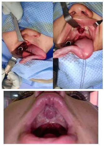
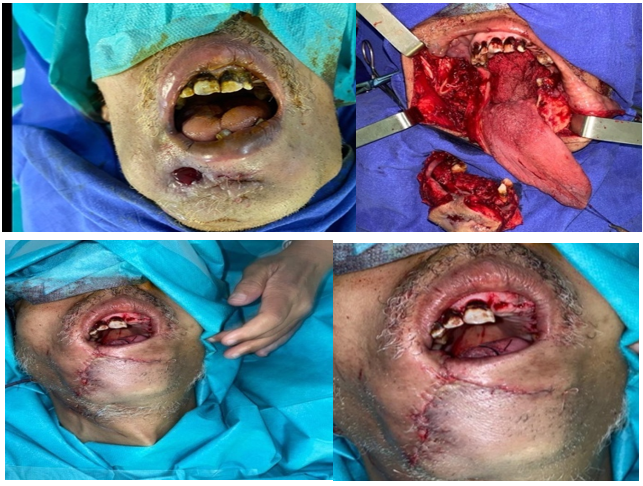
We used a tongue flap:1-Marginolingual with distal pedicle for patients with palatal SDB. 2- Marginolingual with proximal pedicle for patients with maxillary SDB. 3-Ventral for patients with mandibular SDB.
The length of the flap was designed so that 1-2cm of extra tissue covered the posterioredge of the PDS; the wich was dictated by the width of the defect plus 20%.
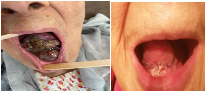
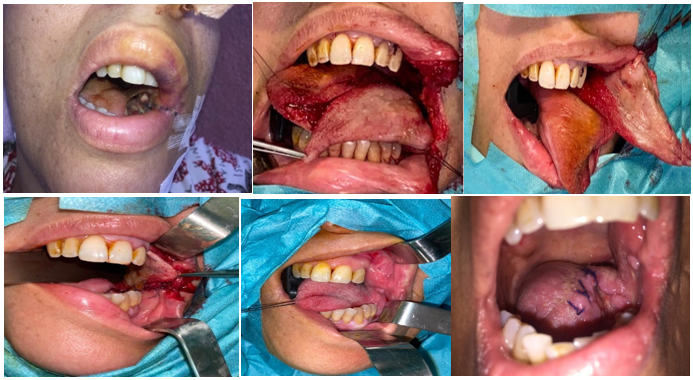
In our study, we used tongueflaps 8.4 to 25 mm wide and 3cm to 4cm long. (Figures 2-10)

Figure 4 12 mm FLP in a patient from our training program: before, during and after reconstruction with a distal margino-lingual flap.

Figure 6 Pelvi labial and mandibular PDS ant post-tumor excision for squamous cell carcinoma: before, after excision and reconstruction with a ventral tongueflap.

Figure 7 Pelvi mandibular PDS in a patient before tumor removal and after reconstruction with a tongueflap with a margino lingual pedicle.

Figure 8 PDS internal jugal intermaxillary commissure and floor before excision, intraoperatively and after reconstruction with a proximal-pedicled margino lingual tongueflap.
The tongue is a musculo-mucosal organ that forms part of the floor of the oral cavity and part of the anterior border of the oropharynx. Made up of 17 or more muscles and innervated by 5 pairs of cranial nerves, where as the face is only innervated by 2, it is quite simply an exceptional and completeorgan, as it performs multiple motor, sensory and sensoryfunctions. A perfect understanding of its anatomy, bio dynamics and growth provides a better understanding of the etiopathogenesis and processes involved in maxillofacial deformities, occlusion, elocution, swallowing and oral disorders.2 With its perfectly symmetrical muscular, nervous and vascular lingual component, thisis of considerable clinical importance in surgical techniques such as glossotomy, flaps and frenectomy, for example.3
In the literature, the age range for substance loss varies from 6 to 60 years,4 with a clear male predominance.5 In ourseries, therewas a predominance of females, with a sex ratio of 0.33.
Because of the tongue'srich blood supply and flexible nature, tongue blades can be taken from the dorsal, lateral or ventral surfaces of the tongue.
Numerous authors have propose différent techniques for closing endobuccal substance losses: composite flaps with bone or cartilage graft,6 remote flaps (free for arm flap,7 free facial flap8) or, more recently, expansion with Foley probes, used by Abram0.9
The advantages of using a tongueflap lie in it ssimplicity and efficacy,10 the reliability of the richly vascularized flap, the few after-effects on the tongue and the short duration of general anesthesia, and the possibility of combining it with other reconstruction techniques (bonegrafts, etc.).
The disadvantages of these techniques are minimal compare with the advantages, which are: Two-stage surgery; disco fort lasting round 2 to 3 seks due to limited mouth opening and reduced lingual mobility; the presence of a nasogastric tube for aroundtwoweeks; and hospitalization lasting around 3 weeks, with the resulting conflict. This last problem can be partially solved by organizing a day hospitalization, or by educating the patient and his or her family so that theycan return home and befollowed up on an out patient basis during the intervening period.
This technique appears to offer an alternative to some of the more complexmethods, which are associated with higher postoperative morbidity, greater over allcost and heaviersequelae.
The distal11 or proximal marginolingualflapis the most widely used, but other types of tongueflap are also possible: the distal bi-pedicled hammer head flap, the dorsal flap12,13 and the ventral flap.
The technique of closing endobuccal substance loss using a tongueflapis simple and reliable, with satisfactory results. In our opinion, itis the technique of choice, an alternative to more complex techniques that are sometimes more harmful to the patient.
None.
Author declare that there is no conflicts of interest.

©2023 Benzenzoum. This is an open access article distributed under the terms of the, which permits unrestricted use, distribution, and build upon your work non-commercially.