International Journal of
eISSN: 2574-8084


Short Communication Volume 5 Issue 6
Department of Oncology, Russia
Correspondence: Shaposhnikov VI, Department of Oncology with a course of radiation Diagnostics and therapy the NIGHT, Kuban Medical Institute, Krasnodar, Russia
Received: October 16, 2018 | Published: November 19, 2018
Citation: Ivanovich SV. Imaging diagnosis of mediastinal tumors and retroperitoneal space. Int J Radiol Radiat Ther. 2018;5(6):326-328. DOI: 10.15406/ijrrt.2018.05.00187
Carried out a comparative analysis of methods of radiodiagnosis Mediastinal tumors and retroperitoneal space for 50 years, starting from the usual x-ray CT and finishing against the backdrop of the introduction of gas into the mediastinum and retroperitoneal space. Stresses that gazokontrastnye overcome the difficulties of methods to detect sprouting of malignant tumors in the surrounding organs and tissues, which reduces the percentage complete false operations that only reinforce local growth and metastasis carcinoma. This allows you to immediately generate the development of algorithm of radiation treatment method. Stresses the added value of pnevmomediastinografii to clarify the nature of the invasive growth of cancer of the esophagus, and pneumoperitoneum-pancreas. These methods are important in the diagnosis and other Neoplasms.
Keywords: mediastinum, retroperitoneal space, tumors, invasion, radiology
Evaluate the diagnostic capabilities of x-ray techniques, which are most often applied in the clinic in the diagnosis of cancer of the esophagus and pancreas.
While working in various oncological hospitals in Kazakhstan, Ukraine and Russia took place to study diagnostic possibilities also go other x-ray method. If Kazakhstan (Kzyl-Orda regional Oncology Center) it was a normal x-ray and grafia, Ukraine (Kyiv n/and rentgenoradiologicheskij and the Cancer Institute, Kharkiv Research Institute of General and emergency surgery) already applied tomografo grafija, rentgeno kinematografija, double and triple contrast abdominal Neoplasm’s after the introduction of gas into the retroperitoneal space and mediastinum. In the Krasnodar region (krajonkodispanser, regional hospital, and emergency hospital) added to computed tomography. I must say that in all of these medical institutions concentrated a large number of patients with cancer of different localizations. This allowed us to make an objective assessment of all the listed beam diagnostic methods. Oppose them to each other, it would be a mistake and with economic and practical point of view. Should rationally use each of them by necessity.1–3 Gazokontrastnye methods are particularly important for the diagnosis of germination of cancer of the esophagus, pancreas, colon in the adjacent organs and tissues, as well as for identifying Mediastinum and retroperitoneal sarcomas space.2–5 Aggressiveness of esophageal cancer esophageal localization features explained in the mediastinum, close contact with zhiznennovazhnymi authorities, resulting in the rapid germination of cancer beyond its own tissues as well as metastasizing.1–7 Early detection saves patient tumors are sprouting not only from vain operations accelerates Cancer Metastasis, but allows timely resort to radiotherapy treatment and thereby prolong his life.4
The number of cancer patients treated in medical institutions referred to above, amounts to thousands, and in one article reflecting all nuances of radiation Diagnostics of all disease diseases is impossible. So for a detailed analysis of the effectiveness of radiation means selected only one disease is a cancer of the esophagus. The same cancer of the pancreas is considered only in the light of modern achievements of radio diagnostics.
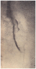
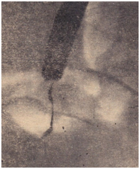
Of the 711 patients referred to a medical institution 18 of age to the age of 93. The youngest of them was a resident of Kazakhstan. Men were almost 2.5 times more than women. All of them were in a State of dramatic mental oppression, up to complete indifference to their fate. And, as a rule, it is easy to agree on any medical manipulations. During the initial screening and diagnosis of esophagus picture was installed at 591(83.1%) patient. But these techniques allowed catching only the brightest causes dysphagia is the nature and kind of narrowing of the authority inherent in one form or another, does not reflect the nature of the tumor growing in the adjacent organs and tissues. Before the advent of computer tomography, it couldn't be produced using gazokontrastnyh methods. The most common routes of gas in the mediastinum (usually carbon dioxide) are methods of Reeves and Kazan. When the first of them, gas in volume 1500-2000ml is injected into the retroperitoneal space using prikopchikovoj puncture. To do this, under local anesthesia and controlled by thumb, halfway between the tailbone and the anus is embedded in the needle behind prjamokishechnuju fiber 5-6 depth cm. On it slowly injected specified gas volume and patient forced to walk. Via 30-40 minutes mezhfascialnym gas spaces of retroperitoneal fiber penetrates the back mediastinum, shrouding the esophagus. The second gas (500-1000ml) injected into the anterior mediastinum puncture via jugular clipping. He then spreads throughout the sredosteniju. Beam study of esophagus, pancreas, and other organs and tissues are carried out via 30-40 minutes after this manipulation. The lower third of the esophagus cancer further resort to the introduction of gas (up to 1000ml) into the free abdomen, IE the laying of pneumoperitoneum, followed by a double or a triple contrasting his clearance. When double-patient took only barium suspension, while triple-he still fanned by those inside through a thin probe. These methods have dramatically intensified the contrast abdominal esophagus and Cardiac of the stomach, and in the application of computer tomography allow to clearly define and tumor invasion into adjacent tissues. These techniques were performed at 260 patients. . Of these, 184(70.7%) It was determined that the tumor operabelna and radical surgery was performed (Figure 1 & Figure 2).8–11

Figure 1 is represented by tomogramma the middle third of esophagus cancer struck. Study performed after the introduction of gas into the mediastinum. It is clearly visible that the tumor could grow into the surrounding tissue.

Figure 2 Radiographs of the esophagus. Cancer of the lower third of the body. The study made after the imposition of pneumoperitoneum. It is clearly visible that the tumor will not germinate in the diaphragm and liver. Correct diagnosis was confirmed during surgery.
Currently, the leading role in the diagnosis of pancreatic cancer is owned by computed tomography. But, as previously speculated on the existence of the disease is based on observation of changes the contours of the stomach and duodenum when performing radiography of these bodies, i.e. their deformation by compression by the tumor. For this reason, tissue contrast enhancement-by introducing gas zabrjushinno and vnutribruchinno, is of great diagnostic value. However, without a Visual inspection of this gland and biopsy to resort to radical surgery is dangerous and with legal and moral side. Indeed, the increase in size of the body may be and in chronic psevdotumoroznom acute pancreatitis in which iron is not removed. Earlier, to avoid severe errors, sick even offered trial laparotomy. Currently replaced by laparoscopy. Computed tomography (CT), after the introduction of the gas in the specified space, was performed at 37 patients with suspected pancreatic cancer. That it has good information, verified by the example of operative treatment 13(35.1%) these patients-they have during the operation confirmed the tumor operabelnost, which was supposed to be following the execution of this type of radiation survey. The remaining 24(64.9%) patients experienced clear signs of cancer neoperabelnosti of that body.
The results of the 260 patients, which was introduced by gas or in the mediastinum, or into the abdominal cavity, with the subsequent, or dual (introduction only barium masses in the lumen of the esophagus) or triple (introduction of barium and with inflation lumen body inside using probe) have been obtained the following results. At 27(13.8%) from 260 to exclude the germination of esophageal cancer in adjacent organs and tissues, and then perform the radical surgery. At 16(6.1%) patients with complaints bring on dysphagia, install benign nature of this symptom, including: 8-deviation of the esophagus, 3-like constriction, 4-diverticula. Thus, modern beam methods of diagnosis are an effective tool in identifying the causes of human frailty and to develop proper algorithm for his treatment.
Currently, determining operability cancerous tumors of the esophagus and pancreas is the most informative computer tomography, but it should be conducted after the introduction of gas into the mediastinum and zabrjushinnuju fiber. This increases the contrast of fabrics and allows you to define their mobility, which is an essential factor in the success of the surgery.
None.
The author declares that there is no conflict of interest.

©2018 Ivanovich. This is an open access article distributed under the terms of the, which permits unrestricted use, distribution, and build upon your work non-commercially.