International Journal of
eISSN: 2574-8084


Case Report Volume 13 Issue 1

1Wayne state University School of Medicine, USA
2Atlanta Health and Welfare Centre for Women, India
Correspondence: Sunil Palchaudhuri Ph.D, D.Sc (Fulbright Scholar), Wayne state University School of Medicine, USA
Received: November 06, 2025 | Published: January 20, 2026
Citation: Palchaudhuri S. E. coli C in early log phase adsorbs the phage phiX174 but the male E. coli K-12 sends bio-signal to attract the male-specific phage M13. Int J Radiol Radiat Ther. 2026;13(1):4-8. DOI: 10.15406/ijrrt.2026.13.00445
Both bacteriophage phiX174 and male- specific bacteriophage M13 are the non- living but their host bacteria E. coli C or the E. coli K-12 are the living. All the viruses do not contain single stranded DNA genome but many viruses have double stranded DNA genome and RNA genome but supercoiled. The non-living does never mean dead. phiX174 and the male specific phage M13 with their single stranded circular DNA genome need to enter the live E. coli C and the live E. coli K 12(male) for their genetic continuity. The E coli C adsorbs the phage phiX174 with spikes if biologically ready to act as surrogate mother! The phiX174 genome after its arrival in the bacterial outer membrane of E. coli C the adhesion zone is formed and the phage genome multiplies for genetic continuity. The E. coli K-12 male uses its sex pili to attract the phage M13 using bio-signaling. Subsequently F- pili retracts into the E. coli K-12 cytoplasm with the phage M13 attached to such F pili. In 1946 Lederberg and his mentor Dr Tatum first observed the maleness in E. coli K-12. In fact, they were looking for such a maleness when the E. coli K-12 population were exposed to X-irradiation and wanted to confirm the donor ability to gene transfer by their intimate contact (conjugation). However, genesis of such F factor was never a serendipity. Knowledge of molecular biology confirmed that these two E. coli strains have the identical DNA sequence but the E. coli K-12 is little longer than the E. coli C chromosome for the presence of sex factor (F). Origin of replication is at the 84.5 min in the entire E. coli chromosome (100 minute long). The E. coli K-12 has a sex factor F (100Kb) as an integral part of the chromosome and therefore E. coli K-12 is little longer than the E. coli C chromosome. Morphologically these two E. coli strains K-12 and C differ, cylindrical and spherical (Fig 1). The cylindrical E. coli K-12 is 2.5um long and spherical E. coli C of 0.2um diameter. F factor caries three mobile DNA elements IS2, IS3 and Tn1000. The copy number is controlled by the transposon Tn1000 (5.7Kb). The Tn1000 is indispensable for maintaining its single chromosome (E. coli C or E. coli K-12). E. coli sex factor F has three replicons RepF1A and RepF1B (incomplete?) and the F1C replicon but F1C remains non- functional by the physical insertion of Tn1000. The E. coli K-12 chromosome has four mobile DNA elements IS1, IS2, IS3 and Tn1000 but the single origin of replication.
Keywords: Hfr E.coli K-12 male with its sex factor F integrated into the host E.coli K-12 (chromosome), mobile DNA element Tn1000 controls the replicon, phage phiX174 with single stranded circular DNA genome, male specific phage M13 also with single stranded circular DNA genome. All the members of E. coli C even in the early growth phase but all the individuals are not ready to act as surrogate mother
All the materials and methods have been clearly described in my articles already published in the two internationally reputed Journals Biochimica Biophysica Acta and Journal of Molecular Biology.1,2
In my PhD thesis I tried to differentiate between E. coli C and the E. coli C infected with phiX174 by using different doses of X-irradiation, at a particular dose the DNA replication of E. coli C stopped but the multiplication of phage phiX174 genome continued using the same host E.coli C because the DNA length of E. coli C is much larger than the phiX174 genome.3 Evidently, the phiX174 genome used the same E.coli C to multiply and form mature phage particles and released in the growth medium. Many years later I noticed the plaques formed in E. coli C are turbid if incubated for longer period few large bacterial colonies appear even in the lysates. Define cross-feeding: nutrients released by the lysis of the sensitive E. coli C may feed the few who are still alive and outgrow in the lysates. At the same time E. coli K-12 lysogen shows confluent lysis but without any turbid plaques.
At a particular dose of X-irradiation the E. coli C chromosome (double stranded DNA bio-macromolecule) stopped its replication but the multiplication of bacteriophage phiX174 continued. I defined the difference between E. coli C replicon and phage phiX174 replicon.
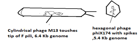
Figure 1 Phage M13 (cylindrical, 6.4 Kb) in E. coli K-12 Hfr male and the male specific phage phiX174 (hexagonal, 5.4 Kb) in E. coli C female.
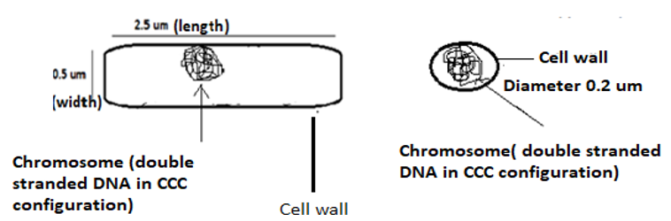
Figure 2 E. coli K-12(cylindrical shape, 2.5 um long), E. coli C (spherical, 0.2um diameter).
Dr. Arthur Kornberg shared Nobel Prize with Dr. Severo Ochoa in 1960 for his discovery of DNA replication with the replicative form (RF) of bacteriophage phiX174 but it was not clear how the phage phiX174 genome (CCC DNA) becomes its RF form (double stranded DNA.4 He used E. coli but without mentioning the difference between E. coli C and E. coli K-12!4 Bacteriophage phiX174 has a single stranded DNA genome but needs to be converted into double RF DNA after its entry into host bacterium E. coli C but cannot enter the E. coli K-12. In the recent years I have recognized the bio-communication (or bio-signals) between the living E. coli C and the non-living phage phiX174 (virus). What is more there are several spikes on the surface of the phage and used for the safe delivery of the phiX174 single stranded circular DNA genome into the site of E. coli where membranes and cell wall are fused.5-7 This fusion site is known as adhesion zone. However, the phage spike is activated after its contact with the live E. coli C (log phase) but never with E. coli K-12 (Palchaudhuri S unpublished data). The adhesion zone is not any permanent structure of E.coli C. The phiX174 uses spikes not only for a bio-signal but also for the safe entry of genome into the outer membrane. In 1960 Arthur Kornberg used E. coli C and phage phiX174 to understand DNA replication, although without the knowledge of different DNA polymerases.6-8 In my thesis work I knew that the polymerase A1 is the only DNA polymerase responsible for DNA replication but in the following years Dr Kornberg recognized the presence of few others DNA polymerases polA2-polA5).9
The bacteriophage phiX174 (virus) is a single covalently closed circular chromosome (CCC) of length 5.4 Kb that prevails in its hexagonal protein coat (Figure 3).9 This phage has several spikes apparently to arrive in the outer membrane for the delivery of phiX174 genome into the E. coli C adhesion zone (Palchaudhuri S). After interaction with E. coli C, a temporary structure is created where E. coli C cell wall and its membranes (outer and inner) are all fused together (fusion zone). I infected E. coli C with phiX174 and I observed the lysis of E. coli C. In the solid agar medium, I tried to understand what happened to turbid culture of E. coli C. If the lysate is spread in nutrient agar medium (solid) incubated for 48 hours few large bacterial colonies grew in the plaques but the nutrients used for the cross-feeding of E. coli C still alive.10 What is more I verified there were not any other bacterial and fungal contaminants.
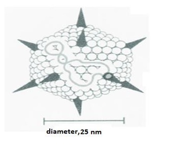
Figure 3 Phage phiX174 (5.4 Kb genome) with spikes gets stuck in the outer membrane of E.coli C but its genome forms double stranded RF to replicate without the RNA primer.
Exposure to X-irradiation helps me to understand why the E. coli C chromosome (4737 Kb) is affected at a dose when the phage genome (5.4 Kb) remains functional being in the same cytoplasm.1 Interestingly E. coli C unlike E. coli K- 12 is lacking sex factor (F factor) but E. coli C can use xylitol -five carbon low calorie sugar-alcohol, neither sugar nor alcohol. Gram-positive Streptococcus pneumoniae (Figure 4) and Gram-negative E. coli C both have xylitol operon but such a homology helps them grow in three phases pre competent, competent, and post competent.6 Morphologically they may differ but their growth pattern does not. Interestingly both E. coli C and Streptococcus pneumoniae dimensionally appears to be similar. E. coli C is not a pathogen but Streptococcus pneumoniae is a serious human pathogen. There is a cleavage in Streptococcus pneumoniae which appears to be diplococcic but it is a single bacterium never the two. E. coli C does not show any cleavage but it differs from E. coli K-12 male (Hfr,F+ and F primes). Figure 4 demonstrate the morphological differences among the three: two Gram-negative E. coli C (spherical), E. coli K-12(cylindrical) and Gram-positive Streptococcus pneumoniae diplococcic but never the two. The DNA sequence of E.coli K-12 and E.coli C chromosomes do not differ very much but the presence of sex factor as an integral part of E.coli K-12 chromosome. Phage M13 (6.4Kb) and phiX174 (5.4Kb) are able to distinguish between E.coli K-12 male and C. I think E. coli C may act as surrogate mother. Phage M13 attaches to the tip of type-IV pili of E. coli K-12 males.
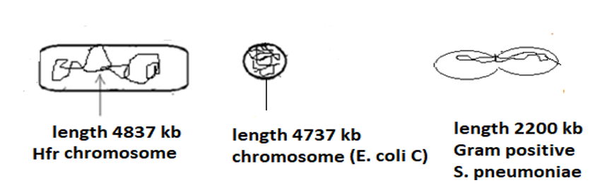
Figure 4 Morphological comparison among E. coli C, E. coli K-12 and S. pneumoniae.
Tripti Bhattacharya works as my academic assistant.
None.

©2026 Palchaudhuri. This is an open access article distributed under the terms of the, which permits unrestricted use, distribution, and build upon your work non-commercially.