International Journal of
eISSN: 2574-8084


Case Report Volume 12 Issue 3
Department of General Surgery, Hospital de Braga, Braga, Portugal
Correspondence: Ana Costa Sousa, General Surgery Resident, Hospital de Braga, Portugal
Received: June 15, 2025 | Published: August 4, 2025
Citation: Sousa AC, Rodrigues J, Antunes A, et al. De Garengeot hernia: a rare clinical entity with appendicitis. Int J Radiol Radiat Ther. 2025;12(3):62-63. DOI: 10.15406/ijrrt.2025.12.00423
De Garengeot hernia is an extremely rare condition characterized by the presence of the vermiform appendix within the femoral hernia sac, accounting for 0.5%–5% of femoral hernias and with appendicitis occurring in only 0.08%–0.13% of cases. It poses a diagnostic challenge due to its nonspecific symptoms, often mimicking incarcerated femoral hernias, and is usually identified intraoperatively. Here, we report the case of an 80-year-old woman presenting with a painful, irreducible right groin mass. The physical examination revealed the presence of an incarcerated femoral hernia, and the ultrasound findings were consistent. During surgery, it was identified that the incarcerated content was the ileocecal appendix. This case underscores the importance of imaging, such as CT, in improving preoperative diagnosis and highlights the need for prompt surgical intervention to prevent complications. Increased awareness of this rare condition is essential for advancing diagnostic and therapeutic strategies.
Keywords: De Garengeot hernia, femoral hernia, appendicitis, surgical management, incarcerated hernia
De Garengeot hernia is an exceedingly rare condition, defined by the presence of the vermiform appendix within the femoral hernia sac. This entity accounts for 0.5 to 5% of all femoral hernias, with associated appendicitis being particularly uncommon, representing only 0.08 to 0.13% of De Garengeot hernia cases.1,2 The condition was first described in 1731 by French surgeon René Jacques Croissant De Garengeot, and since then, its infrequency has presented diagnostic challenges, with most cases being identified intraoperatively.3
The clinicopathological mechanisms behind De Garengeot hernia remain incompletely understood, though certain factors, including connective tissue disorders, advanced age, multiparity, pelvic appendix, and a mobile cecum, are thought to contribute to its development.4,5 Due to the rarity of this presentation, no formal guidelines exist on the optimal surgical approach, leaving management largely at the discretion of the operating surgeon.6
In this report, we present a rare case of De Garengeot hernia with concurrent appendicitis, aiming to contribute to the limited literature on this uncommon clinical entity and provide insights into its diagnosis and management.
An 80-year-old woman presented to the emergency department with a painful swelling in the right inguinal region, which had persisted for approximately one week. She denied any bowel movement irregularities, nausea, or vomiting. The patient reported feeling chills, though no fever had been documented. Her past medical history included vulvar carcinoma and a left oophorectomy.
On physical examination, a bulging mass was noted in the right inguinal area, located below to the inguinal ligament. The mass measured approximately 8 cm in diameter, was firm and non-reducible on palpation, with no Inflammatory skin changes. Laboratory tests showed a leukocytosis and elevated C-reactive protein (CRP).
An ultrasound of the region identified a right-sided, non-reducible inguinal hernia but provided no further details. Based on the clinical findings, The patient underwent emergency surgery with a preliminary diagnosis of incarcerated femoral hernia.
During surgery, an inguinotomy was performed, confirming the presence of an incarcerated femoral hernia. Dissection and opening of the hernia sac revealed an edematous and inflamed vermiform appendix (Figure 1). Following identification of the cecum, an appendectomy was performed. The femoral hernia was then repaired with a plug-type hernioplasty (Figure 2). The procedure was completed in 40 minutes without any complications.
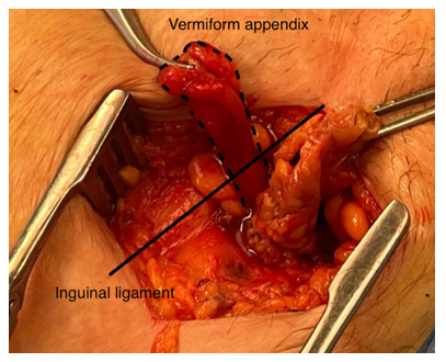
Figure 1 Intraoperative image showing the incarcerated vermiform appendix within the femoral hernia sac.
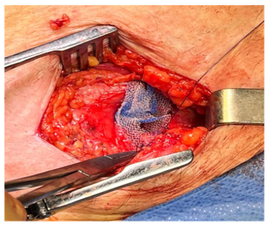
Figure 2 Placement of a plug-type mesh prosthesis following appendectomy and hernia sac excision.
The patient’s postoperative course was favorable, with no complications, and she was discharged on the first postoperative day. At her three-week follow-up appointment, she was in good general health and showed no signs of complications.
Histopathological examination confirmed the presence of a gangrenous appendix consistent with acute appendicitis, with no evidence of malignancy.
Femoral hernias are a rare type of groin hernia, accounting for less than 5% of all hernias, with a female-to-male ratio of 3:1 and increasing incidence in elderly, postmenopausal women.7,8 De Garengeot hernias, characterized by the presence of the vermiform appendix in the femoral hernia sac, are even rarer, with a reported incidence of 0.5%–5% of femoral hernias and inflamed appendices present in only 0.08%–0.13% of cases.2,4 Despite its first description in 1731 by Rene Jacques Croissant De Garengeot, fewer than 100 cases have been documented since 1960, and it remains a diagnostic challenge.3,5
De Garengeot hernias often present as painful, irreducible groin masses, mimicking incarcerated or strangulated femoral hernias. Imaging, particularly CT scans, has proven valuable in identifying the appendix within the hernia sac, with reported sensitivities and specificities of 98%.9 However, the diagnosis is often confirmed intraoperatively, as was the case here. In our patient, only an ultrasound was performed preoperatively, which, although helpful, was insufficient to identify the appendix in the hernia sac. This limitation further compounded the diagnostic challenge inherent to this rare condition. Given that the diagnosis of femoral hernia is often clinical and typically leads to surgical exploration regardless of sac content, intraoperative identification of De Garengeot hernia remains common. Moreover, ultrasound and plain X-rays generally play a limited role, serving primarily to exclude other differential diagnoses such as lymphadenopathy or lipomas.10
The etiology of De Garengeot hernias is debated. Proposed mechanisms include anatomical variations such as a low-lying cecum or pelvic appendix, leading to increased susceptibility to herniation.11 Contributing factors such as advanced age, multiparity, chronic cough, and smoking, which raise intra-abdominal pressure, have also been implicated.4 Although not well established in the literature, prior pelvic surgery—such as in our patient, who had undergone a left-sided pelvic procedure—may potentially alter local anatomy or intra-abdominal dynamics, contributing indirectly to hernia formation. A systematic review reported that up to 33% of male patients and 4.4% of female patients with De Garengeot hernias had a history of prior right-sided inguinal hernia repair, suggesting a possible association between previous surgical interventions and the development of this rare entity.12 Strangulation of the appendix within the femoral canal may lead to ischemia, necrosis, and inflammation, as observed in this case.
Treatment requires prompt surgical intervention. Appendectomy is performed to address the inflamed or necrotic appendix, followed by hernia repair.13 While mesh repair is typically avoided in contaminated fields, primary tissue repair is often preferred to minimize infection risks. In selected cases, laparoscopic techniques can be used, allowing for appendectomy and hernia repair with minimal invasiveness, though this approach remains limited by diagnostic ambiguity and case rarity.14
De Garengeothernias area rareand challengingcondition, often diagnosed intraoperativelydue to their nonspecific presentation and the rarity of appendicitis within a femoral hernia. Prompt surgical intervention, including appendectomy and hernia repair, is essential to prevent complications such as necrosis, perforation, and infection. Imaging techniques like CT scans can aid in preoperative diagnosis, improving surgical planning. Despite advancements in diagnostics and management, there is no standardized treatment protocol due to the limited number of reported cases. Increased awareness and reporting of De Garengeot hernias are crucial for advancing understanding and optimizing outcomes.
None.
The authors declare no conflict of interest.
Informed consent was obtained from the patient.

©2025 Sousa, et al. This is an open access article distributed under the terms of the, which permits unrestricted use, distribution, and build upon your work non-commercially.