International Journal of
eISSN: 2574-8084


Case Report Volume 9 Issue 4
Department of Clinical Radiology, University of MunichGrosshadern Campus, Munich, Germany
Correspondence: Prof. Dr. Dirk-André Clevert, Department of Clinical Radiology, University of Munich-Grosshadern Campus Munich, Germany, Tel +49 89 4400-76640, Fax +4989 4400- 78832
Received: October 14, 2022 | Published: October 28, 2022
Citation: Grossklags G, Clevert DA. Contrast-enhanced ultrasound (CEUS) in the diagnosis and therapy monitoring of a renal artery pseudoaneurysm (RAP) after Biopsy. A case report. Int J Radiol Radiat Ther. 2022;9(4):134-136 DOI: 10.15406/ijrrt.2022.09.00336
We present an uncommon renal artery pseudoaneurysm (RAP) after renal biopsy. The diagnosis was made with contrast-enhanced ultrasound (CEUS), followed by coil embolization therapy, and the subsequent therapy monitoring with CEUS highlighting its applicability.
Keywords: coil embolization, contrast-enhanced ultrasound, pseudorenal aneurysm, renal biopsy
Renal biopsies have been performed for many decades and are considered as a procedure with a low-risk factor.1 Although quite infrequent, one of the possible complications is a renal pseudoaneurysm (RAP). The diagnosis of RAP is a clinical challenge, considering the broad range of symptoms and the risk of rupture, which is a life-threating condition demanding prompt diagnosis and therapy.
A 69-year-old Caucasian male patient underwent renal biopsy due to proteinuria (>3g/d) and elevated creatinine levels (2.5 mg/dl).
The patient had a past medical history of Endovascular Aneurysm Repair (EVAR) due to Infrarenal Abdominal Aortic Aneurysm (AAA) 2 months ago, 4 kg weight gain in the last 2 weeks before his admission and 2+ symmetric pedal edema.
On examination he presented weight: 72 kg, height: 1.76 mts, body-mass index 23.2 kg/m2, blood pressure 127/74 mmHg right arm, 122/63 mmHg left arm, heart rate 80 bpm.
Urinalysis: 3+ proteinuria, 1+ hematuria, pH 6.5, urine specific gravity: 1.020 g/ml, 24-hour urine volume: 2600 ml. He had never smoked, and denied use of drugs and alcohol abuse.
An ultrasound-guided percutaneous renal biopsy was indicated, in order to establish the underlying cause of the worsening renal function.
The biopsy of his left kidney was successfully taken and 45 min later, the patient referred to sharp pain when breathing in.
A baseline sonographic follow-up was performed, and depicted a hypo-anechoic lesion adjacent to the left kidney. (Figure 1)
The Color Doppler and Micro-flow Imaging examinations depicted a ying-yang swirling motion blood flow within the lesion. Because an active bleeding could not be excluded by color Doppler, the examination was complemented with CEUS. (Figure 2)
CEUS depicted contrast-filling of a pseudoaneurysm in the lower pole of the left kidney measuring 22 x 13 mm, additional CEUS detected the feeding vessel of the pseudoaneurysm. A pararenal hematoma was also demarcated. (Figure 3)

Figure 3 Side by side mode with contrast-enhanced ultrasound (a) and B-mode (b); Demonstration of RAP (arrow) and the feeding vessel.
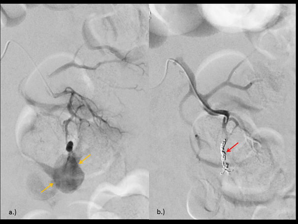
The patient underwent endovascular selective embolization with coil. The digital subtraction angiography (DSA) confirmed the presence of an arterial branch supplying the pseudoaneurysm in the lower pole of the left kidney, followed by coil placement. (Figure 4)

Figure 4 (a) Digital subtraction angiography (DSA) showing the presence of an arterial branch supplying the RAP (yellow arrow) in the lower pole of the left kidney. (b) DSA after coiling procedure (red arrow).
The next day, a control with CEUS was performed, demonstrating the total absence of flow within the aneurysm and normal perfusion of the rest of renal parenchyma. The B-mode image shows a hyperechogenic focal area with acoustical shadow caused by deposited coil. (Figure 5 & 6)
Renal biopsies have been performed for many decades and are considered as a procedure with a low-risk factor.1 Although quite infrequent, one of the possible complications is a renal pseudoaneurysm (RAP). The diagnosis of RAP is a clinical challenge, considering the broad range of symptoms and the risk of rupture, which is a life-threating condition demanding prompt diagnosis and therapy.
The diagnosis of RAPs could be effectively depicted on color Doppler, showing the distinctive swirling motion blood flow with yin-yang sign.2 However, the advantages of CEUS are the diagnosis of extravasation in regard of hematoma and the number of the feeding vessels, together with real-time diagnosis of active bleeding.3
The use of techniques with low-mechanical-index real-time contrast-enhanced ultrasound (CEUS) performed with SonoVue®, a contrast agent of second generation, enable a better and accurate depiction of macrovasculature and microvasculature. SonoVue® is strictly intravascular and is eliminated through breathing, which makes its use feasible in patients with deterioration of kidney function. Postinterventional CEUS could confirm the complete excluding of the aneurysm and could detect renal perfusion defects after intervention.
In our case, CEUS also served as a therapy monitoring alternative, avoiding unnecessary exposure to ionizing radiation and because of the kidney disease of the patient. CEUS responds to a clinical demand in cases where standard imaging modalities do not apply.4 Additionally, CEUS can identify hematuria, helping in the diagnosis and follow-up of a RAP.5
Prior blood tests are not needed, and there is no evidence of adverse effects on renal nor thyroid function. However, there are limitations associated with CEUS. As it is operator-dependent, it requires sufficient knowledge and training in CEUS and the proper ultrasound system soft- and hardware.q6
CEUS is a highly sensitive tool for the real-time diagnosis, characterization, and monitoring of RAPs and coil embolization. Follow-up post interventional confirmed success of treatment and could detect complications like renal infarctions after intervention.
None.
None.

©2022 Grossklags, et al. This is an open access article distributed under the terms of the, which permits unrestricted use, distribution, and build upon your work non-commercially.