International Journal of
eISSN: 2574-8084


Case Report Volume 2 Issue 2
1Department of Radiology, Qi Lu Hospital of Shandong University, China
2Department of Radiology, University of Tokyo, Japan
Correspondence: Shigeru Kiryu, Department of Radiology, Institute of Medical Science, University of Tokyo, 4-6-1 Shirokanedai, Minato-ku, Tokyo 108-8639, Japan, Tel 81-3-3443-8111, Fax 81-3-5449-5746
Received: November 08, 2016 | Published: February 6, 2017
Citation: Wang F, Kiryu S, Akai H, et al. Assessment of the hepatic vasculature in omphalopagus conjoined twins using contrast-enhanced dynamic computed tomography. Int J Radiol Radiat Ther. 2017;2(2):29-31. DOI: 10.15406/ijrrt.2017.02.00017
Eight-hour-old female symmetrical conjoined twins were transferred to our hospital to assess for surgical separation. Imaging modalities were used to assess the anatomy in detail. Contrast agent was administrated to one baby, and dynamic contrast-enhanced computed tomography (CT) was performed. The livers of the two twins were fused, and there was portal venous communication in the early phase. Crossover circulation was observed in the delayed phase. Most of the other organs were found to be independent according to CT, so separation was considered feasible. The surgical separation was carried out successfully.
Keywords: conjoined twins, ct, dynamic study, contrast agent, imaging, circulation, anatomy
US, ultrasound; CT, computed tomography, MPVR, multi-projection volume reconstruction, MRI, magnetic resonance imaging
Conjoined twins are very rare, with an incidence of approximately 1per 250,000 live births, and there is a 3:1 female predominance.1‒3 The classification of conjoined twins is based on the most prominent site of union: the thoracopagus (thorax), omphalopagus (abdomen), pygopagus (sacrum), ischiopagus (pelvis), craniopagus (skull), cephalopagus (face), and rachipagus (back).1,4,5 Conjoined twins often exhibit multiple anomalies and organ sharing. Detailed preoperative imaging facilitates accurate study of the anatomy. We describe a case of omphalopagus conjoined twins evaluated by imaging modalities to identify abnormalities and plan the separation.
Eight-hour-old female symmetrical conjoined twins were transferred to the hospital for evaluation and consideration for surgical separation. The conjoined twins were delivered at term by cesarean section without a prenatal diagnosis. Their combined weight at admission was 2.3kg. The physical examination showed healthy reaction activity. The twins were joined in a face-to-face fashion from the xiphoid process to the umbilicus at the midline. To assess the anatomy in detail, the twins were evaluated using conventional x-rays, ultrasound (US), barium studies, and computed tomography (CT).
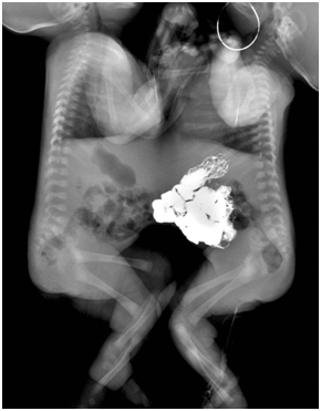
The twins were joined from the lower chest to the umbilicus area. There was no bony connection on conventional x-rays. The hearts and lungs were visible above the conjoined diaphragm. The left- and right-side twins were called Twin A and Twin B, respectively. Barium sulfate was given to Twin A via a nasogastric tube. Serial images showed no evidence of digestive tract communication, and only loops of the upper small bowel of Twin A were seen to project into the abdominal cavity of Twin B through an abdominal wall defect (Figure 1). US showed that the livers of the twins were fused, and the gallbladder and portal vein of Twin B were clearly seen, while those of Twin A were invisible due to intestinal gas interference (not shown). Based on the echo findings, a CT study was designed.

Figure 1 For barium studies, the baby on the right side was given barium sulfate. Barium sulfate was observed only in the intestines of one baby, and no evidence of digestive tract communication was seen.
CT was performed during natural sleep without sedation. A 256-slice MDCT scanner (Brilliance iCT, Philips Healthcare, Cleveland, OH, USA) was used at a 0.8mm collimation and 3mm slice thickness. Dynamic contrast-enhanced CT of the liver was performed to assess the hemodynamics of the fused liver. Iodinated contrast agent (Ultravist 370, Bayer Schering, Berlin, Germany) was administered into the cephalic vein of Twin A at a dose of 2mL/kg combined weight. Early- and delayed-phase images were acquired. Contrast agent was administrated to Twin A only. The detailed anatomy was assessed using axial, multiplanar reconstruction, and multi-projection volume reconstruction (MPVR) images on a workstation.
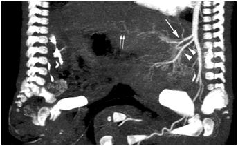
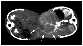
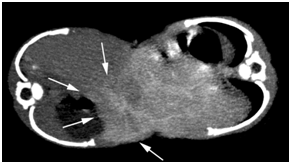
In the early phase, the anatomy of the arterial vasculature was delineated clearly. Due to rapid blood circulation, the portal vein was also demonstrated. In Twin A, the celiac trunk and superior mesenteric artery branched off the abdominal aorta in the proper order (Figure 2). No blood flow from Twin A’s superior mesenteric artery was observed in Twin B, ruling out small intestinal conjunction. The proper anatomy of the branches of the celiac artery and portal vein in Twin A was confirmed. The fused lobe of the liver in Twin B showed an enhanced portal vein that communicated with that of Twin A (Figure 3). In Twin B, the fused lobe was enhanced secondary to crossover circulation in the delayed phase (Figure 4). The fusion area was seen as an irregular strip. No definite enhancement in the other area of liver in Twin B was seen. The entire liver of Twin A was enhanced.

Figure 2 Sagittal MPVR image of early-phase CT shows the celiac trunk (arrow) and superior mesenteric artery (arrowhead). Portal venous communication with the other baby was seen (double arrows).

Figure 3 Axial MPVR image of early-phase CT shows portal venous communication with the other baby (arrows).

Figure 4 Axial MPVR image of delayed-phase CT shows that the fused lobe of the liver in Twin B was enhanced secondary to crossover circulation in the delayed phase (arrows).
Delayed images showed four contrast-enhanced kidneys. Twin A’s kidneys were enhanced normally, and contrast medium was seen in Twin B’s renal pelvises. The two ureters of each twin drained into their respective bladders. Each twin had a pancreas and spleen in the normal position. Each twin had an independent stomach, duodenum, small bowel, colon, and rectum. Therefore, most organs were independent, except the liver, and separation was considered feasible.
After the imaging evaluation, the surgical separation was performed by a team of pediatric surgeons. The fused lower sternum was divided, and the peritoneum was opened. Livers fused by normal parenchymal tissue were found, and the fused area of the livers was separated using an electrotome, with the fused surface measuring approximately 8×5cm. The fusion area formed an irregular strip, which correlated well with the CT findings. Connecting vessels in the cut surfaces of the livers were noted, and several major vessels were ligated meticulously to minimize blood loss. The twins were separated completely, and the abdominal wall was closed in layers. The total operating time was 1hour and 45minutes. During the operation, blood loss of approximately 20mL occurred. The postoperative course was smooth, and the patients were discharged 14days postoperatively. The twins are thriving.
Conjoined twins result from failed separation of the embryonic plate during the third week of gestation.6,7 Omphalopagus conjoined twins comprise 35% of all conjoined twins and are usually fused ventrally in the umbilical area, commonly involving the lower thorax but never involving the heart.1,4 Liver fusion is found in approximately 80% of omphalopagus conjoined twins, with a common biliary tree in 20% of cases.8 The stomachs and proximal small intestines are often separated, and the distal ileum and colon are shared in approximately one-third of cases, with separate rectums.1,8,9
A careful investigation of the anatomy and blood supply in a shared liver is crucial before considering surgical separation. In our case, portal venous communication was found on dynamic CT. The preoperative information on hepatic vascular communication is important for surgical separation, because the liver is a hypervascular organ. Thanks to its improved resolution and multiplanar reconstruction ability, CT is a sensitive method used to define the liver anatomy and vasculature and reliably assess the visceral joining and magnitude of the shunt. However, because of the radiation exposure, the use of CT should be considered carefully. Techniques to reduce the radiation, such as iterative reconstruction, should be applied if possible. Magnetic resonance imaging (MRI) is an alternative modality for assessing the vasculature in conjoined twins; however, it may be difficult to obtain detailed information in this case, due to the limited numbers of protons in small bodies.
Several imaging modalities can be used to evaluate the anatomy of conjoined twins. Conventional x-rays provide a general assessment of the conjoined area. Abdominal US is usually used to assess the liver and document the presence and number of other abdominal organs. However, midline abdominal conjunction may make US assessment unreliable. Therefore, other tomographic imaging methods play a role in the evaluation of conjoined twins.
In conclusion, we present a case of omphalopagus conjoined twins and show the usefulness of dynamic CT for the assessment of hepatic vascular anomalies. Detailed hepatic vascular information was obtained successfully using CT.
None.
Author declares that there is no conflict of interest.

©2017 Wang, et al. This is an open access article distributed under the terms of the, which permits unrestricted use, distribution, and build upon your work non-commercially.