International Journal of
eISSN: 2574-8084


Case Report Volume 13 Issue 1
Department of General Surgery, Hospital de Braga, Braga, Portugal
Correspondence: Ana Costa Sousa, Department of General Surgery, Hospital de Braga, Braga, Portugal
Received: December 21, 2025 | Published: January 5, 2026
Citation: Sousa AC, Rodrigues J, Pereira A, et al. Adult ileo-ileal intussusception: a rare cause of small bowel obstruction. Int J Radiol Radiat Ther. 2026;13(1):1-26. DOI: 10.15406/ijrrt.2026.13.00444
Intussusception is a rare cause of abdominal pathology in adults and is most often associated with an underlying pathological lead point, in contrast to pediatric cases. In the adult population, the small bowel is the most frequently involved segment, and clinical presentation is typically nonspecific and subacute, which may delay diagnosis. We report the case of a 42-year-old male patient who presented to the emergency department with a three-week history of intermittent abdominal pain associated with dyspeptic symptoms. Abdominal ultrasound and contrast-enhanced computed tomography demonstrated a target-like mass consistent with ileo-ileal intussusception. Following partial spontaneous symptom improvement, elective surgical management was undertaken. The patient underwent laparoscopic segmental enterectomy with side-to-side isoperistaltic anastomosis without intraoperative complications. Postoperative recovery was uneventful, and the patient was discharged on postoperative day five. Histopathological examination revealed an ileal lipoma as the pathological lead point. This case highlights the diagnostic challenge of adult intussusception, underscores the value of cross-sectional imaging, and supports surgical resection as the definitive diagnostic and therapeutic approach to exclude underlying malignancy.
Keywords: adult intussusception, small bowel obstruction, lipoma, case report
Intussusception is defined as the invagination of one segment of the gastrointestinal tract into the lumen of an adjacent segment and represents an uncommon cause of abdominal pathology in adults. Unlike pediatric intussusception, which is often idiopathic, adult cases are usually associated with an identifiable pathological lead point, ranging from benign lesions to malignant neoplasms. In the adult population, the small bowel is the most frequently affected site, with ileo-ileal intussusception being particularly rare. Clinical presentation is often nonspecific and insidious, commonly manifesting as intermittent abdominal pain, dyspeptic symptoms, or subocclusive episodes, which can delay diagnosis and complicate the differential diagnosis with other causes of abdominal pain. Cross-sectional imaging, particularly contrast-enhanced computed tomography, plays a crucial role in the preoperative diagnosis by demonstrating characteristic findings such as the “target” or “sausage-shaped” mass.
Surgical management is generally recommended in adult intussusception, as it allows both definitive treatment and histopathological assessment of the underlying lesion, which is essential for excluding malignancy.
This report describes a rare case of adult ileo-ileal intussusception caused by an ileal lipoma, highlighting the diagnostic challenges and surgical management of this uncommon condition.
A 42-year-old male patient, an active smoker, with a past surgical history of right inguinal hernia repair and varicose vein surgery, presented to the emergency department with a three-week history of intermittent abdominal pain, predominantly epigastric, associated with nocturnal heartburn, eructation, postprandial fullness, and nocturnal awakening due to pain. Bowel habits were preserved, with no nausea, vomiting, gastrointestinal bleeding, or weight loss.
On examination, the patient was haemodynamically stable and eupnoeic. The abdomen was soft and non-distended, without guarding or peritoneal signs, although mild, non-tender fullness was noted in the right iliac fossa. Laboratory investigations revealed haemoglobin of 16 g/dL, leukocytosis of 13.3 × 10⁹/L with neutrophilia (86%), and a C-reactive protein level of 0.6 mg/dL.
Abdominal ultrasound demonstrated a target-like intra-abdominal mass measuring approximately 95 × 40 mm, without free fluid or bowel dilatation. Contrast-enhanced computed tomography confirmed an ileo-ileal intussusception located in the distal ileum, with convergence of mesenteric vessels and mesenteric fat, and no evidence of bowel ischemia or significant obstruction (Figure 1). Associated pathology, including Meckel’s diverticulum, could not be excluded.
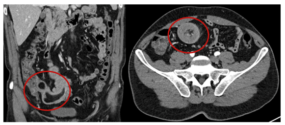
Figure 1 Contrast-enhanced abdominal CT scan. Coronal (left) and axial (right) images reveal a target-like mass with mesenteric fat and vessels within the intussuscepted bowel segment (red circles), compatible with ileo-ileal intussusception.
Following partial spontaneous symptom improvement, elective laparoscopic surgery was planned. Intraoperatively, an ileo-ileal intussusception caused by a polypoid lesion in the terminal ileum was identified. Reduction was achieved, followed by laparoscopic segmental enterectomy and side-to-side isoperistaltic anastomosis using a linear stapler. The specimen was retrieved through an enlarged umbilical incision, confirming a polypoid intraluminal mass with adequate resection margins (Figure 2). Estimated blood loss was 20 mL, and no intraoperative complications occurred.
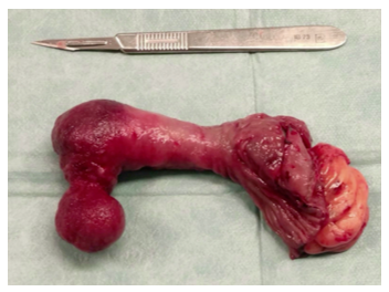
Figure 2 Resected ileal segment with an intraluminal polypoid mass.
Postoperative recovery was uneventful, and the patient was discharged on postoperative day five. Histopathological examination revealed a well-circumscribed ileal lipoma composed of mature adipose tissue, without atypia or malignancy, confirming it as the pathological lead point for the intussusception.
Intussusception in adults is a rare clinical entity, accounting for approximately 1–5% of all cases of intestinal obstruction and less than 5% of all intussusceptions.1,2 In contrast to pediatric intussusception, which is often idiopathic, adult intussusception is associated with an identifiable pathological lead point in up to 70–90% of cases, necessitating a different diagnostic and therapeutic approach.3,4 The small bowel is the most frequently involved segment in adults, with ileo-ileal intussusception representing a particularly uncommon subtype.5
Clinical presentation in adults is typically nonspecific and frequently subacute or chronic, characterized by intermittent abdominal pain, dyspeptic symptoms, nausea, or episodic subocclusive features.6 This vague symptomatology often mimics functional gastrointestinal disorders or more common causes of abdominal pain, contributing to delayed diagnosis, as observed in the present case. Preservation of bowel transit and spontaneous symptom fluctuation, particularly in cases caused by benign lesions, further complicates early recognition.7
Computed tomography is widely regarded as the imaging modality of choice for diagnosing adult intussusception, with reported sensitivities exceeding 80%.8 Typical CT findings include the “target” or “sausage-shaped” mass, bowel-within-bowel configuration, and convergence of mesenteric vessels, which may also provide clues regarding the underlying lead point and associated complications such as ischemia or obstruction.9 In the present case, CT imaging was pivotal in establishing the diagnosis and guiding surgical planning, despite the absence of overt obstructive signs.
Benign lesions, including lipomas, represent a well-recognised cause of small bowel intussusception in adults and are often associated with a more indolent and intermittent clinical course. These lesions may remain asymptomatic until they reach sufficient size to cause luminal compromise or act as a lead point for intussusception, as demonstrated in the present case.
The optimal surgical management of adult intussusception remains a subject of debate, particularly regarding the role of pre-resection reduction. While reduction may be considered in selected cases of small bowel intussusception without suspicion of malignancy, surgical resection is widely recommended to allow definitive diagnosis and exclusion of neoplasia.3 In cases caused by benign small bowel lesions, laparoscopic segmental resection with primary anastomosis has been shown to be safe and effective, offering the benefits of minimally invasive surgery, including reduced postoperative pain and shorter hospital stay.10
In the present case, laparoscopic reduction followed by segmental enterectomy and primary anastomosis allowed definitive treatment, histopathological diagnosis, and exclusion of malignancy, with an uneventful postoperative course. This approach is consistent with current recommendations favouring surgical intervention in adult intussusception to ensure both therapeutic and diagnostic adequacy.
Adult ileo-ileal intussusception is a rare clinical entity with a frequently nonspecific and subacute presentation, which may delay diagnosis. Cross-sectional imaging, particularly computed tomography, plays a pivotal role in preoperative identification of this condition. Surgical management remains the cornerstone of treatment in adult patients, allowing definitive resolution of symptoms and histopathological evaluation of the underlying cause. This case highlights ileal lipoma as an uncommon but important benign lead point for small bowel intussusception and supports laparoscopic resection as a safe and effective therapeutic approach.
None.
The authors declare that there is no conflict of interest.

©2026 Sousa, et al. This is an open access article distributed under the terms of the, which permits unrestricted use, distribution, and build upon your work non-commercially.