International Journal of
eISSN: 2577-8269


Research Article Volume 3 Issue 6
1Emergency Department, ISSSTE Regional Hostipal Monterrey, Graduate from Universidad de Monterrey, México
2Medstudent at intership year, ISSSTE Regional Hostipal Monterrey, Student at Universidad de Monterrey, México
Correspondence: Rolando Martínez Hernández, Emergency Department, ISSSTE Regional Hostipal Monterrey, Graduate from Universidad de Monterrey, Monterrey, Nuevo León, México
Received: August 19, 2019 | Published: November 7, 2019
Citation: Martínez-Hernández R, Montoya-Alarcón P. Relationship between ureterovesical flow and the size of ureteral lites measured with doppler ultrasound. Int J Fam Commun Med. 2019;3(6):263-266. DOI: 10.15406/ijfcm.2019.03.00167
Background: The prevalence of lithiasis in the urinary tract is estimated at 12% of the general population in industrialized countries, of which 2.3% of the population that suffers it with a picture of renal colic. Radiological studies have been the tool used to establish the diagnosis of this pathology. The Gold Standard is computed tomography because it has a sensitivity of 96% - 98% and a specificity of 95% -98%. We currently know that Ultrasound is a first-line tool and with technological advances has allowed us to identify almost all of this pathology, visualizing the renal anatomical alterations, ureters and the difference between the pathological and non-pathological bladder ureter jet.
Material and methods: Analytical study, observations, prospective and correlation. The sample consists of patients older than 18 years who enter the emergency department of the Regional Hospital of Monterrey with the diagnosis of ureteral lithiasis, which has been performed Doppler Ultrasound and Computed Tomography, with entering the department of urology in the study period, and that meet the inclusion criteria in the period from March 2018 to November 2018.
Results: Of the patients found, the majority were women, with 16 samples having an average of 72.7% of the general population. An almost similar relationship was found in the distribution of affected ureters, being mainly the right ureter.
Discussion and conclusion: The use of Doppler ultrasound for the detection and measurement of Ureterovesical flow proved to be an effective method to correlate the size of the stone when it occurs in the uretero. Ultrasound has been placed as a form of current diagnosis for ureteral lithiasis, contrary to what was previously believed because the BMI, age and other factors affect the image at the time of diagnosing the lithium, but new forms have been found to relate this pathology as they are through the degree of hydronephrosis.It was shown that the larger the size of the stone, the lower the ureterovesical flow and vice versa the smaller the size of the stone, the greater the ureterovesical flow.
Keywords: urolithiasis, doppler ultrasound, hydronephrosis, calculi
Ureteral flow is defined as the flow of urine that passes through the ureterovesical orifice into the bladder.1,2 The prevalence of calculus in the urinary tract is estimated at 12% of the general population in industrialized countries, of which 2.3% of the population that suffers from it have a renal colic disorder.2–6 Radiological studies have been the tool used to establish the diagnosis of this pathology, although we know simple abdominal radiography has shown a low sensitivity and specificity to diagnose this pathology,7 we know that the gold standard is computed tomography because since it has a sensitivity of 96% - 98% and a specificity of 95%-98%, nowadays the low-dose computed tomography (1.5-5 millisiverts) is the best way to detect the urinary tract stones.8–10 We know that the CT scan has disadvantages such as the high cost to carry them out and the high exposure to radiation.11,12 We currently know that Ultrasound is a first-line tool for the study of this pathology, since it has a sensitivity of 40%-90% and a specificity of 79%-100%.10–16 But with the technological advances in this ultrasound area has allowed us to identify almost completely said pathology, visualizing renal anatomical alterations, ureters and the difference between the pathological and non-pathological bladder ureter jet.17–24
This protocol aims to study the existence of a correlation between the size of the ureteral calculus and the bladder ureter jet, because as mentioned above, a distortion in this flow has been reported when this pathology occurs.Technological and methodological advances in the field of radiology have allowed us to diagnose ureteral stones with great certainty by means of Computed Tomography as a gold standard, and by ultrasound as a first-line diagnosis, but sometimes there is no means to be able to make the diagnosis in certain units since they do not have CT, we believe that the definitive diagnosis of this disease can be made and its size can be calculated only with simple use of ultrasound and its derived methods such as Doppler.
Analytical study, observations, prospective and correlation. The sample consists of patients over 18 years old who enter through the emergency department of the ISSSTE Monterrey Regional Hospital with the diagnosis of ureteral lithiasis, who have undergone Doppler Ultrasound and Computed Tomography, who enter the urology department during the study period, and that meet the inclusion criteria in the period from March 2018 to November 2018.
Inclusion criteria
Exclusion criteria
Elimination criteria
Doppler Ultrasound Technique: The patient is hydrated orally with 1000 ml of water. The reno-bladder study was performed with a PROSOUND ALPHA6 ultrasound using the convex transducer, using 3.5 MHz. Bladder volume, parenchymal thickness and hydronephrosis grade were evaluated by grayscale. When presenting a bladder with a volume of 110 ml, at least 15 minutes of continuous real time should be observed and the holes in the axial plane were observed and recorded simultaneously by means of color Doppler ultrasound. The red flow system assigned to the flow direction that approaches the transducer. From then on, the measurements of the ureterovesical flow were recorded in centimeters over a second.
Urinary Tract Computed Tomography Technique: Cuts of the images of UroTac with Split bolus technique were taken, to patients who previously had an ultrasound. Once at the tomograph table, with the patient supine, a Scouty is performed an acquisition without contrast from the T12 vertebral level up to 2 cm below the pubic symphysis. With a reconstruction of the cuts of 1-2 mm, taking axial, sagittal and coronal measurements, recording the length of the three cuts mentioned above. Subsequently, it will be saved in the “DICOM Viewer” system and then re-evaluated.
Ethical aspect
The present study is considered low risk since it does not threaten the human or social integrity of patients or their families and is within the margins stipulated in the Regulations of the General Health Law on Health Research, Regulation of Biomedical research by the Mexican Health Code article 1-14, as well as all international laws and agreements such as the Declaration of Helsinki and subsequent amendments. Because it is considered without risk, an informed consent letter is not required. The confidentiality of the information obtained is guaranteed so that work will be carried out with identification of the cases by means of designation of numerical code. The anonymity of patients will be maintained in sessions, congresses, publications and manuscripts that are generated from the study, always respecting their identity.
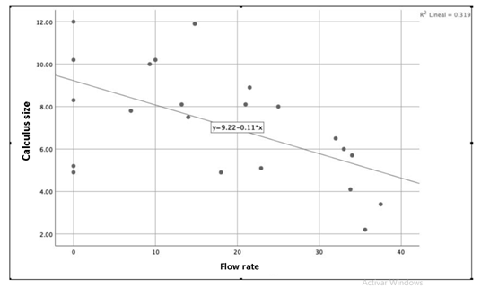
They were 23 patients who met the inclusion criteria. In total, data were collected from 25 patients; however 2 were cataloged with exclusion criteria because they could not have a CT scan. All the patients admitted by the emergency department over 18 years of age were captured, of which the youngest patient was 24 years old and the oldest one was 81 years old, presenting an average of 38 years. Of the patients found, the majority were women (16 patients), having an average of 72.7% of the general population (Table 1). The presence of ureterovesical flow was found by Doppler ultrasound in 17 patients having an average of 77.3%, the rest of the patients could not detect the ureterovesical jet, of which the smallest calculus was 4.9 MM, and the largest it was measured at 12 MM (Table 2). An almost similar relationship was found in the distribution of the affected ureters, mainly the right ureter with a total of 12 patients, presenting a percentage of 54.5% of all registered patients. In this study, no patients were found to have both affected ureters (Table 3). As a final result, using a variable correlation plot by drawing a line from which it goes from the upper left to the lower right. This graph gives us a directly proportional relationship between the size of the calculus and the ureterovesical flow present measured by USG Doppler, of which the data found on the right side expresses a larger calculus which are positioned at the bottom of the graphic, which means that it has a lower bladder urethral flow, as the data on the size of the calculus are shifted to the left of the graph, the bladder urethral flow score is in a higher position which expresses that the smaller the size of the calculus is greater ureterovesical flow will present (Figure 1).

Figure 1 Indicates the inversely proportional relationship between the size of the coastline and the presence of the flow. The larger the size of the coastline, the lower the presence of flow.
Gender |
|||
Frequency |
Percentage |
||
Men |
6 |
27.3 |
27.30% |
Women |
16 |
72.7 |
72.70% |
22 |
100 |
100% |
Table 1 Indicate the frequency by sex
Affected ureter |
||
Frequency |
Porctage |
|
Right |
12 |
54.50% |
Left |
10 |
45.50% |
22 |
100% |
|
Table 2 Indicates the presence of the flow
Affected ureter |
||
Frequency |
Porctage |
|
Right |
12 |
54.50% |
Left |
10 |
45.50% |
22 |
100% |
|
Table 3 Indicates the ureter that was most frequently affected
The use of Doppler Ultrasound for the detection and measurement of Ureterovesical flow proved to be an effective method to correlate the size of the calculus when it occurs in the uretero.The European Urology Association mentions that CT scan has positioned itself as the gold standard for the diagnosis of this pathology since it is the only imaging method capable of fully visualizing the structure of the ureter, while ultrasound has become First-line diagnosis because it is difficult to visualize the middle portion that is where most of the calculi are, because the image is interrupted by the presence of intestinal gas.Ultrasound has been placed as a current diagnostic form for ureteral lithiasis, contrary to what was previously believed because the BMI and age among other factors, affect the image at the time of diagnosing the calculus, but new ways have been found to relate this pathology as they are through the degree of hydronephrosis which does not speak of a total obstruction or almost entirely causing very little flow to pass through the light. Another way in which we relate it is through the pain present in renal colic and the distortion of the ureterovesical flow of the affected and non-pathological ureter.In the present case, all the patients who were admitted to the emergency department due to a probable diagnosis of ureteral lithiasis confirmed their diagnosis, though. The literature tells us that there is no prevalence in age and sex, but it could be found that the pathology occurs more in women than in men and the age at which it occurs most is the 4 and 5 decade of life, being the right ureter the most affected.
We can conclude through this study that urolithiasis is a disease that has no priority in the presentation since it can appear in the same way in men and women and at any age. It was shown that the larger the size of the calculus, the smaller the ureterovesical flow and vice versa the smaller the size of the calculus, the greater the ureterovesical flow.
None.
None.
The author declares there is no conflict of interest.

©2019 Martínez-Hernández, et al. This is an open access article distributed under the terms of the, which permits unrestricted use, distribution, and build upon your work non-commercially.